. 2024 Apr 23;43(4):113976.
doi: 10.1016/j.celrep.2024.113976. Epub 2024 Mar 19.

Stem-loop-induced ribosome queuing in the uORF2/ATF4 overlap fine-tunes stress-induced human ATF4 translational control

Affiliations

Stem-loop-induced ribosome queuing in the uORF2/ATF4 overlap fine-tunes stress-induced human ATF4 translational control

Anna M Smirnova et al. Cell Rep. .

Abstract

Activating transcription factor 4 (ATF4) is a master transcriptional regulator of the integrated stress response, leading cells toward adaptation or death. ATF4's induction under stress was thought to be due to delayed translation reinitiation, where the reinitiation-permissive upstream open reading frame 1 (uORF1) plays a key role. Accumulating evidence challenging this mechanism as the sole source of ATF4 translation control prompted us to investigate additional regulatory routes. We identified a highly conserved stem-loop in the uORF2/ATF4 overlap, immediately preceded by a near-cognate CUG, which introduces another layer of regulation in the form of ribosome queuing. These elements explain how the inhibitory uORF2 can be translated under stress, confirming prior observations but contradicting the original regulatory model. We also identified two highly conserved, potentially modified adenines performing antagonistic roles. Finally, we demonstrated that the canonical ATF4 translation start site is substantially leaky scanned. Thus, ATF4's translational control is more complex than originally described, underpinning its key role in diverse biological processes.

Keywords: ATF4; CP: Molecular biology; integrated stress response; ribosome; ribosome queuing; translation reinitiation; translational control; unfolded protein response.


Conflict of interest statement

Declaration of interests The authors declare no competing interests.

Figures
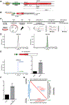
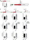
Figure 1.
Figure 1.
Revisiting translational control of human ATF4: Reporters and experimental setup (A) Schematic showing the 5′ end of the mRNA encoding human activating transcription factor 4 (ATF4; NM_182810.2, transcript variant 2), featuring the color-coded Start-stop (St-st) element (yellow), REI-permissive (green) 3-codon uORF1, and inhibitory uORF2 (red) overlapping with the beginning of the main ATF4 ORF, frames 0 and 1. Distances are given in nucleotides. (B) Schematic of the WT CMV-driven ATF4-HA-tagged construct; the HA tag was placed immediately upstream of the ATF4 stop codon. (C) Experimental workflow described in the form of a timeline diagram. PN, Jess protein normalization detection module. (D) All HEK293T cell lysates were subjected to protein separation and immunodetection using the Jess system. The signal, detected in the capillary, is represented as an electropherogram (a single peak of the ATF4-HA tag full-length protein, size of 53 kDa, left) and was automatically quantified (right). Expression of the WT construct under 3 h of Tg stress conditions 8 h post transfection, detected by anti-mouse HA tag antibodies, is shown. (E) Stress-induced upregulation of ATF4-HA protein expression under Tg stress (blue) compared with non-stress conditions (green). Quantified “fold induction” data were plotted (n = 17, right). The differences between experimental groups were tested by a t test. Variables are presented as mean ± SD, and p < 0.05 was considered statistically significant (*p < 0.05, **p < 0.01, ***p < 0.001, ****p < 0.0001). (F) Stress-induced upregulation of the ATF4-HA protein expression from the WT reporter under tunicamycin stress compared with non-stress conditions (set to 1). Quantified “fold induction” data were plotted (n = 3) and analyzed as in (E). (G) Experimental setup illustrating determination of fold change values when comparing (1) each mutant reporter construct vs. the WT construct separately under non-stress (top horizontal arrow) and Tg stress conditions (bottom horizontal arrow) and (2) the fold induction expression of a mutant under stress with the same mutant under non-stress (right vertical arrow) or the WT under stress vs. no stress (blue left vertical arrow). The red diagonal arrow indicates calculated fold induction changes of a given mutant under stress vs. WT under non-stress conditions; i.e., how much each mutant increases or decreases the ~5.2-fold induction of the WT reporter.
Figure 2.
Figure 2.
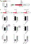
Sequence analysis of the 5′ UTR of the human ATF4 mRNA and ribosome queuing model expanding the mode of ATF4 translational control (A) Schematic of previously unknown, bioinformatically predicted, potential regulatory features within the 5′ UTR of the ATF4 mRNA and beginning of the ATF4 main ORF; point mutations are depicted. For details, please see the main text. (B) Model of the ribosome queuing mechanism under non-stress vs. stress condition employing SL3 and near-cognate CUG as an additional layer of ATF4 translational control. For details, please see the main text. Created with BioRender.
Figure 3.
Figure 3.
SL3 delays the flow of ribosomes in the uORF2/ATF4 overlap (A) Same as Figure 1E for better comparison. (B) Same as Figure 1E except that the SL3 ATF4 mutant constructs depicted at the top of the corresponding panels were subjected to Jess analyses. Relative ATF4-HA protein expression levels were plotted as ratios of values obtained with an indicated mutant construct vs. the WT set to 1 under “non-stress” (row 1) and “Tg stress” (row 2) conditions; “fold-induction” plots (row 3) depict ratios of Tg stress vs. non-stress values obtained with a given mutant construct. The differences between experimental groups were tested by the t test, except wt-SL3Mut−1 Tg stress and d2-SL3Mut−1 non-stress, where a Mann-Whitney test was used (n ≥ 3).
Figure 4.
Figure 4.
SL3 genetically interacts with the upstream CUG near-cognate codon (A) Same as Figure 1E for better comparison. (B) Same as Figure 3B except that the CUG to CUA and SL3Mut−1 ATF4 mutant constructs depicted at the top of the corresponding panels were subjected to Jess analyses (n ≥ 3).
Figure 5.
Figure 5.
Ribosome queuing and substantial leaky scanning at AUG1 of ATF4 contributes to its overall translational control (A–D) Same as in Figure 3B except that the 63 c-Myc tag insertion in frame with ATF4 (A), combined with the SL3Mut−1 mutation (B), in the otherwise WT construct (A and B) vs. the construct lacking uORF2 (C and D), all depicted at the top of the corresponding panels, were subjected to Jess analyses. The differences between experimental groups were tested by a t test, except d2_ins_SL3Mut−1 Tg stress, where a Mann-Whitney test was used (n ≥ 4). (E) The first AUG of the ATF4 ORF is robustly leaky scanned. The electropherograms of the ATF4 construct bearing the 63 c-Myc tag in-frame insertion probed with the anti-c-Myc (left) and anti-HA (right) antibodies are shown. For details, see the main text.
Figure 6.
Figure 6.
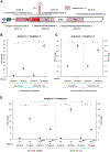
Ribosome protection assay demonstrating that SL3 pauses ribosomes and prompts their queuing under both non-stress and stress conditions (A) Schematic showing the ATF4 mRNA with the sequences of three primer pairs amplifying three different amplicons (A1–A3; also indicated in Figure S6E): 1a-1b (the latter shown in the 3′ to 5′ direction for better illustrative purposes) for the putative queuing fragment and 2a-2b and 3a-3b for two control fragments downstream of SL3 used in the ribosome protection assay. (B) HEK293T cells were cross-linked with formaldehyde (HCHO) and then subjected to the ribosome protection assay as described in STAR Methods. qPCR product levels of the recovered putative queuing region (A1) are normalized to the region immediately downstream of SL3 (A2) as well as to the internal RNA isolation control (SPIKE) with the non-stress values set to 1. Results are representative of three independent replicates, and values are expressed as mean ± SD. Statistical significance was assessed using unpaired two-sided t test (*p < 0.01, **p < 0.001) with Bonferroni correction. (C) HEK293T cells were treated with cycloheximide (a non-cross-linking agent) and then subjected to the ribosome-protection assay as described in STAR Methods. Results from three independent replicates were analyzed as described in (B) with non-stress values set to 1 (*p < 0.01, **p < 0.001). (D) HEK293T cells were transiently transfected with plasmids carrying either WT or SL3-mutated (in SL3Mut−1) ATF4 reporters and treated as described in (B). Results from three independent replicates were analyzed as described in (B) with the WT values set to 1 (*p < 0.01, **p < 0.001, ***p < 0.0001).
Figure 7.
Figure 7.
mRNA methylation further fine tunes ATF4 translation (A) Same as Figure 1E for better comparison. (B) Same as Figure 3B except that A235G (left) and A326G either alone (center) or in combination with SL3Mut−1 (right) ATF4 mutant constructs depicted at the top of the corresponding panels were subjected to Jess analyses (n ≥ 3). (C) mRNA fragments prepared from either HEK293T (left) or HeLa (right) cells carrying the A235G and A326G mutations were subjected to the T3 ligation assay as described in the main text. The normalized percentage of unmodified A235 or A326 bases of the ATF4 mRNA expressed in mock-treated vs. Tg-treated cells, with the latter set to 100%, was plotted as shown (n ≥ 4).

Update of

References

    1. Wek RC, Jiang HY, and Anthony TG (2006). Coping with stress: eIF2 kinases and translational control. Biochem. Soc. Trans 34, 7–11. 10.1042/BST20060007. - DOI - PubMed
    1. Pakos-Zebrucka K, Koryga I, Mnich K, Ljujic M, Samali A, and Gorman AM (2016). The integrated stress response. EMBO Rep. 17, 1374–1395. 10.15252/embr.201642195. - DOI - PMC - PubMed
    1. Denoyelle C, Abou-Rjaily G, Bezrookove V, Verhaegen. M, Johnson TM, Fullen DR, Pointer JN, Gruber SB, Su LD, Nikiforov MA, et al. (2006). Anti-oncogenic role of the endoplasmic reticulum differentially activated by mutations in the MAPK pathway. Nat. Cell Biol 8, 1053–1063. 10.1038/ncb1471. - DOI - PubMed
    1. Donnelly N, Gorman AM, Gupta S, and Samali A (2013). The eIF2αlpha kinases: their structures and functions. Cell. Mol. Life Sci 70, 3493–3511. 10.1007/s00018-012-1252-6. - DOI - PMC - PubMed
    1. Valášek LS (2012). Ribozoomin’ – Translation Initiation from the Perspective of the Ribosome-bound Eukaryotic Initiation Factors (eIFs). Curr. Protein Pept. Sci 13, 305–330. - PMC - PubMed

Publication types

Substances

LinkOut - more resources