. 2024 Mar 26;43(3):113924.
doi: 10.1016/j.celrep.2024.113924. Epub 2024 Mar 18.

SUMOylation controls Hu antigen R posttranscriptional activity in liver cancer

Affiliations

SUMOylation controls Hu antigen R posttranscriptional activity in liver cancer

Sofia Lachiondo-Ortega et al. Cell Rep. .

Abstract

The posttranslational modification of proteins critically influences many biological processes and is a key mechanism that regulates the function of the RNA-binding protein Hu antigen R (HuR), a hub in liver cancer. Here, we show that HuR is SUMOylated in the tumor sections of patients with hepatocellular carcinoma in contrast to the surrounding tissue, as well as in human cell line and mouse models of the disease. SUMOylation of HuR promotes major cancer hallmarks, namely proliferation and invasion, whereas the absence of HuR SUMOylation results in a senescent phenotype with dysfunctional mitochondria and endoplasmic reticulum. Mechanistically, SUMOylation induces a structural rearrangement of the RNA recognition motifs that modulates HuR binding affinity to its target RNAs, further modifying the transcriptomic profile toward hepatic tumor progression. Overall, SUMOylation constitutes a mechanism of HuR regulation that could be potentially exploited as a therapeutic strategy for liver cancer.

Keywords: CP: Cancer; CP: Molecular biology; ELAVL1; HCC; PTMs; SUMO; senescence.


Conflict of interest statement

Declaration of interests The authors declare no competing interests.

Figures
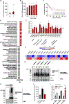
Figure 1.
Figure 1.. HuR SUMOylation is increased in human HCC
(A and B) ELAVL1 mRNA expression levels (A) in the T (n = 368) and NT (n = 50) tissue of patients with HCC, and (B) at the different stages (I–IV) of the disease. (C) Survival curve of liver cancer patients with high (n = 180) and low (n = 190) ELAVL1 mRNA expression levels. (D) Enriched molecular processes after performing a GSEA according to ELAVL1 mRNA expression in HCC patients. (E) mRNA expression levels of the main components of the SUMO pathway in the T (n = 370) of HCC patients relative to the NT tissue (n = 50). (F) Heatmap representing R2 values obtained from Pearson correlation studies on ELAVL1 mRNA expression and the canonical SUMOylation pathway members in paired T and ST liver samples from a cohort of patients with HCC (n = 86). (G–J) Enrichment, identification, and quantification of the SUMO-interacting proteome from non-tumoral and tumoral (G) human (n = 5) and (H) mouse liver tissue (n = 10) and (I and J) human cell line protein extracts by means of GST-SUBEs pull-down technology in combination with (G and H) western blotting and (I and J) LC-MS/MS. Data in (A–E) were obtained from TCGA Research Network. Data in (A), (B), (E), (I), and (J) are presented as the mean ± SD of at least three biological replicates within one representative experiment. *p < 0.05, **p < 0.01, and ***p < 0.001, two-tailed t test vs. NT (A, B, and E), THLE-2 (I), or GST (J). If not indicated otherwise, the differences were not significant. In (H), western blots are representative of at least three biological replicates. See also Figure S1.
Figure 2.
Figure 2.. HuR is mainly modified by SUMO2/3 at lysines in position 120 and 182 inducing a structural rearrangement of the RRM 1 and 2 so as to modulate its intrinsic RNA-binding ability
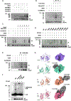
(A–D) Modified V5-HuR protein enrichment after transient transfection of plasmids expressing the different (A) SUMO, (B) PIAS, and (C) SENP isoforms as well as (D) all the lysine-to-arginine HuR mutants contained in the RRM1–2 domains, and subsequent nickel-histidine affinity purification, relative to total V5-HuR protein expression levels in the mouse liver progenitor MLP-29 cell line. (E) Modified V5-HuR protein enrichment after co-transfection of plasmids expressing UBC9, SUMO2/3, and WT HuR or the SUMOylation double mutant, and subsequent nickel-histidine affinity purification, relative to total V5-HuR protein expression levels in the human hepatoma HuH-7 cell line. (F) SUMOylated HuR enrichment after transient transfection of WT HuR and the different SUMOylation mutants and subsequent GST-SUBEs protein pull-down, relative to total V5-HuR protein expression levels in the HuH-7 cell line. (G) Ribbon and surface representations of the HuR RRM1–2 protein construct and the different single and doubly SUMOylated species (K120, K182, and K120/182). RRM2 domains were kept in a fixed position in all representations to show them in the same orientation. The rotation angles of RRM1 relative to RRM2 (using the WT conformation as a reference) are depicted on the left of SUMOylated HuR models, and SUMOylated sites are highlighted with dashed circles on the ribbon structures. Models correspond to the structures with the lowest root-mean-square deviation (RMSD) with respect to the last 10-ns average coordinates in each MD trajectory. In (A–F), western blots are representative of at least three biological replicates within one representative experiment. In (D) and (E), the entire blot image was digitally processed to eliminate irrelevant lanes. See also Figures S2–S6 and Table S3.
Figure 3.
Figure 3.. SUMOylation of HuR promotes the main cancer hallmarks and avoids palbociclib-induced senescence in human hepatoma cells
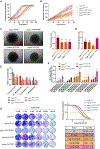
(A and B) Cell (A) proliferation and (B) scratch-wound healing process of HuH-7 cell lines stably expressing WT HuR and the different SUMOylation mutants analyzed in the IncuCyte system. (C) Representative pictures of 3D spheroids from HuH-7 cell lines stably expressing WT HuR and the SUMOylation mutant species embedded on a collagen type I matrix for 48 h, and quantification of the relative invasive area. (D) Percentage of apoptosis detected in the WT HuR and the indicated SUMOylation mutant HuH-7 cell variants by flow-cytometry analysis after FITC-annexin V and viability stainings, relative to STS-treated HuH-7 cells. (E) Quantification of cell proliferation in the HuH-7 cell lines stably expressing WT HuR and the different SUMOylation mutants after an acute 3-day treatment with a range of palbociclib concentrations analyzed by crystal violet staining. (F) Quantification of relative senescence in the HuH-7 cell lines stably expressing the WT and the K120/182R, K120R, K182R HuR mutant species after chronic 2-week treatment with a range of palbociclib concentrations analyzed by β-galactosidase staining. (G) Crystal violet staining of colonies from HuH-7 cell lines stably expressing WT HuR and the SUMOylation mutants treated with the indicated doses of palbociclib for 10 days, and generation of dose-response curves for calculation of IC50 values. In (A–F), data are presented as the mean ± SD of at least three biological replicates within one representative experiment. *p < 0.05, **p < 0.01, and ***p < 0.001, two-tailed t test vs. HuH-7WT HuR. If not indicated otherwise, the differences were not significant. In (C) and (G), images are representative of at least three biological replicates within one representative experiment. See also Figures S7–S9.
Figure 4.
Figure 4.. SUMOylated HuR evades palbociclib-mediated senescence by increasing HuR and global SUMOylation levels in human hepatoma cells
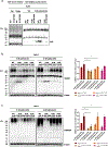
(A) Enrichment of SUMOylated HuR in the HuH-7WT HuR and HuH-7HuR [K120/182R] cells treated with 5 nM palbociclib for 2 weeks by means of protein pull-down with GST control and SUBEs in combination with western blotting analysis. (B and C) (B) SUMO1 and (C) SUMO2/3 protein expression levels and quantification in the HuH-7cell lines stably expressing WT HuR and the K120/182R, K120R, K182R HuR SUMOylation mutant species treated with 5 nM palbociclib for 2 weeks. In (A–C), western blots are representative of at least three biological replicates within one representative experiment. In (B) and (C), data are presented as the mean ± SD of at least three biological replicates within one representative experiment. *p < 0.05 and **p < 0.01, two-tailed t test vs. 0 nM palbociclib or HuH-7WT HuR. If not indicated otherwise, the differences were not significant.
Figure 5.
Figure 5.. Absence of HuR SUMOylation generates a senescent phenotype with compromised mitochondria and ER in human hepatoma cells
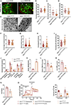
(A–D) (A) Representative images of TOM20 immunofluorescent detection and estimation of the (B) mitochondrial content and mitochondrial network (C) AR and (D) F in HuH-7WT HuR and HuH-7HuR [K120/182R] cells. (E–I) (E) Representative TEM images of the mitochondrial and ER ultrastructure and determination of the (F) mitochondrial area relative to cell area, (G) mitochondrial AR, (H) ER area relative to cell area, and (I) ER area relative to mitochondrial area in HuH-7WT HuR and HuH-7HuR [K120/182R] cells. (J) Seahorse-based quantification of mitochondrial respiration parameters in the HuH-7WT HuR and HuH-7HuR [K120/182R] cells. (K–N) Quantification of (K) MitoSOX red mitochondrial superoxide indicator, (L) TMRE staining for the assessment of mitochondrial membrane potential, (M) total ATP content, and (N) NAD+/NADH levels in the HuH-7WT HuR and HuH-7HuR [K120/182R] cells. (O) Representative curves and quantification of cytosolic Ca2+ levels with Fura-2AM fluorescent probe labeling in the HuH-7WT HuR and HuH-7HuR [K120/182R] cells after stimulating Ca2+ release from the ER with thapsigargin and ATP, and addition of extracellular Ca2+. In (A) and (E), images are representative of at least three biological replicates within one representative experiment. Scale bars represent 5 μm in (A) and 1 μm in (E). In (B–D) and (F–O), data are presented as the mean ± SD of at least three biological replicates within one representative experiment. *p < 0.05, **p < 0.01, ***p < 0.001, and ****p < 0.0001, two-tailed t test. If not indicated otherwise, the differences were not significant. See also Figure S10.
Figure 6.
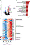
Figure 6.. SUMOylation modulates HuR RNA-binding affinity to confer a tumoral phenotype in human hepatoma cells
(A) Volcano plot representing all of the identified RNAs bound to V5-tagged HuR after RIP-seq in the HuH-7WT HuR and HuH-7HuR [K120/182R] cells. Blue and red indicate the transcripts showing more than a 1.5-fold change enrichment and padj < 0.05 in the WT and K120/182R HuR expressing HuH-7 cells, respectively, after performing a background correction with the corresponding input fraction. Black represents RNAs showing no statistically significant enrichment. (B) List of the top most statistically significant canonical pathways identified by IPA software and associated with the enrichment of transcripts most significantly bound to V5-tagged WT and K120/182R HuR in the HuH-7 cell line. (C) Heatmap representing the significantly enriched RNAs related with mitochondrial membrane potential and permeability, cell-cycle control DDR, Ubl-PTMs, cholesterol synthesis via mevalonate, ER stress pathway, and Ca2+ signaling in the V5-bound and input fractions of HuH-7WT HuR and HuH-7HuR [K120/182R] cells after performing RIP-seq.
Figure 7.
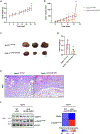
Figure 7.. Xenograft tumors from human hepatoma cells lacking HuR SUMOylation sites show delayed growth and expression of senescence protein markers in mice
(A and B) (A) Body weight and (B) tumor volume evolution over time after the subcutaneous injection of HuH-7WT HuR and HuH-7HuR [K120/182R] cells in each flank of NSG mice (n = 6). (C–F) (C) Macroscopic appearance, (D) weight, (E) H&E staining, and (F) CCND1 and p-H2AXSer139 protein expression levels and quantification relative to β-actin in the human xenograft tumors derived from HuH-7WT HuR and HuH-7HuR [K120/182R] cells 4 weeks after implantation in NSG mice (n = 6). In (C), (E), and (F), images are representative of at least three biological replicates within one representative experiment. Scale bars represent 1 cm in (C) and 200 μm in (E). In (A), (B), (D), and (F), data are presented as the mean ± SD of at least three biological replicates within one representative experiment. *p < 0.05, **p < 0.01, and ***p < 0.001, two-tailed t test. If not indicated otherwise, the differences were not significant.

References

    1. Craig AJ, von Felden J, Garcia-Lezana T, Sarcognato S, and Villanueva A (2020). Tumour evolution in hepatocellular carcinoma. Nat. Rev. Gastroenterol. Hepatol. 17, 139–152. 10.1038/s41575-019-0229-4. - DOI - PubMed
    1. Faivre S, Rimassa L, and Finn RS (2020). Molecular therapies for HCC: Looking outside the box. J. Hepatol. 72, 342–352. 10.1016/j.jhep.2019.09.010. - DOI - PubMed
    1. Seeler J-S, and Dejean A (2017). SUMO and the robustness of cancer. Nat. Rev. Cancer 17, 184–197. 10.1038/nrc.2016.143. - DOI - PubMed
    1. Vertegaal ACO (2022). Signalling mechanisms and cellular functions of SUMO. Nat. Rev. Mol. Cell Biol. 23, 715–731. 10.1038/s41580-022-00500-y. - DOI - PubMed
    1. Bouchard D, Wang W, Yang W-C, He S, Garcia A, and Matunis MJ (2021). SUMO paralogue–specific functions revealed through systematic analysis of human knockout cell lines and gene expression data. Mol. Biol. Cell 32, 1849–1866. 10.1091/mbc.E21-01-0031. - DOI - PMC - PubMed

Publication types