. 2024 Apr 3;16(13):16861-16879.
doi: 10.1021/acsami.4c01334. Epub 2024 Mar 20.

Co-immobilization of Ciprofloxacin and Chlorhexidine as a Broad-Spectrum Antimicrobial Dual-Drug Coating for Poly(vinyl chloride) (PVC)-Based Endotracheal Tubes

Affiliations

Co-immobilization of Ciprofloxacin and Chlorhexidine as a Broad-Spectrum Antimicrobial Dual-Drug Coating for Poly(vinyl chloride) (PVC)-Based Endotracheal Tubes

Diana Filipa Alves et al. ACS Appl Mater Interfaces. .

Erratum in

Abstract

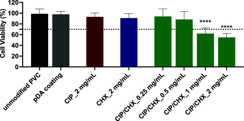
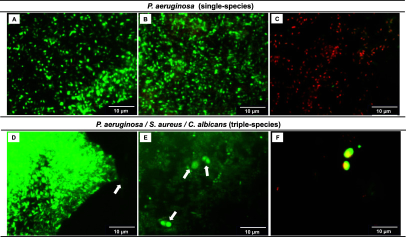
The endotracheal tube (ETT) affords support for intubated patients, but the increasing incidence of ventilator-associated pneumonia (VAP) is jeopardizing its application. ETT surfaces promote (poly)microbial colonization and biofilm formation, with a heavy burden for VAP. Devising safe, broad-spectrum antimicrobial materials to tackle the ETT bioburden is needful. Herein, we immobilized ciprofloxacin (CIP) and/or chlorhexidine (CHX), through polydopamine (pDA)-based functionalization, onto poly(vinyl chloride) (PVC) surfaces. These surfaces were characterized regarding physicochemical properties and challenged with single and polymicrobial cultures of VAP-relevant bacteria (Pseudomonas aeruginosa, Acinetobacter baumannii, Klebsiella pneumoniae, Staphylococcus aureus, Staphylococcus epidermidis) and fungi (Candida albicans). The coatings imparted PVC surfaces with a homogeneous morphology, varied wettability, and low roughness. The antimicrobial immobilization via pDA chemistry was still evidenced by infrared spectroscopy. Coated surfaces exhibited sustained CIP/CHX release, retaining prolonged (10 days) activity. CIP/CHX-coated surfaces evidencing no A549 lung cell toxicity displayed better antibiofilm outcomes than CIP or CHX coatings, preventing bacterial attachment by 4.1-7.2 Log10 CFU/mL and modestly distressingC. albicans. Their antibiofilm effectiveness was endured toward polymicrobial consortia, substantially inhibiting the adhesion of the bacterial populations (up to 8 Log10 CFU/mL) within the consortia in dual- and even inP. aeruginosa/S. aureus/C. albicans triple-species biofilms while affecting fungal adhesion by 2.7 Log10 CFU/mL (dual consortia) and 1 Log10 CFU/mL (triple consortia). The potential of the dual-drug coating strategy in preventing triple-species adhesion and impairing bacterial viability was still strengthened by live/dead microscopy. The pDA-assisted CIP/CHX co-immobilization holds a safe and robust broad-spectrum antimicrobial coating strategy for PVC-ETTs, with the promise laying in reducing VAP incidence.

Keywords: co-immobilization; dual-drug coating; endotracheal tube; polymicrobial biofilm; ventilator-associated pneumonia.


Conflict of interest statement

The authors declare no competing financial interest.

Figures

Scheme 1
Scheme 1. Scheme of the Methodology Used in the PVC Functionalization with CIP, CHX and CHX-CIP
(A) PVC immersion in an alkaline solution (pH 8.5) of dopamine (at 2 mg/mL) resulted in its polymerization and subsequent deposition of an adhesive film called polydopamine (pDA). (B) Antimicrobial immobilization was performed by immersion of PVC on dopamine (at 2 mg/mL) combined with each or both compounds (at 0.25 mg/L to 2 mg/mL), resulting in CIP-, CHX-, or CIP/CHX-modified PVC surfaces upon dopamine polymerization and antimicrobial(s) immobilization. All pDA, CIP, and CHX chemical structures were designed using the ChemDraw Professional for Mac (version 16.0.1.4, CambridgeSoft).
Figure 1
Figure 1
Surface morphology obtained by SEM: (A) unmodified PVC surface, (B) pDA-coated surface, (C) PVC surface modified with CHX at 0.5 mg/mL or with (D) CHX at 2 mg/mL, (E) CIP at 0.5 mg/mL, (F) CIP at 2 mg/mL, (G) CIP/CHX at 0.5 mg/mL, and (H) CIP/CHX at 2 mg/mL. The scale bars in the insets and respective images indicate 2 and 10 μm, respectively.
Figure 2
Figure 2
Surface roughness and wettability. (A) Average roughness and (B) static water contact angles of PVC surfaces before and after immobilization with CIP, CHX, or CIP/CHX at concentrations of 0.5 and 2 mg/mL. Unmodified PVC and pDA-coated surfaces were used for comparison. The results are shown as mean ± SDs. (****) p < 0.0001, (**) p < 0.01,vs unmodified PVC, one-way ANOVA, Tukey'′s multiple comparison test.
Figure 3
Figure 3
ATR-FTIR spectra of PVC surfaces before and after immobilization with CIP, CHX, or CIP/CHX at concentrations of 0.5 and 2 mg/mL.
Figure 4
Figure 4
Antimicrobials released from coated surfaces. (A) Cumulative release of CIP and (B) of CHX from PVC surfaces functionalized with each compound at 0.5 and 2 mg/mL. Panel (C) depicts the outcomes from the contact antimicrobial activity and the release of CIP or CHX-immobilized alone, and CIP/CHX co-immobilized on PVC surfaces at 0.5 and 2 mg/mL, at days 4 and 10 against K. pneumoniae. Bacterial growth after 24 h contact with modified surfaces is indicated at the bottom right of each image, where “+” is indicative of visible bacterial growth and “–” means no visible growth observed. The release of antimicrobials on solid agar was evaluated by the presence or absence of an inhibition zone. The average length of inhibition zones was determined using ImageJ and is presented in mm. pDA-coated surfaces were used for comparison.
Figure 5
Figure 5
Cell cytotoxicity. Viability of A549 lung epithelial cells after indirect contact with PVC surfaces functionalized with CIP or CHX at the highest concentration tested (2 mg/mL) and with CIP/CHX at 2 mg/mL and lower concentrations (1 mg/mL, 0.5 mg/mL, and 0.25 mg/mL). Unmodified PVC and pDA-coated surfaces were used for comparison. A threshold for cell toxicity of 70% viability was used, which is indicated by a dotted line. The results are shown as mean ± SDs. (****) p < 0.0001, vs unmodified PVC, one-way ANOVA, Tukey’s multiple comparison test.
Figure 6
Figure 6
Efficacy against single-species biofilms. PVC surfaces modified with CIP and CHX were challenged with single-species biofilms formed byP. aeruginosa (A),A. baumannii (B),K. pneumoniae (C),S. aureus (D),S. epidermidis (E), andC. albicans (F). Unmodified PVC and pDA-coated surfaces were used for comparison. The threshold value marked by the dotted line represents the detection limit of CFU counting. The results are shown as mean ± SDs. (****) p < 0.0001, (*) p < 0.05 vs unmodified PVC, one-way ANOVA, Tukey’s multiple comparison test. (****) p < 0.0001, vs unmodified PVC, one-way ANOVA, Tukey’s multiple comparison test.
Figure 7
Figure 7
Efficacy against dual-species biofilms. Effect of PVC surfaces before and after pDA-based coating and immobilization of CIP and/or CHX against dual-species biofilms ofP. aeruginosa andA. baumannii (A),P. aeruginosa andK. pneumoniae (B),P. aeruginosa andS. aureus (C),P. aeruginosa andS. epidermidis (D), andP. aeruginosa andC. albicans (E). Unmodified PVC and pDA-coated surfaces were used for comparison. The threshold value marked by the dotted line represents the detection limit of CFU counting. The results are shown as mean ± SDs. (****) p < 0.0001 vs P. aeruginosa growth on unmodified PVC and (##) p < 0.01 or (####) p < 0.0001 vs other than P. aeruginosa species growth on unmodified PVC, two-way ANOVA, Tukey′s multiple comparison test.
Figure 8
Figure 8
Efficacy against triple-species biofilms. Effect of PVC surfaces before and after pDA-based coating and immobilization of CIP and/or CHX against triple-species biofilms ofP. aeruginosa,S. aureus, andC. albicans. Unmodified PVC and pDA-coated surfaces were used for comparison. The threshold value marked by the dotted line represents the detection limit of CFU counting. The results are shown as mean ± SDs. (****) p < 0.0001 vs P. aeruginosa growth on unmodified PVC; (####) p < 0.0001 vs S. aureus species growth on unmodified PVC; and () p < 0.05, (∂∂) p < 0.01, and (∂∂∂∂) p < 0.0001 vs C. albicans species growth on unmodified PVC two-way ANOVA, Tukey′s multiple comparison test.
Figure 9
Figure 9
Biofilm viability. On the top row,P. aeruginosa single-species biofilms developed on uncoated PVC (A), pDA-coated PVC (B), and CIP/CHX-modified surfaces (C). On the bottom row,P. aeruginosa,S. aureus, andC. albicans triple-species biofilms developed on uncoated PVC (D), pDA-coated PVC (E), and CIP/CHX-modified surfaces (F). Biofilms were stained with LIVE/DEAD BacLight Bacterial Viability system, where live cells were stained with the green fluorescent dye SYTO 9, and compromised/damaged cells were labeled in red by the PI stain.

References

    1. Haas C. F.; Eakin R. M.; Konkle M. A.; Blank R. Endotracheal Tubes: Old and New. Respir. Care 2014, 59 (6), 933–952. 10.4187/respcare.02868. - DOI - PubMed
    1. Ippolito M.; Misseri G.; Catalisano G.; Marino C.; Ingoglia G.; Alessi M.; Consiglio E.; Gregoretti C.; Giarratano A.; Cortegiani A. Ventilator-Associated Pneumonia in Patients with Covid-19: A Systematic Review and Meta-Analysis. Antibiotics 2021, 10 (5), 54510.3390/antibiotics10050545. - DOI - PMC - PubMed
    1. Pinciroli R.; Mietto C.; Berra L. Respiratory Therapy Device Modifications to Prevent Ventilator-Associated Pneumonia. Curr. Opin. Infect. Dis 2013, 26 (2), 175–183. 10.1097/QCO.0b013e32835d3349. - DOI - PubMed
    1. Rouzé A.; Martin-Loeches I.; Nseir S. Airway Devices in Ventilator-Associated Pneumonia Pathogenesis and Prevention. Clin. Chest Med. 2018, 39 (4), 775–783. 10.1016/j.ccm.2018.08.001. - DOI - PubMed
    1. Vandecandelaere I.; Coenye T. Microbial Composition and Antibiotic Resistance of Biofilms Recovered from Endotracheal Tubes of Mechanically Ventilated Patients. Adv. Exp. Med. Biol. 2015, 830, 137–155. 10.1007/978-3-319-11038-7_9. - DOI - PubMed

LinkOut - more resources