Comparative Study
. 2025 May 6;50(5):429-436.
doi: 10.1136/rapm-2024-105417.

Fluoroscopy-guided high-intensity focused ultrasound ablation of the lumbar medial branch nerves: dose escalation study and comparison with radiofrequency ablation in a porcine model

Affiliations
Comparative Study

Fluoroscopy-guided high-intensity focused ultrasound ablation of the lumbar medial branch nerves: dose escalation study and comparison with radiofrequency ablation in a porcine model

Michael Gofeld et al. Reg Anesth Pain Med. .

Abstract

Background: Radiofrequency ablation (RFA) is a common method for alleviating chronic back pain by targeting and ablating of facet joint sensory nerves. High-intensity focused ultrasound (HIFU) is an emerging, non-invasive, image-guided technology capable of providing thermal tissue ablation. While HIFU shows promise as a potentially superior option for ablating sensory nerves, its efficacy needs validation and comparison with existing methods.

Methods: Nine adult pigs underwent fluoroscopy-guided HIFU ablation of eight lumbar medial branch nerves, with varying acoustic energy levels: 1000 (N=3), 1500 (N=3), or 2000 (N=3) joules (J). An additional three animals underwent standard RFA (two 90 s long lesions at 80°C) of the same eight nerves. Following 2 days of neurobehavioral observation, all 12 animals were sacrificed. The targeted tissue was excised and subjected to macropathology and micropathology, with a primary focus on the medial branch nerves.

Results: The percentage of ablated nerves with HIFU was 71%, 86%, and 96% for 1000 J, 1500 J, and 2000 J, respectively. In contrast, RFA achieved a 50% ablation rate. No significant adverse events occurred during the procedure or follow-up period.

Conclusions: These findings suggest that HIFU may be more effective than RFA in inducing thermal necrosis of the nerve.

Keywords: Animal Experimentation; Back Pain; Methods; Pain Management; TECHNOLOGY.


Conflict of interest statement

Competing interests: AH and RA are the founders of FUSMobile and hold ordinary options in the company. J-FA is a member of FUSMobile’s scientific advisory board and holds ordinary options in the company. SL and EM hold shares and ordinary options in the company. SM holds ordinary options in the company. MG holds ordinary options in the company and is a consultant to the company. TT has no interests to declare.

Figures

Figure 1
Figure 1. (A) Lumbar spondylosis: the model demonstrates facet degeneration in red. The proximal medial branch nerve supplies sensory innervation to the ipsilateral facet joint just inferior to the neural foramen (black arrow) while the distal medial branch nerve innervates the facet joint more inferiorly (black arrowhead). (B) Disposable coupling gel pad. (C) Cradle. (D) Neurolyzer positioner, which includes the therapeutic transducer inside the cradle along with the aiming system; central rings for target localization (black arrow) and adjacent top (white) and side (blue) camera boxes for assisting with right-to-left and superior-inferior aiming. (E) AP fluoroscopic image confirms HIFU targeting. The pink dot overlays the target location of the medial branch nerve along the lateral aspect of the pedicle inside the two concentric green rings. (F) Lateral fluoroscopic image confirms that the red lines from the margins of the HIFU transducer converge at the simulated target location. The green lines delineate the depth of the treatment envelope. (G) Fluoroscopic imaging of lateral view and (H) AP views confirmed cannulae positions prior to RFA. AP, anterior–posterior; HIFU, high-intensity focused ultrasound; RFA, radiofrequency ablation.
Figure 2
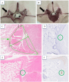
Figure 2. Macropathology demonstrates lesion formation at the proximal medial branch nerve location following (A) HIFU and (B) RFA (black arrows). Histology demonstrates the HIFU lesion on H&E stain (C) creates a large area of necrosis along the bony surface (outlined in green) with the proximal medial branch nerve located along the bony surface within the area of necrosis (green arrow) confirmed using MBP stain (small green circle, (D). The RFA lesion on H&E stain (E) reveals necrosis in the soft tissues near the bone with the proximal medial branch nerve (green circle) located outside and medial to the area of necrosis confirmed using MBP stain (small green circle, (F). HIFU, high-intensity focused ultrasound; MBP, myelin basic protein; RFA, radiofrequency ablation.
Figure 3
Figure 3. (A) Hemorrhage, possibly due to mechanical trauma in the paraspinal muscles (black arrow) following radiofrequency ablation (RFA). (B) Macropathology showing an area of RFA ablation near the bone (black rectangle) and (C) its corresponding micro pathology showing a viable medial branch nerve (green arrowhead) outside RFA-ablated tissue (outlined in green). (D) Macropathology of an area of high-intensity focused ultrasound (HIFU) ablation along the bony surface (black rectangle) and (E) its corresponding micropathology showing an ablated medial branch nerve (green arrow) within HIFU-ablated tissue (outlined in green).
Figure 4
Figure 4. Comparison of lesion width on histology (blue boxes) for RFA and various HIFU energy levels compared with k-wave simulated HIFU lesions (green boxes). One simulation was performed per porcine lumbar spinal level per side, mimicking the GLP study. GLP, Good Laboratory Practice; HIFU, high-intensity focused ultrasound; RFA, radiofrequency ablation.

Comment in

References

    1. Deyo RA, Phillips WR. Low back pain. A primary care challenge. Spine (Phila Pa 1976) 1996;21:2826–32. doi: 10.1097/00007632-199612150-00003. - DOI - PubMed
    1. Cohen SP, Bhaskar A, Bhatia A, et al. Consensus practice guidelines on interventions for lumbar facet joint pain from a Multispecialty, international working group. Reg Anesth Pain Med. 2020;45:424–67. doi: 10.1136/rapm-2019-101243. - DOI - PMC - PubMed
    1. Cohen SP, Raja SN. Pathogenesis, diagnosis, and treatment of lumbar Zygapophysial (facet) joint pain. Anesthesiology. 2007;106:591–614. doi: 10.1097/00000542-200703000-00024. - DOI - PubMed
    1. Fitzgibbon DR, Posner KL, Domino KB, et al. Chronic pain management: American society of Anesthesiologists closed claims project. Anesthesiology. 2004;100:98–105. doi: 10.1097/00000542-200401000-00018. - DOI - PubMed
    1. Barbieri M, Bellini M. Radiofrequency Neurotomy for the treatment of chronic pain: interference with Implantable medical devices. Anaesthesiol Intensive Ther. 2014;46:162–5. doi: 10.5603/AIT.2014.0029. - DOI - PubMed

Publication types

MeSH terms

LinkOut - more resources