. 2024 Mar 19;12(3):e008402.
doi: 10.1136/jitc-2023-008402.

Circular RNA as a source of neoantigens for cancer vaccines

Affiliations

Circular RNA as a source of neoantigens for cancer vaccines

Yi Ren et al. J Immunother Cancer. .

Abstract

Background: The effectiveness of somatic neoantigen-based immunotherapy is often hindered by the limited number of mutations in tumors with low to moderate mutation burden. Focusing on microsatellite-stable colorectal cancer (CRC), this study investigates the potential of tumor-associated circular RNAs (circRNAs) as an alternative source of neoepitopes in CRC.

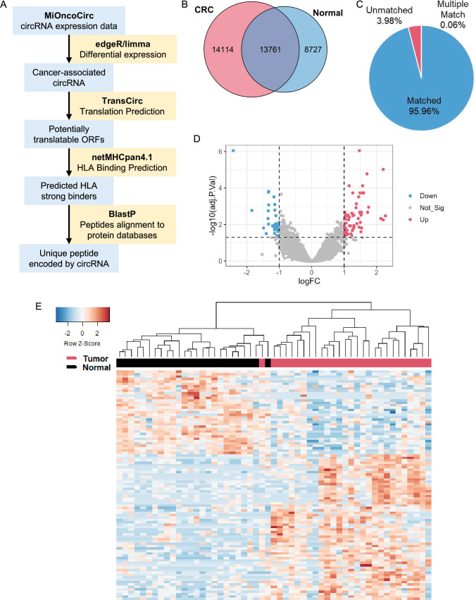
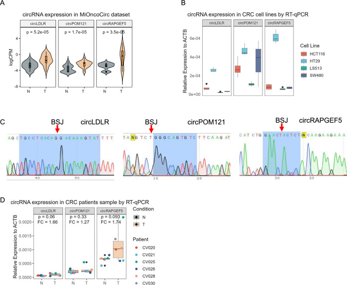
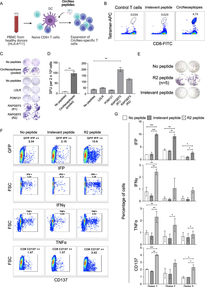
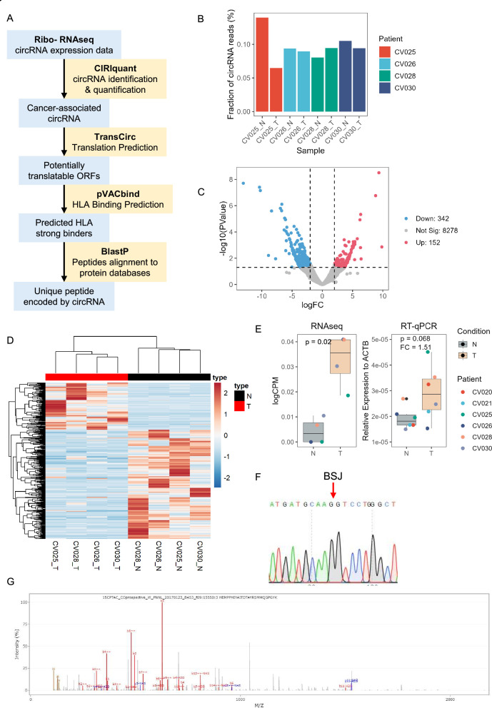
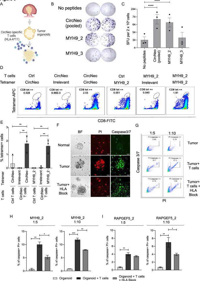
Methods: Tumor-associated circRNAs in CRC were identified using the MiOncoCirc database and ribo-depletion RNA sequencing of paired clinical normal and tumor samples. Candidate circRNA expression was validated by quantitative real-time PCR (RT-qPCR) using divergent primers. TransCirc database was used for translation prediction. Human leukocyte antigen binding affinity of open reading frames from potentially translatable circRNA was predicted using pVACtools. Strong binders from messenger RNA-encoded proteins were excluded using BlastP. The immunogenicity of the candidate antigens was functionally validated through stimulation of naïve CD8+ T cells against the predicted neoepitopes and subsequent analysis of the T cells through enzyme-linked immunospot (ELISpot) assay, intracellular cytokine staining (ICS) and granzyme B (GZMB) reporter. The cytotoxicity of T cells trained with antigen peptides was further tested using patient-derived organoids.

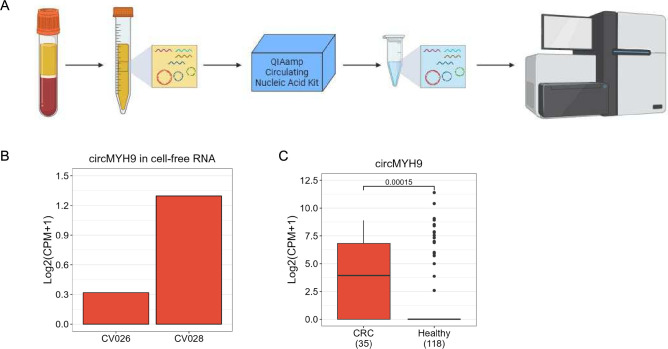
Results: We identified a neoepitope from circRAPGEF5 that is upregulated in CRC tumor samples from MiOncoCirc database, and two neoepitopes from circMYH9, which is upregulated across various tumor samples from our matched clinical samples. The translation potential of candidate peptides was supported by Clinical Proteomic Tumor Analysis Consortium database using PepQuery. The candidate peptides elicited antigen-specific T cells response and expansion, evidenced by various assays including ELISpot, ICS and GZMB reporter. Furthermore, T cells trained with circMYH9 peptides were able to specifically target and eliminate tumor-derived organoids but not match normal organoids. This observation underscores the potential of circRNAs as a source of immunogenic neoantigens. Lastly, circMYH9 was enriched in the liquid biopsies of patients with CRC, thus enabling a detection-to-vaccination treatment strategy for patients with CRC.

Conclusions: Our findings underscore the feasibility of tumor-associated circRNAs as an alternative source of neoantigens for cancer vaccines targeting tumors with moderate mutation levels.

Keywords: circular RNA; colorectal cancer; neoantigen.


Conflict of interest statement

Competing interests: No, there are no competing interests.

Figures

Figure 1
Figure 1
circRNA expression in MiOncoCirc CRC samples. (A) circRNA neoantigen prediction pipeline. circRNA expression data were acquired from MiOncoCirc and differentially expressed circRNA were analyzed using R package edgeR. circRNA were then mapped to TransCirc database for ORF and translation prediction. HLA-A*11:01 binding affinity of potentially translatable ORFs were predicted using netMHCpan4.1 and shortlisted strong binders were aligned to NCBI protein database using BlastP to screen for novel peptides. (B) circRNA detected in CRC and normal samples. Only overlapped circRNA were kept for downstream analyses (C) fraction of circRNA mapped to TransCirc. The genomic locations of circRNA were used as identifiers to map to TransCirc database. Only uniquely matched circRNA were kept. (D) Volcano plot of differentially expressed circRNA. (E) Expression of differentially expressed circRNA in tumor and normal samples. circRNA, circular RNA; CRC, colorectal cancer; HLA, human leukocyte antigen; ORF, open reading frame.
Figure 2
Figure 2
Shortlisted circRNA expression in CRC cell lines and clinical samples. (A) Expression of shortlisted circRNA in CRC (n=28) and normal (n=25) samples in MiOncoCirc data set. (B) Expression of shortlisted circRNA in CRC cell lines by RT-qPCR using divergent primers (n=4 in HCT116 and LS513, n=3 in SW480 and HT29). (C) Sanger sequencing of qPCR products showing the back splicing junction of circRNA. (D) Relative expression of shortlisted circRNA in six pairs of tumor and normal samples from patients with CRC by RT-qPCR. Empirical Bayes moderated t-statistics from edgeR was used in (A). The paired two-sided Student’s t-test was used in (D). The boxes in boxplots represented the median ±1 quartile, with the whiskers extending to the largest or smallest non-outlier value within 1.5 times the IQR from the third quartile and first quartile, respectively. BSJ, back-splicing junction; circRNA, circular RNA; CRC, colorectal cancer; RT-qPCR, quantitative real-time PCR; ACTB, actin beta.
Figure 3
Figure 3
Immunogenicity validation of shortlisted circRNA neoantigens predicted from MiOncoCirc database. (A) Schematics depicting the flow of training HLA-A*11+ naïve CD8+ T cells and the subsequent immunogenicity validation experiments. Created with BioRender.com. (B) Pooled tetramer and CD8 staining of the circRNA neoantigens specific T cells. (C) ELISpot characterization of IFN-γ secretion by circRNA neoantigens specific T cells after stimulation with HLA-A*11:01 expressing aAPCs, in the absence or presence of pooled peptides and as well as individual peptides encoding the circRNA neoantigens. Images were taken and spots were counted using the CTL ImmunoSpot analyzer. (D) Quantification of the number of IFN-γ spots per 20,000 CD8+ T cells in. Two-tailed unpaired Student’s t-test was used when comparing two experimental groups (pooled ELISpot) and one-way ANOVA was used when comparing five experimental groups (individual ELISpot) **p<0.01. (E) ELISpot quantification of IFN-γ secretion by RAPGEF5_2 (R2) neoantigen peptides specific T cells after stimulation with HLA-A*11:01 expressing aAPCs, in the absence (top panel) or presence (middle panel) of R2 peptide or the irrelevant EBV peptide (bottom panel). (F) Representative FACS plots of granzyme B reporter validation (first row), ICS of IFN-γ (second row), ICS of TNF-α (third row) and expression of cluster of differentiation 137 (fourth row) in R2 specific T cells in the absence of peptides (first column), presence of irrelevant EBV peptides (second column) and the presence of R2 peptide (third column). (G) Quantification of the representative FACS plots in (F) for trained T cells from three different independent healthy donors. n=3 biological samples per group, each experiment was performed with R2 neoantigen-specific T cells trained independently from three different healthy donors. One-way ANOVA was used for multiple experimental groups with p value adjustment. Data is shown as mean±SEM. *P<0.05; **p<0.01; ***p<0.001. aAPC, artificial antigen-presenting cells; ANOVA, analysis of variance; circRNA, circular RNA; DC, dendritic cell; EBV, Epstein-Barr virus; ELISpot, enzyme-linked immunospot; GFP, green fluorescent protein; HLA, human leukocyte antigen; ICS, intracellular cytokine staining; IFN, interferon; IFP, infrared fluorescent protein; PBMC, peripheral blood mononuclear cell; SFU, spot-forming units; TNF, tumor necrosis factor; FSC, forward scatter; FACS, fluorescence-activated cell sorting; FITC, fluorescein isothiocyanate.
Figure 4
Figure 4
circRNA expression in paired tumor and normal samples from patients with CRC. (A) circRNA neoantigen prediction pipeline. RNA from three pairs of tumor and normal samples were sent for ribosome-depletion RNA-seq. circRNA were identified and quantified using CIRIquant. circRNA were then mapped to TransCirc database for ORF and translation prediction. HLA binding affinity of potentially translatable ORFs were predicted using pVACbind, which employed four algorithms for HLA binding affinity prediction, to increase the sensitivity and specificity. Shortlisted strong binders were aligned to NCBI protein database using BlastP to screen for novel peptides. (B) Fraction of total mapped RNA and circRNA identified from each sample (n=1). (C) Volcano plot of differentially expressed circRNA. (D) Expression of differentially expressed circRNAs in tumor and normal samples from RNA-seq data. (E) Expression of circMYH9. Left, expression data from RNA-seq. Right, Relative expression in six pairs of tumor and normal samples from patients with CRC by RT-qPCR. (F) Sanger sequencing result showing the back splicing junction of circMYH9 from RT-qPCR amplicon. (G) Representative PSM of circMYH9 peptide SMMQGPGYK. Empirical Bayes moderated t-statistics from edgeR and the paired two-sided Student’s t-test were used in (E) for RNA-seq and RT-qPCR, respectively. The boxes in boxplots represented the median ±1 quartile, with the whiskers extending to the largest or smallest non-outlier value within 1.5 times the IQR from the third quartile and first quartile, respectively. BSJ, back-splicing junction; circRNA, circular RNA; CRC, colorectal cancer; HLA, human leukocyte antigen; ORF, open reading frame; RNA-seq, RNA sequencing; RT-qPCR, quantitative real-time PCR; CPM, counts per million; ACTB, actin beta.
Figure 5
Figure 5
Immunogenicity of shortlisted potential circular RNA neoantigens predicted from patients with CRC. (A) Schematics depicting antitumor effects of trained antigen-specific HLA-A*11 CD8+ T cells on patient-derived organoids. Created with BioRender.com. (B) Enzyme-linked immunospot characterization of IFN-γ secretion by circRNA neoantigens specific T cells after stimulation with HLA-A*11:01 expressing artificial antigen-presenting cells, in the absence (first row) or presence (second row) of CircNeo peptides (MYH9_2 & MYH9_3 pool), as well as individual peptides encoding MYH9_2 (third row) or MYH9_3 (fourth) neoepitopes. Images were taken and spots were counted using the CTL ImmunoSpot analyzer. (C) Quantification of the number of IFN-γ spots per 20,000 CD8+ T cells in. n=3 biological samples per group, each experiment was performed using independently trained T cells from three different healthy donors. One-way ANOVA was used for multiple experimental groups with p value adjustment. Data is shown as mean±SEM. *P<0.0001. (D) Tetramer staining showing the percentage of the control T cells specific to CircNeo peptides (first panel) or the MYH9_2 peptide (fourth panel), CircNeo trained T cells specific to the irrelevant EBV peptide (second panel) or CircNeo peptides (third panel), as well as MYH9_2 trained T cells specific to the irrelevant EBV peptide (fifth panel) and MYH9_2 peptide (last panel). (E) Quantification of tetramer percentages from CircNeo and MYH9_2 trained T cells in (D). n=3 biological samples per group, each experiment was performed using independently trained T cells from three different healthy donors. One-way ANOVA was used for multiple experimental groups with p value adjustment. Data is shown as mean±SEM. **P<0.01. (F) Representative images of organoid-neoantigen specific T cells killing assay showing normal organoid co-cultured with MYH9_2 specific T cells (first row), paired tumor organoid co-cultured with MYH9_2 specific T cells (second row) and tumor organoid co-cultured with MYH9_2 specific T cells in the presence of HLA blockade (third row). Scale bar, 100 µm. (G–I) Dose-dependent killing of tumor organoids by MYH9_2 or RAPGEF5_2 trained CD8+ T cells. Tumor organoids were dissociated into single cells or small clusters prior to co-culture with CD8+ T cells at the effector: target ratio 5:1 and 10:1, and analyzed by flow cytometry. The representative flow cytometry plots from circMYH9_2 T cells were shown in (G). n=2. The two-sided Student’s t-test was used to compare between each two groups. Data is shown as mean±SEM. *P<0.05; **p<0.01; ***p<0.001. ANOVA, analysis of variance; APC, antigen presenting cell; circRNA, circular RNA; EBV, Epstein-Barr virus; HLA, human leukocyte antigen; IFN, interferon; PI, propidium iodide; SFU, spot-forming units; FITC, fluorescein isothiocyanate; BF, bright field.
Figure 6
Figure 6
circMYH9 expression in blood samples. (A) Schematics depicting the workflow of cell-free RNA extraction and RNA-seq. Created with BioRender.com. (B) Expression of circMYH9 in two patient with CRC blood samples by RNA-seq (n=1). (C) Expression of circMYH9 in patient with CRC and healthy donor blood samples in exoRbase V.2.0. Wilcoxon’s test was used to compare the expression levels between CRC and the healthy group. CRC, colorectal cancer; RNA-seq, RNA sequencing; CPM, counts per million.

References

    1. Linnemann C, van Buuren MM, Bies L. High-throughput epitope discovery reveals frequent recognition of neo-antigens by CD4+ T cells in human melanoma. Nat Med 2015;21:81–5. 10.1038/nm.3773 - DOI - PubMed
    1. Verdegaal EME, de Miranda NFCC, Visser M, et al. . Neoantigen landscape dynamics during human melanoma-T cell interactions. Nature 2016;536:91–5. 10.1038/nature18945 - DOI - PubMed
    1. Zacharakis N, Chinnasamy H, Black M, et al. . Immune recognition of somatic mutations leading to complete durable regression in metastatic breast cancer. Nat Med 2018;24:724–30. 10.1038/s41591-018-0040-8 - DOI - PMC - PubMed
    1. Popat S, Hubner R, Houlston RS. Systematic review of microsatellite instability and colorectal cancer prognosis. J Clin Oncol 2005;23:609–18. 10.1200/JCO.2005.01.086 - DOI - PubMed
    1. Fabrizio DA, George TJ, Dunne RF, et al. . Beyond microsatellite testing: assessment of tumor mutational burden identifies subsets of colorectal cancer who may respond to immune checkpoint inhibition. J Gastrointest Oncol 2018;9:610–7. 10.21037/jgo.2018.05.06 - DOI - PMC - PubMed

Publication types