. 2024 Mar 20;7(6):e202302366.
doi: 10.26508/lsa.202302366. Print 2024 Jun.

Reverse-engineering the anti-MUC1 antibody 139H2 by mass spectrometry-based de novo sequencing

Affiliations

Reverse-engineering the anti-MUC1 antibody 139H2 by mass spectrometry-based de novo sequencing

Weiwei Peng et al. Life Sci Alliance. .

Abstract

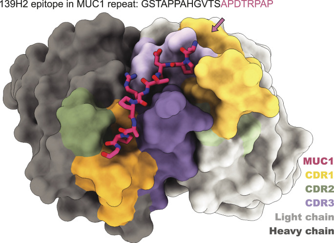
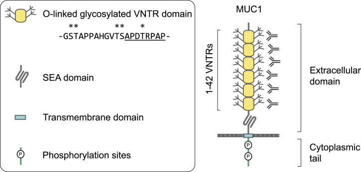
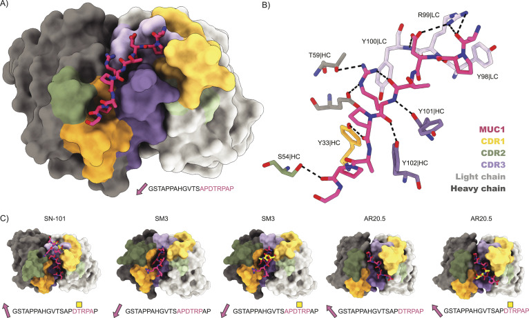
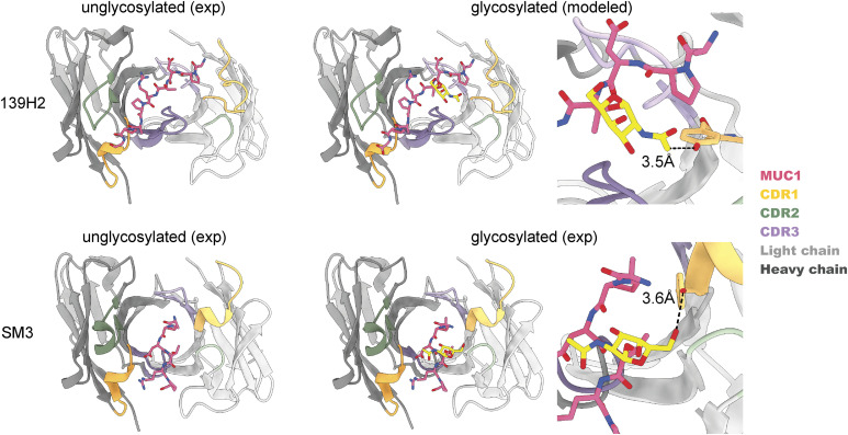
Mucin 1 (MUC1) is a transmembrane mucin expressed at the apical surface of epithelial cells at mucosal surfaces. MUC1 has a barrier function against bacterial invasion and is well known for its aberrant expression and glycosylation in adenocarcinomas. The MUC1 extracellular domain contains a variable number of tandem repeats (VNTR) of 20 amino acids, which are heavily O-linked glycosylated. Monoclonal antibodies against the MUC1 VNTR are powerful research tools with applications in the diagnosis and treatment of MUC1-expressing cancers. Here, we report direct mass spectrometry-based sequencing of anti-MUC1 hybridoma-derived 139H2 IgG, enabling reverse-engineering of the functional recombinant monoclonal antibody. The crystal structure of the 139H2 Fab fragment in complex with the MUC1 epitope was solved, revealing the molecular basis of 139H2 binding specificity to MUC1 and its tolerance to O-glycosylation of the VNTR. The available sequence of 139H2 will allow further development of MUC1-related diagnostic, targeting, and treatment strategies.


Conflict of interest statement

The authors declare that they have no conflict of interest.

Figures

None
Graphical abstract
Figure 1.
Figure 1.. Schematic overview of the MUC1 domain structure.
VNTR, variable number of tandem repeats; SEA, domain name from initial identification in a sperm protein, enterokinase, and agrin.
Figure S1.
Figure S1.. Depth of coverage (total number of overlapping peptides mapped per position) for the variable domains of the 139H2 heavy and light chains.
Figure 2.
Figure 2.. De novo sequencing of the hybridoma 139H2 based on bottom-up proteomics.
The variable region alignment to the inferred germline sequence is shown for both heavy and light chains. Positions with putative somatic hypermutation are highlighted with asterisks (*). The MS/MS spectra supporting the complementarity-determining region are shown beneath the sequence alignment, b/y ions are indicated in blue and red, whereas c/z ions are indicated in green and yellow.
Figure S2.
Figure S2.. Production and purification of reverse engineered 139H2.
(A) Purification of synthetic recombinant 139H2 IgG. (B) Purification of synthetic recombinant 139H2 Fab. Left is the elution profile across the imidazole gradient from the HisTrap purification, the blue line represents the protein absorption at 280 nm (mAU), and the green line represents the percentage of the Buffer B (%); right is the SDS–PAGE of the purified 139H2 product under reducing/non-reducing conditions.
Figure 3.
Figure 3.. Validation of synthetic recombinant 139H2 following the mass spectrometry–derived sequence.
(A) Immunoblot analysis of lysates of intestinal epithelial HT29-MTX and HT29-MTX ∆MUC1 cells with the original hybridoma-derived 139H2 IgG antibody and synthetic recombinant 139H2. (B) Immunofluorescence confocal microscopy imaging of confluent HT29-MTX and HT29-MTX ΔMUC1 monolayers. Cells were stained for nuclei (DAPI, blue) and MUC1 (139H2, green). The signal of the 139H2 Fab was enhanced to compensate for the expected low signal/binding. White scale bars represent 20 μm.
Figure S3.
Figure S3.. Surface plasmon resonance (SPR) quantification of binding affinity for the recombinant full-length 139H2 and its Fab to surface-immobilized MUC1 peptide.
(A) Binding of 139H2 full IgG to surface-immobilized biotinylated MUC1 peptide, analyzed by SPR. Equilibrium binding was fitted using a two-site binding model (right panel), with the respective KD and Bmax values for the high- and low-affinity binding indicated. (B) Binding of 139H2 Fab to surface-immobilized biotinylated MUC1 peptide, analyzed by surface plasmon resonance (SPR). Equilibrium binding was fitted using a one-site binding model (Langmuir isotherm, right panel), with KD and Bmax values indicated.
Figure S4.
Figure S4.. Electron density for the MUC1 peptide bound to 139H2 Fab.
(A) Positive Fo-Fc omit density plotted at 3.0σ (green mesh), after correcting complementarity-determining regions and refinement in REFMAC but excluding placement of the MUC1 peptide, shows well-resolved additional density at the peptide binding site. (B) 2Fo-Fc density plotted at 1.0σ (gray mesh) of the final refined model including water (red spheres) shows a good fit for the modeled MUC1 peptide (green sticks). In both panels, the 139H2 Fab molecule is shown in stick representation, with the light chain colored in light gray, and the heavy chain colored in dark gray.
Figure 4.
Figure 4.. Structure of 139H2 Fab in complex with the MUC1 peptide.
(A) Surface representation of the Fab with complementarity-determining regions highlighted in colors and MUC1 peptide shown as a model. N- to C-terminus direction of MUC1 peptides is shown as a pink arrow. (B) Interactions between 139H2 Fab and MUC1 peptide. (C) Comparison with previously reported structures of monoclonal anti-MUC1 antibodies targeting the variable number of tandem repeats. Glycosylated residues of the epitope are depicted by a yellow square above.
Figure S5.
Figure S5.. Sites of somatic hypermutation in 139H2 framework.
(A) Structure of 139H2 FAB with somatic hypermutations highlighted in red. (B) Hypermutations in heavy chain are organized in a stripe across the beta-sheet, and side chains are oriented to the center of the beta-barrel formed by heavy and light chains. (C) Interaction of Y35 and T97 with N106 and Y111, respectively, tilts CDR3 loop into the position where it can interact with MUC1.
Figure 5.
Figure 5.. Binding of 139H2 to MUC1 reporter constructs with different O-linked glycosylation.
(A) Schematic representation of the MUC1 fragments used. The four fragments used contain seven transmembrane repeats (TR) of MUC1 with 5 O-glycosylation sites with WT Core 2/ST (WT)/DiST/STn/Tn glycan structures. (B) Western blots against the MUC1 WT/DiST/STn/Tn fragments with the 139H2 hybridoma-derived antibody. (C) Western blots against the MUC1 WT/DiST/STn/Tn fragments with the 139H2 reverse-engineered antibody. (D) Western blots against the MUC1 WT/DiST/STn/Tn fragments with an α-His-tag antibody control. (E, F) Western blot band intensities analyzed with Image Lab 6.0 software. Calculated intensity ratios were made relative to the intensity of MUC1-Tn. No significant difference in binding of hybridoma-derived 139H2 or synthetic recombinant 139H2 was observed compared with the 6 α-His-tag antibody control.
Figure S6.
Figure S6.. Comparison of 139H2 and SM3 binding to MUC1.
In SM3, the GalNAc residue makes an additional hydrogen bond with a tyrosine in CDRL1; similar interaction between T4 and GalNAc is predicted to be present also in 139H2.

Similar articles

Cited by

References

    1. Bramwell ME, Wiseman G, Shotton DM (1986) Electron-microscopic studies of the ca antigen, epitectin. J Cell Sci 86: 249–261. 10.1242/jcs.86.1.249 - DOI - PubMed
    1. Frey A, Giannasca KT, Weltzin R, Giannasca PJ, Reggio H, Lencer WI, Neutra MR (1996) Role of the glycocalyx in regulating access of microparticles to apical plasma membranes of intestinal epithelial cells: Implications for microbial attachment and oral vaccine targeting. J Exp Med 184: 1045–1059. 10.1084/jem.184.3.1045 - DOI - PMC - PubMed
    1. Lindén SK, Sheng YH, Every AL, Miles KM, Skoog EC, Florin THJ, Sutton P, McGuckin MA (2009) MUC1 limits helicobacter pylori infection both by steric hindrance and by acting as a releasable decoy. PLoS Pathog 5: e1000617. 10.1371/journal.ppat.1000617 - DOI - PMC - PubMed
    1. McAuley JL, Linden SK, Png CW, King RM, Pennington HL, Gendler SJ, Florin TH, Hill GR, Korolik V, McGuckin MA (2007) MUC1 cell surface mucin is a critical element of the mucosal barrier to infection. J Clin Invest 117: 2313–2324. 10.1172/JCI26705 - DOI - PMC - PubMed
    1. Li X, Bleumink-Pluym NMC, Luijkx YMCA, Wubbolts RW, van Putten JPM, Strijbis K (2019) MUC1 is a receptor for the Salmonella SiiE adhesin that enables apical invasion into enterocytes. PLoS Pathog 15: e1007566. 10.1371/journal.ppat.1007566 - DOI - PMC - PubMed

Associated data