. 2024 Mar 20;14(1):6689.
doi: 10.1038/s41598-024-56848-y.

Oxytoxaceae are prorocentralean rather than peridinialean dinophytes and taxonomic clarification of heterotrophic Oxytoxum lohmannii (≡ "Amphidinium" crassum) by epitypification

Affiliations

Oxytoxaceae are prorocentralean rather than peridinialean dinophytes and taxonomic clarification of heterotrophic Oxytoxum lohmannii (≡ "Amphidinium" crassum) by epitypification

Marc Gottschling et al. Sci Rep. .

Abstract

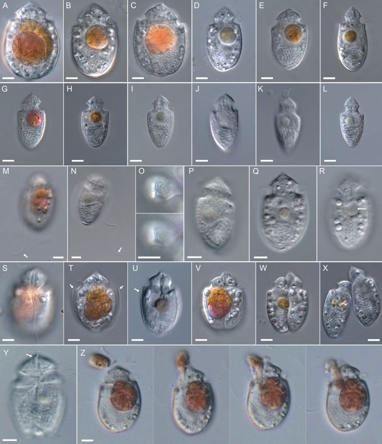
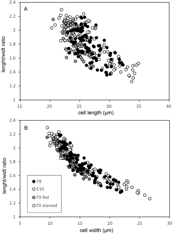
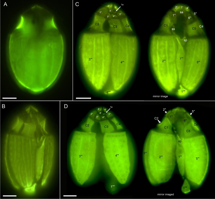
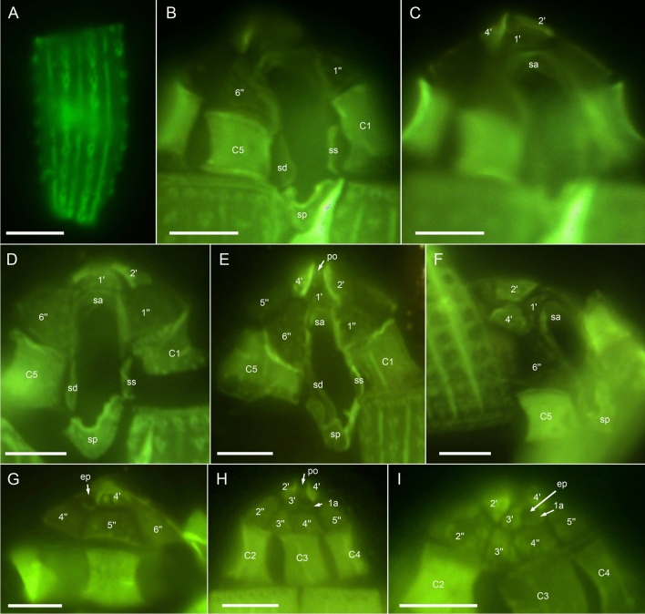
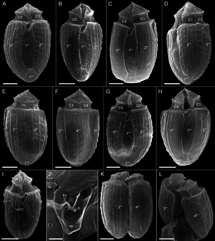
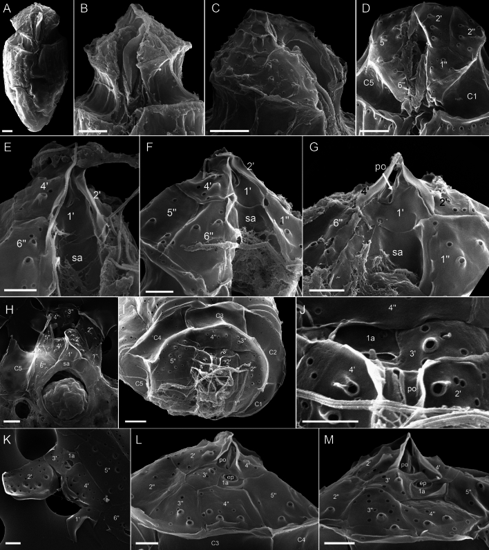
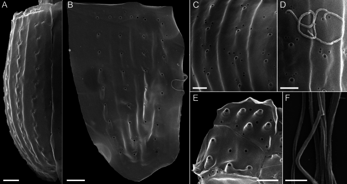
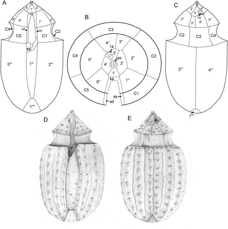
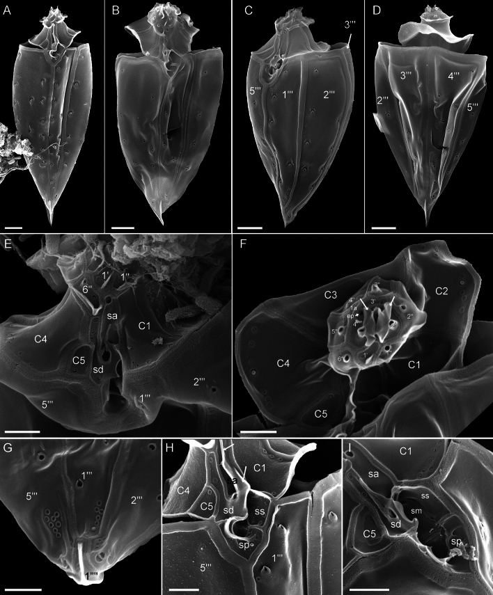
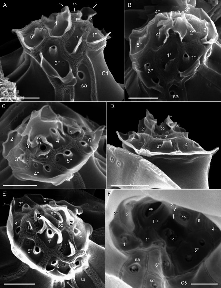
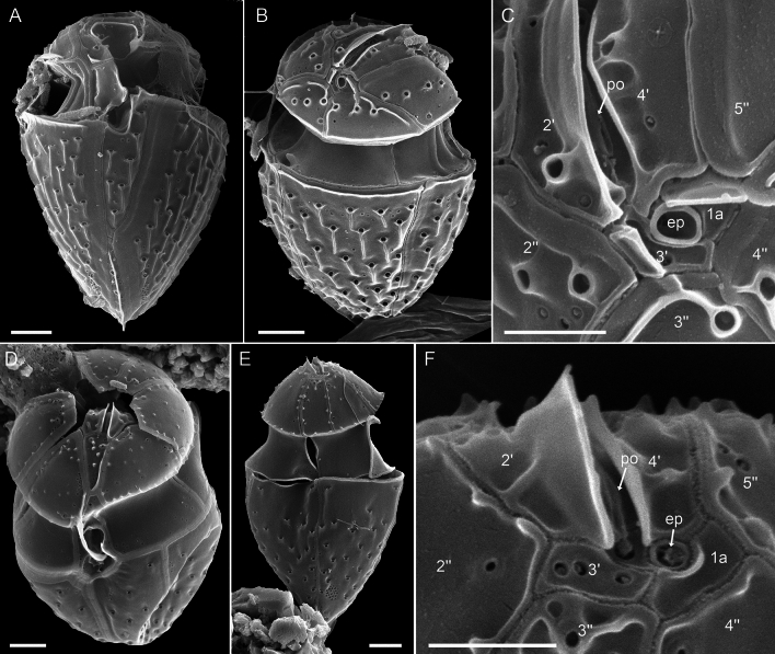
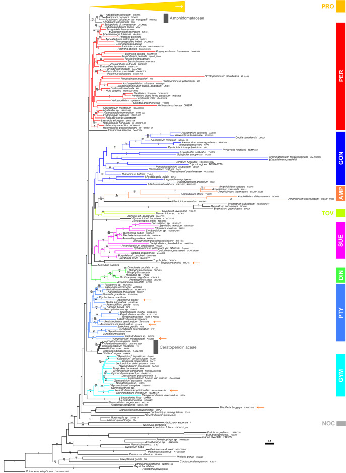
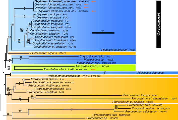
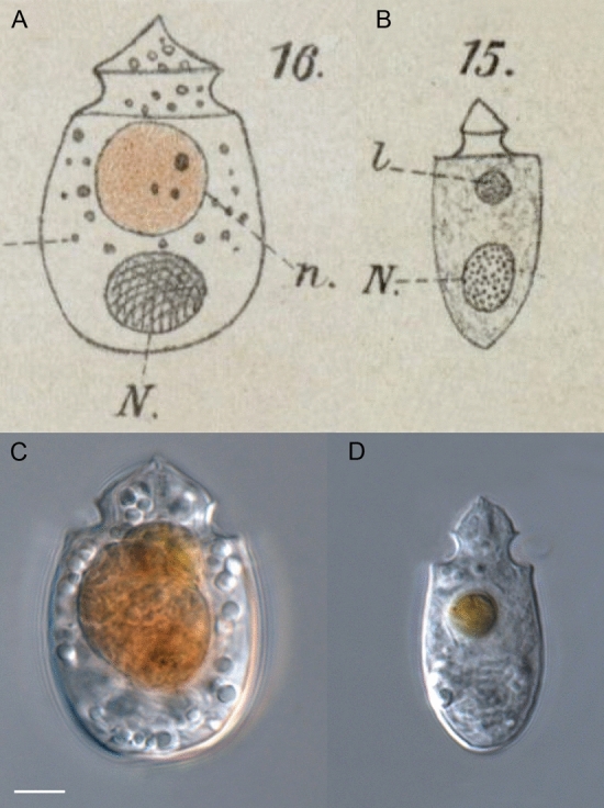
During evolution of Dinophyceae, size reduction of the episome has occurred in several lineages (including unarmoured Amphidiniales and armoured Prorocentrales). One such species is Amphidinium crassum, whose taxonomic identity is elusive though showing morphological similarities with Oxytoxaceae (currently placed in armoured Peridiniales). Plankton samples were taken at the type locality of A. crassum in Kiel Bight (Baltic Sea) in order to establish monoclonal strains. The protist material was examined in detail using light and electron microscopy, and a long (2984 bp) ribosomal RNA sequence gained was part of a taxon sample comprising 206 specimen vouchers and representing the known molecular diversity of Dinophyceae. Cells of A. crassum were ovoid and exhibited a plate pattern po, 4', 1a, 6'', 5c, 4s, 5''', 1''''. In the molecular phylogeny, the species seemed to belong neither to Amphidiniales nor to Peridiniales but to Prorocentrales and clustered with other representatives of Oxytoxaceae. The morphological diversity of Prorocentrales appears thus expanded, and the group may include a number of previously unrecognised representatives unusually having five postcingular and only a single antapical plate. The taxonomic identity of A. crassum is clarified by epitypification, and the species notably exhibits both an apical pore and an additional epithecal pore.


Conflict of interest statement

The authors declare no competing interests.

Figures

Figure 1
Figure 1
Oxytoxum lohmannii (strain K-AC-E10), light microscopy of living cells. (AL) Size and shape of different cells in ventral/dorsal view. (MN) Two different cells indicating length of the longitudinal flagellum (arrow). (O) Two focal planes of an apical view. (P) Cell in lateral view, note the antapical position of the nucleus and the transverse striation of chromosomes. (QR) Two focal planes of the same cell, note the presence of a pusule just below the cingulum (arrow in R). (S) Cell in ventral view, note the insertion of the longitudinal flagellum and the peduncle canal in the central sulcal area and the episome. (TU) Slightly squeezed cells revealed the presence of thecal plates (arrows). (VX) Different stages of vegetative cell division. (Y) Cell in ventral view, note the extruded peduncle (arrow). (Z) Single frames of a cell ingesting a cell of Rhodomonas salina. Scales bars = 5 µm.
Figure 2
Figure 2
Scatterplot of morphometric size measurements of different strains (F8, K-AC-E10, F9 fed and starved) of Oxytoxum lohmannii. (A) Cell length (µm) and (B) cell width (µm) versus length/width ratio.
Figure 3
Figure 3
Oxytoxum lohmannii (strain K-AC-E10), light microscopy of formaldehyde fixed cells stained with Solophenyl Flavine and viewed with epifluorescence and green light excitation. (AB) Cells in ventral view. (CD) Two focal planes each of squeezed cells in dorsal view; note that the corresponding ventral view (right part) is mirror imaged. Plate labels according to the Kofoidean system. Sulcal plate labels: sp, posterior sulcal plate; ss, left sulcal plate. Scale bars = 5 µm.
Figure 4
Figure 4
Oxytoxum lohmannii (strain K-AC-E10), detailed light microscopy view of formaldehyde fixed cells stained with Solophenyl Flavine and viewed with epifluorescence and green light excitation. (A) One of the large postcingular plates, note the large thecal pores and the plate surface ornamentation. (BF) Arrangement of epithecal and sulcal plates in ventral (BE) and lateral (F) view. (GI) Arrangement of epithecal plates in dorsal view. Plate labels according to the Kofoidean system. ep, epithecal pore; Sulcal plate labels: sa, anterior sulcal plate; sd, right sulcal plate; ss, left sulcal plate; sp, posterior sulcal plate. Scale bars = 5 µm.
Figure 5
Figure 5
Oxytoxum lohmannii (strain K-AC-E10), SEM of different cells. (AI) Cells in ventral (AC, GI), in left lateral (D) or in dorsal view (EF); note the peduncle canal in G and H, and the insertion of the longitudinal flagellum in I. (J) Detailed view of the posterior sulcal plate. (KL) Two different cells in dorsal view in division; note that in the epitheca all apical plates are allocated to the left daughter cell. Plate labels according to the Kofoidean system. Sulcal plate labels: sp, posterior sulcal plate. Scale bars = 5 µm (AI, KL) or 1 µm (J).
Figure 6
Figure 6
Oxytoxum lohmannii (strain K-AC-E10), SEM of different cells. (A) Cell with extruded peduncle inside the peduncle canal. (BC) Detailed view of the peduncle canal area in ventral (B) and lateral view (C); note that the peduncle is visible in B. (DG) Detailed view of epitheca and sulcal area in ventral-apical (D) and ventral view (EG). (HJ) Epithecal plates in apical view; note that J is an enlarged view of the cell shown in I. (K) Internal view of apical plates; note that the pore plate probably is disrupted. (LM) Epithecal plates in dorsal view. Plate labels according to the Kofoidean system. ep, epithecal pore; sa, anterior sulcal plate. Scale bars = 2 µm (AD, HI) or 1 µm (FH, JM).
Figure 7
Figure 7
Oxytoxum lohmannii (strain K-AC-E10), SEM. (AD) Thecal pores and surface ornamentation of precingular plates; note that mucocyst-like material is extruded from two of the large pores. (E) Thecal pores on epithecal plates. (F) Bundles of ejected material. Scale bars = 2 µm (AB) or 1 µm (CE) or 0.5 µm (F).
Figure 8
Figure 8
Drawings of Oxytoxum lohmannii plate pattern. (AC) Schematic line drawings of the plate pattern in ventral view (A). (B) Epithecal plates in apical view. (C) Dorsal view. (DE) Schematic drawing of a whole cell in SEM in ventral (D) and dorsal (E) view.
Figure 9
Figure 9
Oxytoxum gladiolus, SEM of field sample specimens. (AD) Cells in ventral (AB), left lateral (C) and dorsal view (D). (E, F) Detailed view of cingulum and epithecal plates in ventral (E) and apical view (F). (G) Antapex in ventral view. (HI) Detailed view of the sulcal area in ventral view. Plate labels according to the Kofoidean system. ep, epithecal pore. Sulcal plate labels: sa, anterior sulcal plate; sd, right sulcal plate; ss, left sulcal plate; sm, median sulcal plate; sp, posterior sulcal plate. Scale bars = 2 µm (AD) or 1 µm (EI).
Figure 10
Figure 10
Oxytoxum gladiolus, SEM of field sample specimen. (AF) Detailed views of epithecal plates in ventral (A), ventral apical (B), right lateral dorsal (C), dorsal (D), right lateral apical (E) and internal view (F). Plate labels according to the Kofoidean system. ep, epithecal pore. Sulcal plate labels: sa, anterior sulcal plate; sd, right sulcal plate. Scale bars = 1 µm (AE) or 0.5 µm (F).
Figure 11
Figure 11
Species of Oxytoxum, SEM of field specimens. (AC) Oxytoxum laticeps; cell in ventral (A) and in dorsal apical view (BC); note that C is a higher magnification view of the cell shown in B. (DF) Oxytoxum sp. 1; cell in ventral apical (D) and dorsal view (EF); note that F is a higher magnification view of the cell shown in E. Plate labels according to the Kofoidean system. ep, epithecal pore. Scale bars = 2 µm (AB, DE) or 1 µm (C, F).
Figure 12
Figure 12
A molecular reference phylogeny recognising major groups of dinophytes. Maximum Likelihood (ML) tree of 207 systematically representative dinophyte sequences (with strain number information) inferred from a rRNA nucleotide alignment (4467 parsimony-informative positions). The numbers on the branches are ML non-parametric bootstrap (above the branch line) and Bayesian probabilities (below the branch line) for the clusters (asterisks indicate maximal support values; values under 50 for the ML bootstrap and 0.90 for Bayesian probability are not shown). Branch lengths are to scale. Orange arrows indicate taxa formerly assigned to Amphidinium but later identified to belong to other lineages. Abbreviations: AMP, Amphidiniales; DIN, Dinophysales; GON, Gonyaulacales; GYM, Gymnodiniales; NOC, Nocticulales; PER, Peridiniales; PRO, Prorocentrales; PTY, Ptychodiscales; SUE, †Suessiales; TOV, Tovelliales.
Figure 13
Figure 13
Phylogenetic sub-tree of prorocentralean dinophytes including Oxytoxaceae. Colours indicate groups with two thecal plates and an epitheca barely identifiable (brownish), a single antapical plate and five postcingular plates (bluish) and a single antapical plate and the postcingular plates split leading to the presence of posterior intercalary plates (greenish).
Figure 14
Figure 14
Comparing the original drawings of Amphidinium crassum (A) and Amphidinium longum (B) and cells of the clonal strain K-AC-E10 of Oxytoxum lohmannii (CD). Scale bar = 5 µm (CD). Abbreviations: n, shiny yellow–brown body; N, nucleus; l, small, light-refracting body.

Similar articles

Cited by

References

    1. Gómez F. A checklist and classification of living dinoflagellates (Dinoflagellata, Alveolata) CICIMAR Oceánides. 2012;27:65–140. doi: 10.37543/oceanides.v27i1.111. - DOI
    1. Takayama H. Apical grooves of unarmored dinoflagellates. Bull. Plankton Soc. Jpn. 1985;32:129–140.
    1. Daugbjerg N, Hansen G, Larsen J, Moestrup Ø. Phylogeny of some of the major genera of dinoflagellates based on ultrastructure and partial LSU rDNA sequence data, including the erection of three new genera of unarmoured dinoflagellates. Phycologia. 2000;39:302–317. doi: 10.2216/i0031-8884-39-4-302.1. - DOI
    1. Janouškovec J, et al. Major transitions in dinoflagellate evolution unveiled by phylotranscriptomics. Proc. Natl. Acad. Sci. USA. 2017;114:E171–E180. doi: 10.1073/pnas.1614842114. - DOI - PMC - PubMed
    1. Price DC, Bhattacharya D. Robust Dinoflagellata phylogeny inferred from public transcriptome databases. J. Phycol. 2017;53:725–729. doi: 10.1111/jpy.12529. - DOI - PubMed