. 2024 Mar 20;14(1):6713.
doi: 10.1038/s41598-024-57330-5.

Non-canonical RNA substrates of Drosha lack many of the conserved features found in primary microRNA stem-loops

Affiliations

Non-canonical RNA substrates of Drosha lack many of the conserved features found in primary microRNA stem-loops

Karen Gu et al. Sci Rep. .

Abstract

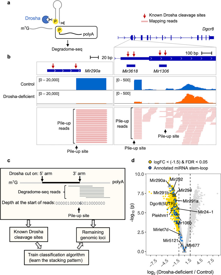
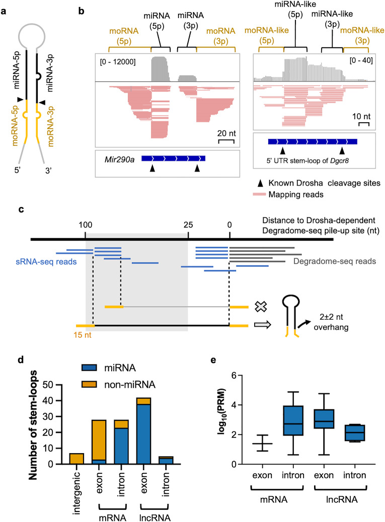
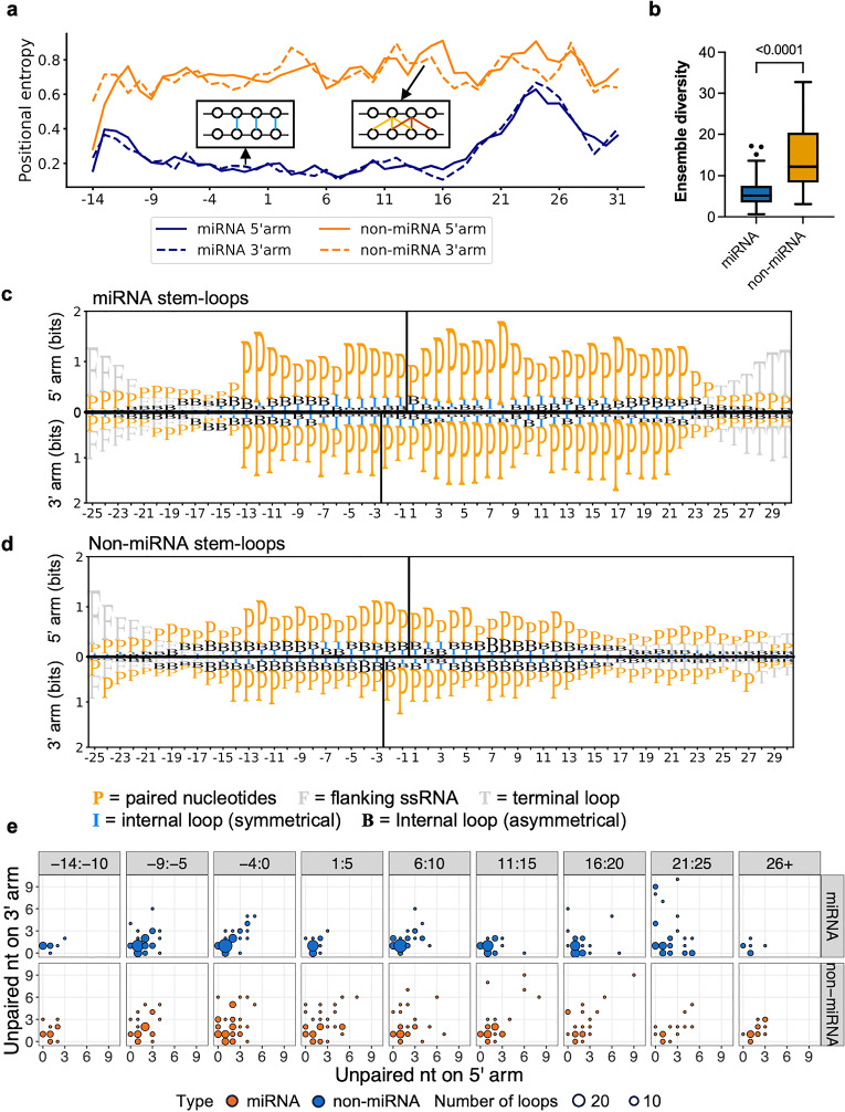
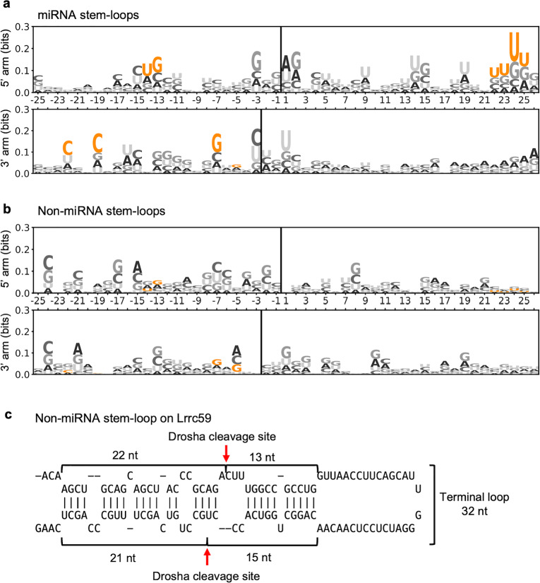
The RNase III enzyme Drosha has a central role in microRNA (miRNA) biogenesis, where it is required to release the stem-loop intermediate from primary (pri)-miRNA transcripts. However, it can also cleave stem-loops embedded within messenger (m)RNAs. This destabilizes the mRNA causing target gene repression and appears to occur primarily in stem cells. While pri-miRNA stem-loops have been extensively studied, such non-canonical substrates of Drosha have yet to be characterized in detail. In this study, we employed high-throughput sequencing to capture all polyA-tailed RNAs that are cleaved by Drosha in mouse embryonic stem cells (ESCs) and compared the features of non-canonical versus miRNA stem-loop substrates. mRNA substrates are less efficiently processed than miRNA stem-loops. Sequence and structural analyses revealed that these mRNA substrates are also less stable and more likely to fold into alternative structures than miRNA stem-loops. Moreover, they lack the sequence and structural motifs found in miRNA stem-loops that are required for precise cleavage. Notably, we discovered a non-canonical Drosha substrate that is cleaved in an inverse manner, which is a process that is normally inhibited by features in miRNA stem-loops. Our study thus provides valuable insights into the recognition of non-canonical targets by Drosha.


Conflict of interest statement

The authors declare no competing interests.

Figures

Figure 1
Figure 1
Analysis pipeline for identifying Drosha-dependent cleavage sites captured by Degradome-seq. (a) Degradome-seq captures polyA-tailed RNA with a 5’P, which is the hallmark of Drosha cleavage. (b) Example of Degradome-seq reads mapping to a miRNA gene (Mir290a) locus (left) and the stem-loops within the 5’UTR and CDS of Dgcr8 (right). Both stem-loops in Dgcr8 are also annotated as miRNAs. The gene/miRNA genomic positions are indicated in the top panel, with identified cleavage sites indicated by red arrows. Read depth for control and Drosha-deficient cells are shown in the next two tracks (representative of two replicates), with the data range for each track indicated in square brackets. The last track shows a collapsed view of aligned reads. (c) Schematic of the pipeline used to identify pile-up sites in Degradome-seq libraries. For each genomic locus, the depth at the start of the Degradome-seq reads was counted. The pile-up at annotated miRNA stem-loops (i.e. known Drosha cleavage sites) was used to train the classification algorithm for identifying the stacking pattern of Drosha-cleaved RNAs. The trained algorithm was then applied to the remaining genomic loci to identify those with similar stacking patterns. (d) Volcano plot of site pile-ups comparing Drosha-deficient and control ESCs. Sites that are significantly decreased (logFC < [− 1.5] and FDR < 0.05, 913 sites) in Drosha-deficient cells are indicated in yellow. Known cleavage sites on annotated miRNA stem-loops are indicated in blue.
Figure 2
Figure 2
Identifying the boundaries of Drosha-cleaved stem-loops using miRNA and moRNA fragments from sRNA-seq. (a) Schematic representation of a miRNA stem-loop structure within the pri-miRNA transcript. Indicated are the positions from where the miRNA (black) and moRNA (yellow) fragments are derived. The Drosha cleavage sites are indicated by the black triangles. (b) sRNA-seq reads mapping to Mir290a and the 5’ UTR stem-loop of Dgcr8. The top panel shows the read depth followed by the aligned sRNA-seq reads in a collapsed view (data range indicated in square brackets). The bottom panel shows the relative position of the stem-loop of Mir290a and Dgcr8. (c) Strategy for identifying Drosha-cleaved stem-loops from sRNA-seq reads. Reads starting or ending between 25 and 100 nt from the Drosha-dependent Degradome-seq pile-up were collected. If the sRNA-seq reads stack at one of the termini and the depth of that terminus is greater than one read per million (RPM), it is considered a potential Drosha cleavage site for the opposite arm of the stem-loop. The RNA sequence between the terminus and the corresponding Degradome-seq pile-up site, flanked by a 15 nt sequence, was then used to predict the stem-loop structure. A stem-loop was considered a Drosha cleavage substrate if the terminus of the sRNA-seq reads and the Degradome-seq pile-up site formed a Drosha signature 3′ 2 ± 2 nt overhang. (d) Genomic locations of Drosha-cleaved stem-loops identified in ESCs. Annotated miRNA stem-loops are indicated in blue and non-miRNA stem-loops are indicated in orange. (e) The levels of annotated miRNAs derived from mRNAs and lncRNAs. The levels of each miRNA in mouse ESCs were obtained from mirGeneDB.
Figure 3
Figure 3
The thermodynamic properties of miRNA and non-miRNA stem-loops. (a,b,c,d) Comparison of the MFEs, stem-loop length, base pairing frequency and maximum stacking of base pairing frequency between Drosha-cleaved annotated miRNA and non-miRNA stem-loops identified in ECSs. Statistical testing: Two-tailed Welch’s t-test. P-values are indicated above the box. (e) The stem-loop in Mir291a and Cad are shown here to illustrate the typical difference between the miRNA and non-miRNA stem-loops. The miRNA sequences are highlighted in blue. The Drosha cleavage sites identified in ESCs are indicated by red arrows. (e,f) Comparison of the base pair composition of lower- and upper-stem of miRNA and non-miRNA stem-loops. The base pair frequency is normalized to the total number of base pairs of the stem. P-values are indicated above the box (two-way ANOVA). For a,b,c,d and f, the box represents the interquartile range (IQR), the whiskers extend to the most extreme data points within 1.5 times the IQR, and outliers are shown as individual points in boxplots. The median is represented by the line within the box.
Figure 4
Figure 4
The structural features of miRNA and non-miRNA stem-loops. (a) Comparing the entropy of Drosha-cleaved annotated miRNA (blue) and non-miRNA (orange) stem-loops identified in ESCs. The 5’ and 3’ arms are plotted in solid and dashed lines, respectively. The x-axis shows the relative position of the stem-loop. Position 1 is the first nucleotide in the upper-stem. (b) Comparison of the ensemble diversity of miRNA and non-miRNA stem-loops. The box represents the IQR, the whiskers extend to the most extreme data points within 1.5 times the IQR, and outliers are shown as individual points. The median is represented by the line within the box. P-values are indicated above the box (independent Welch’s t-test, two-tailed). (c,d) Information bits plot comparing the structure of Drosha-cleaved (c) annotated miRNA and (d) non-miRNA stem-loops identified in ESCs. The 5′ arm is shown in the top panels of (c) and (d), while the 3’ arm is shown in the bottom panels. The position of the Drosha cleavage sites is indicated by a solid line. The x-axis indicates the relative position within the stem-loop, with Drosha cleavage site located between -1 and 1. P = paired nucleotides; F = flanking ssRNA; I = internal loop (symmetrical); B = Internal loop (asymmetrical); T = terminal loop. (e) Balloon plot of the size of internal loops within the stem in each 5 bp window. The size of the internal loop is indicated by the number of unpaired nt on the 5′ arm and 3′ arm. The position of the internal loop is indicated by the first unpaired nt that is closest to the Drosha cleavage site on the 5′ arm. The miRNA stem-loop is shown in the top panel in blue, and the non-miRNA stem-loops are shown in the bottom panel in orange. The size of the balloon corresponds to the number of internal loops that size in that window. Position 1 is the first nucleotide in the upper-stem.
Figure 5
Figure 5
Non-miRNA stem-loops lack known miRNA sequence motifs. Logo of the sequence of Drosha-cleaved (a) annotated miRNA and (b) non-miRNA stem-loops identified in ESCs. The 5′ arm is shown in the top panels of (a) and (b), while the 3’ arm is shown in the bottom panels. The position of the Drosha cleavage site is indicated by a solid line. The x-axis indicates the relative position of the stem-loop. The y-axis shows the information bit of the sequence. Known miRNA motifs that are important for Drosha cleavage are highlighted in orange. (c) The non-miRNA stem-loop in Lrrc59 is likely inversely cleaved due to its large terminal loop and lack of sequence motif. The Drosha cleavage sites are indicated by the red arrows.

Similar articles

Cited by

References

    1. Ameres SL, Zamore PD. Diversifying microRNA sequence and function. Nat. Rev. Mol. Cell Biol. 2013;14:475–488. doi: 10.1038/nrm3611. - DOI - PubMed
    1. Jonas S, Izaurralde E. Towards a molecular understanding of microRNA-mediated gene silencing. Nat. Rev. Genet. 2015;16:421–433. doi: 10.1038/nrg3965. - DOI - PubMed
    1. Bartel DP. Metazoan MicroRNAs. Cell. 2018;173:20–51. doi: 10.1016/j.cell.2018.03.006. - DOI - PMC - PubMed
    1. Ha M, Kim VN. Regulation of microRNA biogenesis. Nat. Rev. Mol. Cell Biol. 2014;15:509–524. doi: 10.1038/nrm3838. - DOI - PubMed
    1. Denli AM, Tops BB, Plasterk RH, Ketting RF, Hannon GJ. Processing of primary microRNAs by the Microprocessor complex. Nature. 2004;432:231–235. doi: 10.1038/nature03049. - DOI - PubMed