. 2024 Mar 20;14(1):6731.
doi: 10.1038/s41598-024-57516-x.

RNA-binding proteins potentially regulate the alternative splicing of cell cycle-associated genes in proliferative diabetic retinopathy

Affiliations

RNA-binding proteins potentially regulate the alternative splicing of cell cycle-associated genes in proliferative diabetic retinopathy

Ning Yang et al. Sci Rep. .

Abstract

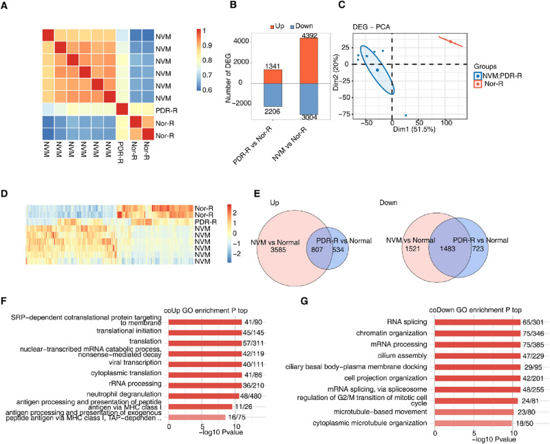
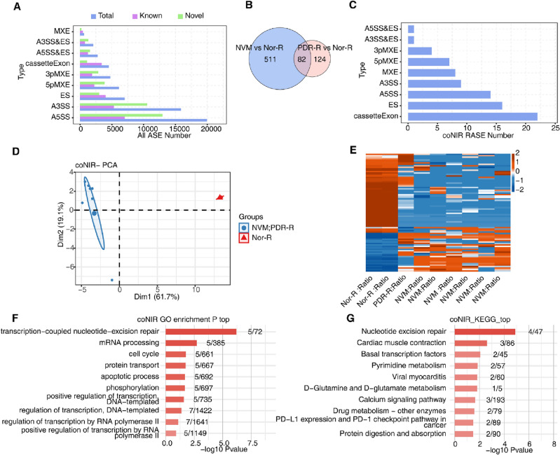
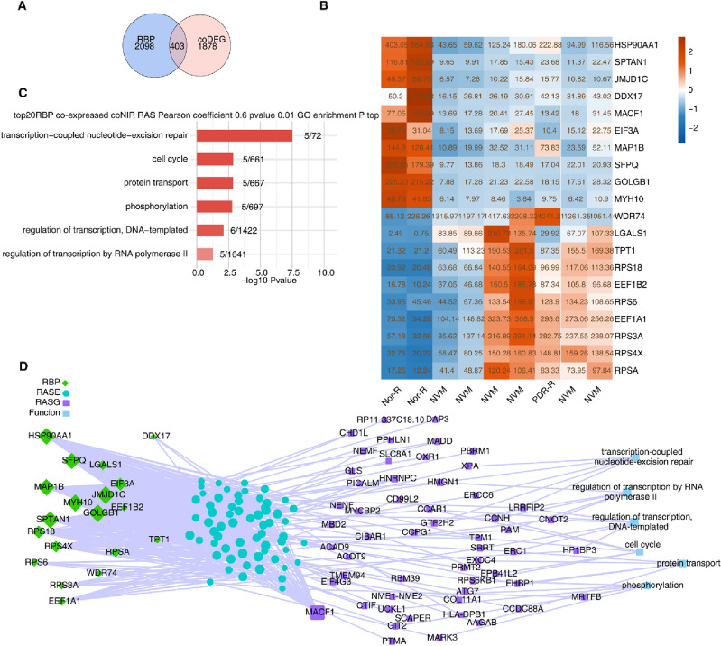
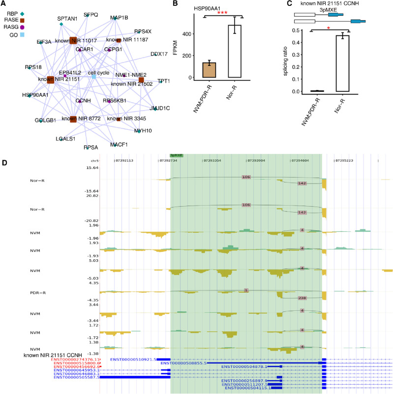
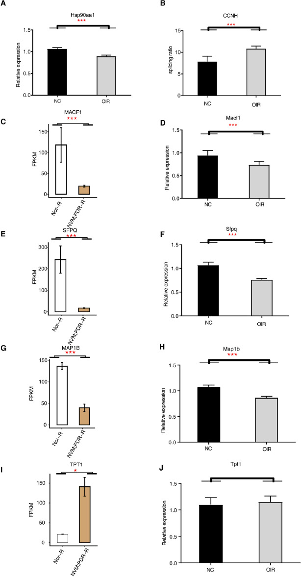
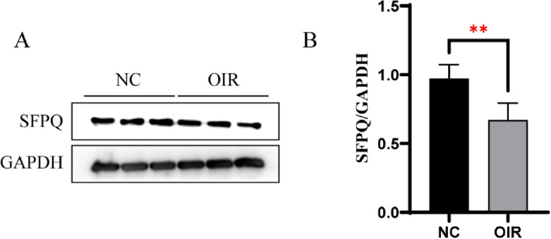
RNA-binding proteins (RBPs) contribute to the pathogenesis of proliferative diabetic retinopathy (PDR) by regulating gene expression through alternative splicing events (ASEs). However, the RBPs differentially expressed in PDR and the underlying mechanisms remain unclear. Thus, this study aimed to identify the differentially expressed genes in the neovascular membranes (NVM) and retinas of patients with PDR. The public transcriptome dataset GSE102485 was downloaded from the Gene Expression Omnibus database, and samples of PDR and normal retinas were analyzed. A mouse model of oxygen-induced retinopathy was used to confirm the results. The top 20 RBPs were screened for co-expression with alternative splicing genes (ASGs). A total of 403 RBPs were abnormally expressed in the NVM and retina samples. Functional analysis demonstrated that the ASGs were enriched in cell cycle pathways. Cell cycle-associated ASEs and an RBP-AS regulatory network, including 15 RBPs and their regulated ASGs, were extracted. Splicing factor proline/glutamine rich (SFPQ), microtubule-associated protein 1 B (MAP1B), heat-shock protein 90-alpha (HSP90AA1), microtubule-actin crosslinking factor 1 (MACF1), and CyclinH (CCNH) expression remarkably differed in the mouse model. This study provides novel insights into the RBP-AS interaction network in PDR and for developing screening and treatment options to prevent diabetic retinopathy-related blindness.


Conflict of interest statement

The authors declare no competing interests.

Figures

Figure 1
Figure 1
Gene expression profile of NVMs and retinas from patients with proliferative diabetic retinopathy. (A) Hierarchical clustering heatmap showing correlation between samples from the NVM, PDR-R, and normal groups based on the FPKM values of all expression genes. (B) Bar plot showing all DEGs between samples from the NVM, PDR-R, and normal groups with DESeq2. False discovery rate ≤ 0.05 and fold-change ≥ 2 or ≤ 0.5. (C) PCA based on FPKM values of all DEGs. The ellipse for each group is the confidence ellipse. (D) Hierarchical clustering heatmap displaying the expression levels of all DEGs. (E) Venn diagram of the gene ID showing the co-upregulated DEGs (left) and co-downregulated DEGs (right) between samples from the NVM and PDR-R groups. (F) Bar plot showing the most enriched GO biological processes of the co-upregulated genes. (G) Bar plot showing the most enriched GO biological processes of the co-downregulated genes. DEGs, differentially expressed genes; FPKM, fragments per kilobase of transcript per million fragments mapped; GO, Gene Ontology; NVM, neovascular membranes; PCA, principal component analysis; PDR-R, retinas from patients with PDR.
Figure 2
Figure 2
Abnormal alternative splicing patterns of genes in NVMs and retinas from patients with PDR. (A) Bar plot showing the number of all significantly regulated ASEs between samples from the NVM, PDR-R, and normal groups. X-axis: ASE numbers. Y-axis: Different types of ASEs. (B) NIR RAS (no-intron) filtering should have detectable splice junctions in all samples, and at least 80% of samples should have ≥ 10 splice junction supporting reads. Venn diagram of the gene ID showing the overlapped RAS (coNIR RAS) between samples from the NVM and PDR-R groups. (C) Bar plot showing the number of all significantly regulated ASEs (coNIR RAS) between NVM and PDR-R and normal samples. X-axis: coNIR RASE number. Y-axis: Different types of ASEs. (D) PCA was based on the splicing ratio of all coNIR RAS. The ellipse for each group was a confidence ellipse. (E) Hierarchical clustering heatmap of all significant coNIR RAS based on splicing ratio. (F) Bar plot showing the most enriched GO biological processes of all significant coNIR RAS. (G) Bar plot showing the most enriched KEGG pathway results for all significant coNIR RAS. ASEs, alternative splicing events; coNIR, co-expressed non-intron retention; GO, Gene Ontology; KEGG, Kyoto Encyclopedia of Genes and Genomes; NVM, neovascular membranes; PCA, principal component analysis; PDR, proliferative diabetic retinopathy; PDR-R, retinas from patients with PDR; RAS, regulated alternative splicing; RASE, regulated alternative splicing events.
Figure 3
Figure 3
Co-expression analysis between RBPs and genes with alternative splicing in neovascular membranes and retinas from patients with proliferative diabetic retinopathy. (A) Venn diagram showing the overlap between co-expressed RBPs and DEGs. (B) Among the upregulated and downregulated coDE RBPs, RBPs with FDR ≤ 0.001 and the top 10 expression levels were screened, respectively, and the heatmap shows the expression levels of the top 20 RBPs. (C) Top 20 RBPs and coNIR RAS were co-expressed. Bar plot showing the most enriched GO biological processes of the top 20 RBP-regulated coNIR RAS. Cutoffs of P-value ≤ 0.01 and Pearson coefficient ≥ 0.6 or ≤  − 0.6 were applied to identify the co-expression pairs. (D) Network diagram showing the pathway of ASEs regulated by the top 10 RBPs. ASEs, alternative splicing events; coNIR, co-expressed non-intron retention; DEGs, differentially expressed genes; FDR, false discovery rate; GO, Gene Ontology; RAS, regulated alternative splicing; RBPs, RNA-binding proteins.
Figure 4
Figure 4
HSP90AA1 regulated CCNH in the cell cycle. (A) Network diagram of co-expression of differential alternative splicing events between RBPs and cell cycle. (BC) Bar diagram showing expression of HSP90AA1 and mutually exclusive 3ʹ-UTRs (3PMXE) event of CCNH. #: not significant, *P < 0.05, **P < 0.01, ***P < 0.001. The exon sequences are denoted by boxes and intron sequences by the horizontal line. (D) Reads distribution chart showing CCNH. CCNH, CyclinH; HSP90AA1, heat-shock protein 90-alpha; RBPs, RNA-binding proteins.
Figure 5
Figure 5
Validation of DE RBPs and ASEs in the OIR mouse model. (A,D,F,H,J) RT-PCR analysis for DE RBPs (Hsp90aa1, Macf1, Sfpq, Map1b, and Tpt1) in the retinas of OIR mice at P17. (B) RT-PCR analysis for DE ASEs (CCNH) in the retinas of OIR mice at P17. (C,E,G,I) Bar diagram showing MACF1, SFPQ, MAP1B, and TPT1. *P < 0.05, ***P < 0.001. ASEs, alternative splicing events; CCNH, CyclinH; DE RBPs, differentially expressed RNA-binding proteins; OIR, oxygen-induced retinopathy; MACF1, microtubule-actin crosslinking factor 1; MAP1B, microtubule-actin crosslinking factor 1; SFPQ, splicing factor proline/glutamine rich; Tpt1, tumor protein translationally controlled 1.
Figure 6
Figure 6
The protein expression of SFPQ by western blot (WB). (A) The representative image of WB. (B)The bar chart shows the protein expression level of SFPQ of the two groups. **P < 0.01. OIR, oxygen-induced retinopathy; SFPQ, splicing factor proline/glutamine rich.

Similar articles

Cited by

References

    1. Wong TY, Cheung CMG, Larsen M, Sharma S, Simó R. Diabetic retinopathy. Nat. Rev. Dis. Primers. 2016;2:16012. doi: 10.1038/nrdp.2016.12. - DOI - PubMed
    1. Antonetti DA, Klein R, Gardner TW. Diabetic retinopathy. N. Engl. J. Med. 2012;366:1227–1239. doi: 10.1056/NEJMra1005073. - DOI - PubMed
    1. Turner M, Díaz-Muñoz MD. RNA-binding proteins control gene expression and cell fate in the immune system. Nat. Immunol. 2018;19:120–129. doi: 10.1038/s41590-017-0028-4. - DOI - PubMed
    1. Holmqvist E, Vogel J. RNA-binding proteins in bacteria. Nat. Rev. Microbiol. 2018;16:601–615. doi: 10.1038/s41579-018-0049-5. - DOI - PubMed
    1. Dash S, et al. The master transcription factor SOX2, mutated in anophthalmia/microphthalmia, is post-transcriptionally regulated by the conserved RNA-binding protein RBM24 in vertebrate eye development. Hum. Mol. Genet. 2020;29:591–604. doi: 10.1093/hmg/ddz278. - DOI - PMC - PubMed

Substances