. 2024 Dec;15(1):2329566.
doi: 10.1080/21505594.2024.2329566. Epub 2024 Mar 20.

Iron depletion has different consequences on the growth and survival of Toxoplasma gondii strains

Affiliations

Iron depletion has different consequences on the growth and survival of Toxoplasma gondii strains

Eléa A Renaud et al. Virulence. 2024 Dec.

Abstract

Toxoplasma gondii is an obligate intracellular parasite responsible for a pathology called toxoplasmosis, which primarily affects immunocompromised individuals and developing foetuses. The parasite can scavenge essential nutrients from its host to support its growth and survival. Among them, iron is one of the most important elements needed to sustain basic cellular functions as it is involved in a number of key metabolic processes, including oxygen transport, redox balance, and electron transport. We evaluated the effects of an iron chelator on the development of several parasite strains and found that they differed in their ability to tolerate iron depletion. The growth of parasites usually associated with a model of acute toxoplasmosis was strongly affected by iron depletion, whereas cystogenic strains were less sensitive as they were able to convert into persisting developmental forms that are associated with the chronic form of the disease. Ultrastructural and biochemical characterization of the impact of iron depletion on parasites also highlighted striking changes in both their metabolism and that of the host, with a marked accumulation of lipid droplets and perturbation of lipid homoeostasis. Overall, our study demonstrates that although acute iron depletion has an important effect on the growth of T. gondii, it has a more profound impact on actively dividing parasites, whereas less metabolically active parasite forms may be able to avoid some of the most detrimental consequences.

Keywords: Acute toxoplasmosis; bradyzoites; chronic toxoplasmosis; cystogenic strains; iron depletion.


Conflict of interest statement

No potential conflict of interest was reported by the author(s).

Figures

Figure 1.
Figure 1.
The iron chelator BPD differentially impacts growth of the type I, II and III strains of T. gondii. (a) Plaque assays were performed in the presence or absence of BPD: parasites were added onto HFF monolayer for 7–10 days and lysis plaques were imaged. Included was a control for which the vehicle (dimethyl sulphoxide) only was used. (b) The half maximal inhibitory concentration (IC50) of BPD for type I, II and III strains of T. gondii was calculated from plaque assays (as described in A) by assessing relative plaque size compared with the vehicle control. Data are from n = 3 independent experiments (except for NED, n = 2). Shown are mean values ± SD. (c) Parasites of the RH and PRU strains were treated for 3 days with 50 µM of BPD, and then treatment continued (+BPD) or the chelator was washed out and parasites were left to grow for another 7 days before imaging of the plaques. (d) Quantification of the relative total lysed area for the RH and the PRU parasites (compared with the vehicle control) in washout assays performed as described in C. Data are mean values ± SD from n = 4 independent experiments. ** denotes p ≤ 0.01, Student’s t‐test.
Figure 2.
Figure 2.
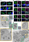
Acute iron chelation by BPD strongly impacts parasite morphology. (a) Immunofluorescence assay showing co-staining of the parasite plasma membrane (“PM,” labelled with the anti-SAG1 antibody) and the apicoplast (“apico,” labelled with the anti-PDH E2 subunit antibody) in parasites from the type I, II and III strains treated or not for 2 days with BPD. Asterisks denote parasites with either nucleus or apicoplast segregation defects. DAPI was used to stain DNA. Scale bar = 10 µm. (b) Immunofluorescence assay showing co-staining of the parasite inner membrane complex (“IMC,” labelled with the anti-IMC3 antibody) and the mitochondrion (“mito,” labelled with the anti-ATPase beta subunit antibody) in parasites from the type I, II and III strains treated or not for 2 days with BPD. DAPI was used to stain DNA. Scale bar = 5 µm. (c) Electron microscopy analysis of the effects of BPD treatment on the RH strain: asterisks denote lipid droplets (LD) which are magnified on selected insets, along with adjacent multilamellar membranes (m). The main inset shows a dividing parasite with duplicated nuclei (N) that displays interrupted inner membrane complex (arrowheads), as well as many vacuoles (V) and membranous structures potentially resembling autophagic vesicles (arrows). Scale bar = 1 µm. (d) electron microscopy analysis of the effects of BPD treatment on the PRU strain: the vacuole contains parasites dividing asynchronously, and displaying structures resembling amylopectin granules (AG, inset), the asterisk denotes a lipid droplet (LD, inset) in a parasite that shows a rather normal aspect for the mitochondrion, rhoptries secretory organelles, apicoplast (M, R and A, in the inset, respectively). Scale bar = 1 µm.
Figure 3.
Figure 3.
Iron deprivation by BPD triggers stage conversion into bradyzoites. (a) Parasites from the type I, II and III strains treated or not for 2 days with BPD were stained for the inner membrane complex (IMC) to outline the parasite shape and co-stained with dolichos biflorus lectin (DBL) to detect the maturation of the parasitophorous vacuole membrane into a cyst wall. DAPI was used to stain DNA. Scale bar = 10 µm. (b) quantification of DBL-positive vacuoles after 2 days of BPD treatment. Data are mean values ± SD from n = 3 independent experiments. At least 50 vacuoles were counted in each experimental condition. ** denotes p ≤ 0.01, Student’s t‐test. (c) type II and III cystogenic strains were kept for 7 days in the presence of BPD or stage conversion was induced by alkaline pH stress for the same duration and co-staining was performed for the cysts wall (DBL) and the late bradyzoite marker P21. (d) quantification of DBL-positive vacuoles after 7 days of BPD treatment or alkaline pH stress. Data are mean values ± SD from n = 3 independent experiments. At least 30 vacuoles were counted in each experimental condition. ns: not statistically significant, Student’s t‐test. (e) quantification of DBL-labelled vacuoles containing P21-positive parasites after 7 days of BPD treatment or alkaline pH stress. Data are mean values ± SD from n = 3 independent experiments. at least 20 vacuoles were counted in each experimental condition. ** denotes p ≤ 0.01, ns: not statistically significant, Student’s t‐test. F. measurement of cyst area after 7 days of BPD treatment or alkaline pH stress. Data are mean values ± SD from n = 3 independent experiments. At least 25 DBL-positive cysts/vacuoles were measured in each experimental condition. *** denotes p ≤ 0.001, ns: not statistically significant, Student’s t‐test.
Figure 4.
Figure 4.
Iron chelation by BPD leads to lipid droplet accumulation in uninfected host cells. (a) Representative transmission electron microscopy micrograph showing the accumulation of lipid droplets (LD) in a fibroblast after 2 days of BPD treatment. ER: endoplasmic reticulum, N: nucleus. Scale bar = 2 µm. (b) fluorescence microscopy picture of fibroblasts either untreated (left) or treated (right) with BPD for 2 days showing that iron chelation leads to an increase in LD, which were labelled with Nile red (NR). DNA was stained with DAPI. Scale bar = 20 µm.
Figure 5.
Figure 5.
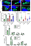
Treatment by BPD affects lipid homoeostasis in T. gondii. (a) Intracellular parasites from the type I, II and III strains were treated or not for 2 days with BPD and stained with nile red (NR) to label the lipid droplets (LD) and counterstained for the inner membrane complex (IMC) protein IMC3 to outline the parasite shape. DNA was stained with DAPI. Scale bar = 10 µm. (b) Quantification of LD numbers per parasite after treatment or not with BPD for 2 days. Data are mean values ± SD from n = 3 independent experiments. At least 280 parasites were counted in each experimental condition. ** denotes p ≤ 0.01, *** denotes p ≤ 0.001, Student’s t‐test. (c) measurement of LD area in parasites after treatment or not with BPD for 2 days. Data are mean values from n = 3 independent experiments. At least 40 LDs were measured in each experimental condition. Symbols are matched between identical experimental groups. **** p ≤ 0.0001, non-parametric Mann-Whitney test. (d) analysis of neutral lipid content in parasites after treatment or not with BPD for 2 days. Chol: cholesterol, DAG: diacylglycerol, Chol Est: cholesteryl esters, TAG: triacylglycerol. Data are mean values ± SD from n = 3 independent experiments. * denotes p ≤ 0.05, Student’s t‐test. (e) analysis of phospholipid content in parasites after treatment or not with BPD for 2 days. PC: phosphatidylcholine, PE: phosphatidylethanolamine, SM: sphingomyelin, PI: phosphatidylinositol, PS: phosphatidylserine. Data are mean values ± SD from n = 3 independent experiments. * denotes p ≤ 0.05, ** denotes p ≤ 0.01, Student’s t‐test.

Similar articles

Cited by

References

    1. Montoya JG, Liesenfeld O.. Toxoplasmosis. Lancet. 2004;363(9425):1965–15. doi: 10.1016/S0140-6736(04)16412-X. - DOI - PubMed
    1. Sanchez SG, Besteiro S. The pathogenicity and virulence of Toxoplasma gondii. Virulence. 2021;12(1):3095–3114. doi: 10.1080/21505594.2021.2012346 - DOI - PMC - PubMed
    1. Clough B, Frickel E-M. The Toxoplasma parasitophorous vacuole: an evolving host–parasite frontier. Trends Parasitol. 2017;33(6):473–488. doi: 10.1016/j.pt.2017.02.007 - DOI - PubMed
    1. Tu V, Tomita T, Sugi T, et al. The Toxoplasma gondii cyst wall interactome. MBio. 2020;11(1):e02699–19. doi: 10.1128/mBio.02699-19. - DOI - PMC - PubMed
    1. Saliba KJ, Kirk K. Nutrient acquisition by intracellular apicomplexan parasites: staying in for dinner. Int J Parasitol. 2001;31(12):1321–1330. doi: 10.1016/S0020-7519(01)00258-2 - DOI - PubMed