. 2024 Mar 7;6(2):fcae083.
doi: 10.1093/braincomms/fcae083. eCollection 2024.

Linking sarcopenia, brain structure and cognitive performance: a large-scale UK Biobank study

Affiliations

Linking sarcopenia, brain structure and cognitive performance: a large-scale UK Biobank study

Tiril P Gurholt et al. Brain Commun. .

Abstract

Sarcopenia refers to age-related loss of muscle mass and function and is related to impaired somatic and brain health, including cognitive decline and Alzheimer's disease. However, the relationships between sarcopenia, brain structure and cognition are poorly understood. Here, we investigate the associations between sarcopenic traits, brain structure and cognitive performance. We included 33 709 UK Biobank participants (54.2% female; age range 44-82 years) with structural and diffusion magnetic resonance imaging, thigh muscle fat infiltration (n = 30 561) from whole-body magnetic resonance imaging (muscle quality indicator) and general cognitive performance as indicated by the first principal component of a principal component analysis across multiple cognitive tests (n = 22 530). Of these, 1703 participants qualified for probable sarcopenia based on low handgrip strength, and we assigned the remaining 32 006 participants to the non-sarcopenia group. We used multiple linear regression to test how sarcopenic traits (probable sarcopenia versus non-sarcopenia and percentage of thigh muscle fat infiltration) relate to cognitive performance and brain structure (cortical thickness and area, white matter fractional anisotropy and deep and lower brain volumes). Next, we used structural equation modelling to test whether brain structure mediated the association between sarcopenic and cognitive traits. We adjusted all statistical analyses for confounders. We show that sarcopenic traits (probable sarcopenia versus non-sarcopenia and muscle fat infiltration) are significantly associated with lower cognitive performance and various brain magnetic resonance imaging measures. In probable sarcopenia, for the included brain regions, we observed widespread significant lower white matter fractional anisotropy (77.1% of tracts), predominantly lower regional brain volumes (61.3% of volumes) and thinner cortical thickness (37.9% of parcellations), with |r| effect sizes in (0.02, 0.06) and P-values in (0.0002, 4.2e-29). In contrast, we observed significant associations between higher muscle fat infiltration and widespread thinner cortical thickness (76.5% of parcellations), lower white matter fractional anisotropy (62.5% of tracts) and predominantly lower brain volumes (35.5% of volumes), with |r| effect sizes in (0.02, 0.07) and P-values in (0.0002, 1.9e-31). The regions showing the most significant effect sizes across the cortex, white matter and volumes were of the sensorimotor system. Structural equation modelling analysis revealed that sensorimotor brain regions mediate the link between sarcopenic and cognitive traits [probable sarcopenia: P-values in (0.0001, 1.0e-11); muscle fat infiltration: P-values in (7.7e-05, 1.7e-12)]. Our findings show significant associations between sarcopenic traits, brain structure and cognitive performance in a middle-aged and older adult population. Mediation analyses suggest that regional brain structure mediates the association between sarcopenic and cognitive traits, with potential implications for dementia development and prevention.

Keywords: T1-weighted MRI; degenerative conditions; ectopic fat; mediator; skeletal muscle.


Conflict of interest statement

O.A.A. has received a speaker's honorarium from Lundbeck, Sunovion, Otsuka and Janssen and is a consultant to Cortechs.ai. J.L. is an employee and shareholder of AMRA Medical AB and has received consulting honorarium/speaking fees from Eli Lilly and BioMarin. The remaining authors declare no conflict of interest.

Figures

Graphical Abstract
Graphical Abstract
Figure 1
Figure 1
Flowchart of participant inclusion process. We performed subsample analyses for the participants with a measure of total thigh MFI in percentage and for those with a measure of general cognitive performance (PC1, first principal component). YTTRIUM, Fast qualitY conTrol meThod foR derIved diffUsion Metrics.
Figure 2
Figure 2
Brain structure differences between individuals with probable sarcopenia versus non-sarcopenia. We used multiple linear regression to obtain the partial correlation r effect size maps for probable sarcopenia (n = 1704) relative to non-sarcopenia (n = 32 171) for (A) cortical thickness, (B) brain volumes and (C) white matter FA. For white matter FA, we additionally display the 95% confidence interval. Significant r effect sizes are |r| in (0.02, 0.06) and P-values in (0.0002, 4.2e−29). We adjusted for sex, age, age2, sex-by-age, sex-by-age2, body mass index, ancestry, metabolic/lifestyle variables, higher education, site, ICV (except FA and cortical thickness) and Euler numbers (T1-weighted MRI). L, left; R, right; r, partial correlation coefficient; Brainstem tracts: CST, corticospinal tract; ML, medial lemniscus; P, pontine; MCP, middle cerebellar peduncle; ICP, inferior cerebellar peduncle; SCP, superior cerebellar peduncle. Projection pathways: CP, cerebral peduncle; ACR, anterior corona radiata; PCR, posterior corona radiata; SCR, superior corona radiata; ALIC, anterior limb of the internal capsule; PLIC, posterior limb of the internal capsule; RLIC, retrolenticular part of the internal capsule; PTR, posterior thalamic radiation. Commissural pathways: BCC, body of corpus callosum; GCC, genu of corpus callosum; SCC, splenium of the corpus callosum; TAP, tapetum. Association pathways: FX, fornix; FXST, fornix stria terminalis; CGC, cingulum cingulate gyrus; CGH, cingulum (hippocampal portion); EC, external capsule; SFO, superior fronto-occipital fasciculus; SLF, superior longitudinal fasciculus; SS, sagittal stratum; UNC, uncinate fasciculus.
Figure 3
Figure 3
Brain structure associations with total MFI. We used multiple linear regression to obtain the partial correlation r effect size maps for total thigh MFI in percentage (n = 30 561) and (A) cortical thickness, (B) brain volumes and (C) white matter FA. For white matter FA, we additionally display the 95% confidence interval. Significant r effect sizes are |r| in (0.02, 0.07) and P-values in (0.0002, 1.9e−31). We adjusted for sex, age, age2, sex-by-age, sex-by-age2, body mass index, ancestry, metabolic/lifestyle variables, higher education, site, ICV (except FA and cortical thickness) and Euler numbers (T1-weighted MRI). FA, fractional anisotropy; ICV, estimated intracranial volume; L, left; R, right; r, partial correlation coefficient; Brainstem tracts: CST, corticospinal tract; ICP, inferior cerebellar peduncle; MCP, middle cerebellar peduncle; ML, medial lemniscus; P, pontine; SCP, superior cerebellar peduncle. Projection pathways: ACR, anterior corona radiata; ALIC, anterior limb of the internal capsule; CP, cerebral peduncle; PCR, posterior corona radiata; PLIC, posterior limb of the internal capsule; RLIC, retrolenticular part of the internal capsule; PTR, posterior thalamic radiation; SCR, superior corona radiata. Commissural pathways: BCC, body of corpus callosum; GCC, genu of corpus callosum; SCC, splenium of the corpus callosum; TAP, tapetum. Association pathways: CGC, cingulum cingulate gyrus; CGH, cingulum (hippocampal portion); EC, external capsule; FX, fornix; FXST, fornix stria terminalis; SFO, superior fronto-occipital fasciculus; SLF, superior longitudinal fasciculus; SS, sagittal stratum; UNC, uncinate fasciculus.
Figure 4
Figure 4
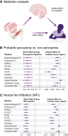
Brain structure mediates the link between sarcopenic traits and cognitive performance. The figure shows (A) an illustration of the mediation analysis (created with BioRender.com); and the results of the SEM mediation analyses for (B) probable sarcopenia (n = 1190) relative to non-sarcopenia (n = 21 340) with significant direct effects in (−0.33, −0.29) [P-values in (1.4e−10, 1.9e−08)] and indirect effects in (−0.04, −0.01) [P-values in (1.0e−11, 0.0001)]; and (C) continuously measured total thigh MFI in percentage (n = 20 850) with significant direct effects in (−0.07, −0.06) [P-values in (0, 2.2e−16)] and indirect effects in (−0.006, −0.002) [P-values in (1.2e−12, 7.7e−05)], in relation to cognitive performance (direct effect) and via mediator (brain phenotype; indirect effect). The direct and indirect effect estimates and corresponding confidence intervals are obtained from the SEM mediation model. We adjusted for sex, age, age2 body mass index, ancestry, metabolic/lifestyle variables, higher education, site, ICV (except FA and cortical thickness) and Euler numbers (T1-weighted MRI). BCC, body of corpus callosum; CP, cerebral peduncle; CT, cortical thickness; FA, fractional anisotropy; GCC, genu of corpus callosum; MFI, muscle fat infiltration; ML, medial lemniscus; SCP, superior cerebellar peduncle; WM, white matter.

References

    1. Cruz-Jentoft AJ, Bahat G, Bauer J, et al. Sarcopenia: Revised European consensus on definition and diagnosis. Age Ageing. 2019;48(1):16–31. - PMC - PubMed
    1. Yuan S, Larsson SC. Epidemiology of sarcopenia: Prevalence, risk factors, and consequences. Metabolism. 2023;144:155533. - PubMed
    1. Dost FS, Ates Bulut E, Dokuzlar O, et al. Sarcopenia is as common in older patients with dementia with Lewy bodies as it is in those with Alzheimer's disease. Geriatr Gerontol Int. 2022;22(5):418–424. - PubMed
    1. Waite SJ, Maitland S, Thomas A, Yarnall AJ. Sarcopenia and frailty in individuals with dementia: A systematic review. Arch Gerontol Geriatr. 2021;92:104268. - PubMed
    1. Arosio B, Calvani R, Ferri E, et al. Sarcopenia and cognitive decline in older adults: Targeting the muscle-brain axis. Nutrients. 2023;15(8):1853. - PMC - PubMed