. 2024 Mar 1;6(2):fcae075.
doi: 10.1093/braincomms/fcae075. eCollection 2024.

In vivo PET classification of tau pathologies in patients with frontotemporal dementia

Affiliations

In vivo PET classification of tau pathologies in patients with frontotemporal dementia

Manabu Kubota et al. Brain Commun. .

Abstract

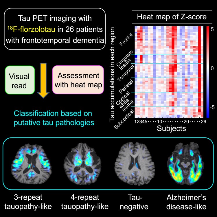
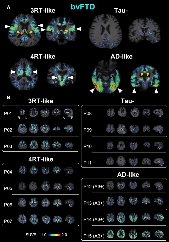
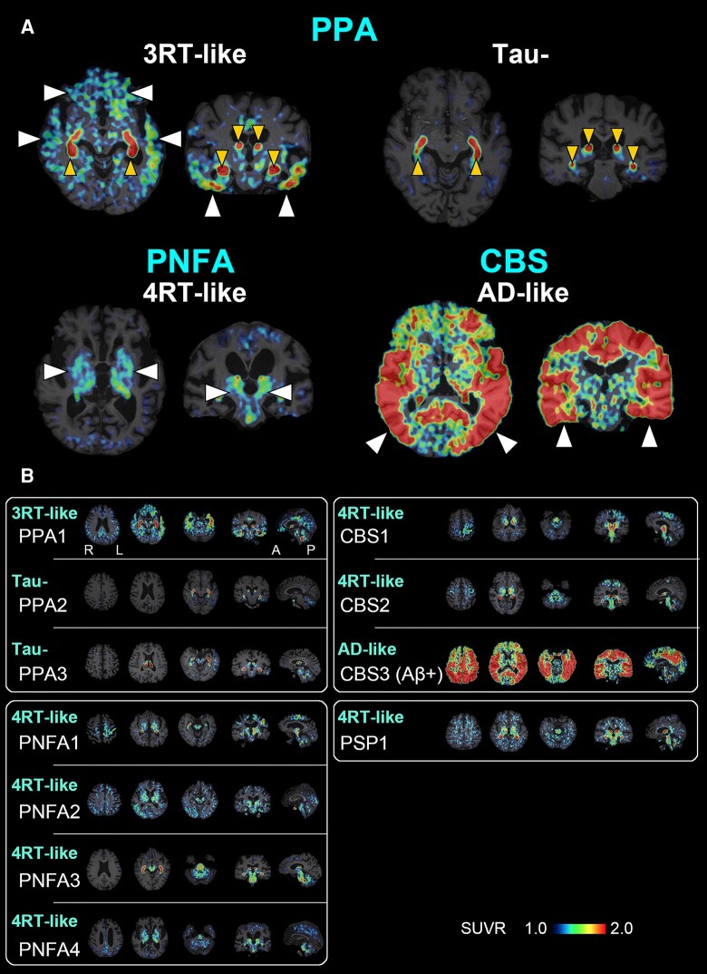
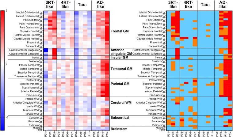
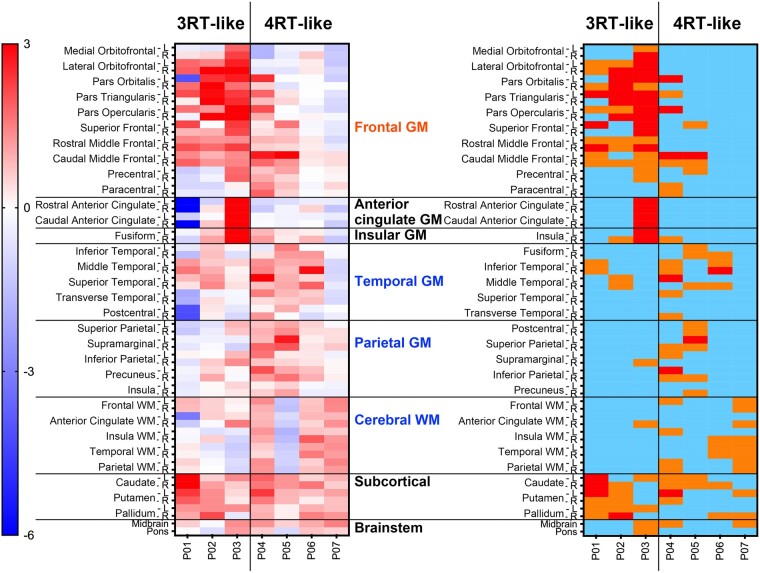
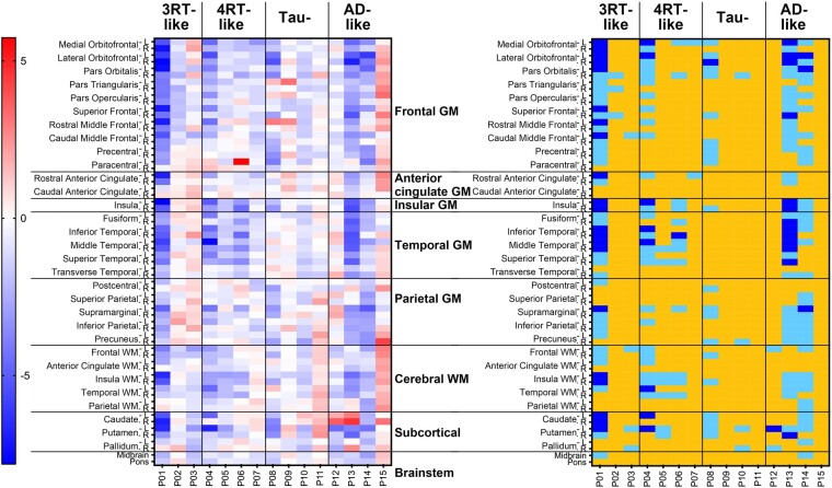
Frontotemporal dementia refers to a group of neurodegenerative disorders with diverse clinical and neuropathological features. In vivo neuropathological assessments of frontotemporal dementia at an individual level have hitherto not been successful. In this study, we aim to classify patients with frontotemporal dementia based on topologies of tau protein aggregates captured by PET with 18F-florzolotau (aka 18F-APN-1607 and 18F-PM-PBB3), which allows high-contrast imaging of diverse tau fibrils in Alzheimer's disease as well as in non-Alzheimer's disease tauopathies. Twenty-six patients with frontotemporal dementia, 15 with behavioural variant frontotemporal dementia and 11 with other frontotemporal dementia phenotypes, and 20 age- and sex-matched healthy controls were included in this study. They underwent PET imaging of amyloid and tau depositions with 11C-PiB and 18F-florzolotau, respectively. By combining visual and quantitative analyses of PET images, the patients with behavioural variant frontotemporal dementia were classified into the following subgroups: (i) predominant tau accumulations in frontotemporal and frontolimbic cortices resembling three-repeat tauopathies (n = 3), (ii) predominant tau accumulations in posterior cortical and subcortical structures indicative of four-repeat tauopathies (n = 4); (iii) amyloid and tau accumulations consistent with Alzheimer's disease (n = 4); and (iv) no overt amyloid and tau pathologies (n = 4). Despite these distinctions, clinical symptoms and localizations of brain atrophy did not significantly differ among the identified behavioural variant frontotemporal dementia subgroups. The patients with other frontotemporal dementia phenotypes were also classified into similar subgroups. The results suggest that PET with 18F-florzolotau potentially allows the classification of each individual with frontotemporal dementia on a neuropathological basis, which might not be possible by symptomatic and volumetric assessments.

Keywords: PET; biomarker; florzolotau; frontotemporal lobar degeneration; tauopathy.


Conflict of interest statement

H.Shimada, M.-R.Z. and M.H. hold patents on compounds related to the present report (JP 5422782/EP 12 884 742.3/CA2894994/HK1208672). All authors declare no competing interests.

Figures

Graphical Abstract
Graphical Abstract
Figure 1
Figure 1
A PET-based subtyping of bvFTD. (A) Representative axial (left) and coronal (right) images of radioactivity SUVRs at 90–110 min after 18F-florzolotau administration. The arrowheads of normal size indicate enhanced parenchymal radioligand retentions characteristic of each putative tau topology subtype. The smaller arrowheads denote radioactivity accumulations in the choroid plexus supposedly unrelated to tau depositions. Radio signal intensification primarily in the frontal and temporal cortices in contrast to relative sparing of posterior and subcortical areas is indicative of Pick’s disease–type 3RT aggregations (P03), while involvements of subcortical regions, including the basal ganglia, thalamus, subthalamic nucleus and brainstem, suggest PSP- or CBD-type 4RT lesions (P04). The negativity for 18F-florzolotau-PET implies non-tau bvFTD exemplified by Type A TDP-43 pathologies (P11). A case with Aβ-PET–positive exhibited limbic and neocortical radioligand accumulations involving temporo-parietal regions and is accordingly granted a clinicopathological phenotype of bvFTD due to Alzheimer’s disease pathologies (P15). (B) A subgrouping of 15 cases with bvFTD by the visual read of individual tau PET images. Patients P01–P03 showed predominant 18F-florzolotau accumulations in the frontotemporal and frontolimbic cortices with minimal posterior cortical involvements suggestive of 3RT pathologies. P01 is a case with autopsy-confirmed Pick’s disease. P04–P07 presented increased radioligand retentions in subcortical structures accompanied by neocortical involvements to varying degrees, implying the presence of 4RT depositions. P08–P11 displayed no overt radioligand accumulations. P12–P15 were Aβ-positive, and the distribution of radio signals followed Braak tau staging consistent with Alzheimer’s disease. In each subject, three axial, one coronal and one sagittal sections providing characteristic information are presented from the left. A, anterior; L, left; P, posterior; R, right.
Figure 2
Figure 2
A PET-based subtyping of PPA, PNFA, CBS and PSP. (A) Representative axial (left) and coronal (right) images of radioactivity SUVRs at 90–110 min after 18F-florzolotau administration. The arrowheads of normal size indicate enhanced parenchymal radioligand retentions characteristic of each putative tau topology subtype. The smaller arrowheads denote radioactivity accumulations in the choroid plexus supposedly unrelated to tau depositions. Radio signal intensification primarily in the frontotemporal regions and less involvement of posterior and subcortical areas are suggestive of Pick’s disease–type 3RT aggregations in a case with PPA (PPA1), while negativity for tau depositions is indicated in another case with PPA (PPA2). Radio signal increases predominantly in subcortical regions implying PSP- or CBD-type 4RT deposits in a case with PNFA (PNFA4). A case with Aβ-PET–positive CBS (CBS3) exhibited widespread and highly intensified limbic and neocortical radioligand accumulations involving temporo-parietal regions, which is indicative of CBS due to Alzheimer’s disease pathologies. (B) The characterization of putative brain pathologies in three PPA (PPA1–3), four PNFA (PNFA1–4), three CBS (CBS1–3) and one PSP (PSP1) patients by the visual read of tau PET images. Cases with PPA were characterized as 3RT-like (PPA1) and tau-negative (PPA2 and 3), whereas all cases with PNFA were categorized as 4RT-like. Image findings in cases with CBS were either 4RT-like (CBS1, 2) or Alzheimer’s disease–like (CBS3), and radio signal distribution in the PSP-FTD case was consistent with 4RT-like pathology. In each subject, three axial, one coronal and one sagittal section providing characteristic information are presented from the left. A, anterior; L, left, P, posterior; R, right.
Figure 3
Figure 3
Heatmaps of Z-scores for regional 18F-florzolotau SUVRs in patients with bvFTD (n = 15). Classification by visual read of tau PET images (see Fig. 1) is indicated on the top. The left panel displays uncategorized Z-scores. The right map presents the following Z-score ranges: light blue, <1; orange, 1–2; red, >2. P01–11 are Aβ-negative, and P12–15 are Aβ-positive.
Figure 4
Figure 4
Heatmaps of Z-scores for regional 18F-florzolotau SUVRs with PVC in patients with bvFTD with 3RT- and 4RT-like radio signal accumulations by visual read (n = 7). Classification by visual read of tau PET images (see Fig. 1) is indicated on the top. The left panel displays uncategorized Z-scores. The right map presents the following Z-score ranges: light blue, <1; orange, 1–2; red, >2. Patients P01–P03, who were indicated as possessing 3RT-like pathologies by visual inspections of images, had 10 or more ROIs with Z-score > 1 within the inferior and middle pre-frontal GM, the inferior, middle and superior temporal GM and the caudate. In contrast, the other four patients (P04–P07) who were visually estimated to possess 4RT-like tau pathologies did not meet the above Z-score criterion for 3RT and had multiple ROIs with Z-score > 1 within pre-central and parietal GM, cerebral WM and the basal ganglia.
Figure 5
Figure 5
Scatter plots of 18F-florzolotau SUVRs with PVC in the striatum against total scores of MMSE and FAB in patients with tau-positive bvFTD (total n = 11). Filled circles indicate patients with Aβ positivity assessed by 11C-PiB (n = 4).
Figure 6
Figure 6
Comparisons of clinical measures among PET-based neuropathological subgroups of bvFTD. By visual read and Z-score mapping of tau PET data, cases with bvFTD were classified into ‘3RT-like’ (Subgroup 1; n = 3), ‘4RT-like’ (Subgroup 2; n = 4), ‘tau-’ (Subgroup 3; n = 4) and ‘Alzheimer’s disease–like’ (Subgroup 4; n = 4) subgroups. Lower scores of MMSE and FAB indicate worse cognition or function, while higher scores of NPI and SRI indicate more severe symptoms. There were no significant subgroup differences in these clinical variables by the Kruskal–Wallis test. Solid symbols denote individual values. NPI and SRI data for one patient in Subgroup 3 are not available.
Figure 7
Figure 7
Heatmaps of Z-scores for regional volumes divided by ICV in patients with bvFTD. Classification by visual read of tau PET images (see Fig. 1) is indicated on the top. The left panel displays uncategorized Z-scores. The right map presents the following Z-score ranges: blue, <−4.5; light blue, −4.5 to −2; yellow, >−2. There are no overt associations between the regionality of the volume loss and putative neuropathological phenotypes in these subjects.

Similar articles

Cited by

  • Neuropathological correlations of 18F-florzolotau PET in a case with pick's disease.
    Suzuki H, Kubota M, Kurose S, Tagai K, Endo H, Onaya M, Yamamoto Y, Sahara N, Ohgidani M, Haga C, Hara H, Akiyama H, Takahata K, Higuchi M. Suzuki H, et al. EJNMMI Res. 2025 Jul 31;15(1):96. doi: 10.1186/s13550-025-01296-6. EJNMMI Res. 2025. PMID: 40745396 Free PMC article.
  • Diverse tau pathologies in late-life mood disorders revealed by PET and autopsy assays.
    Kurose S, Moriguchi S, Kubota M, Tagai K, Momota Y, Ichihashi M, Sano Y, Endo H, Hirata K, Kataoka Y, Goto R, Mashima Y, Yamamoto Y, Suzuki H, Nakajima S, Mizutani M, Sano T, Kawamura K, Zhang MR, Tatebe H, Tokuda T, Onaya M, Mimura M, Sahara N, Takahashi H, Uchida H, Takao M, Meyer JH, Higuchi M, Takahata K. Kurose S, et al. Alzheimers Dement. 2025 Jun;21(6):e70195. doi: 10.1002/alz.70195. Alzheimers Dement. 2025. PMID: 40488253 Free PMC article.

References

    1. Neary D, Snowden JS, Gustafson L, et al. Frontotemporal lobar degeneration: A consensus on clinical diagnostic criteria. Neurology. 1998;51(6):1546–1554. - PubMed
    1. Rabinovici GD, Miller BL. Frontotemporal lobar degeneration: Epidemiology, pathophysiology, diagnosis and management. CNS Drugs. 2010;24(5):375–398. - PMC - PubMed
    1. Seeley WW, Crawford R, Rascovsky K, et al. Frontal paralimbic network atrophy in very mild behavioral variant frontotemporal dementia. Arch Neurol. 2008;65(2):249–255. - PMC - PubMed
    1. Josephs KA, Hodges JR, Snowden JS, et al. Neuropathological background of phenotypical variability in frontotemporal dementia. Acta Neuropathol. 2011;122(2):137–153. - PMC - PubMed
    1. Leuzy A, Chiotis K, Lemoine L, et al. Tau PET imaging in neurodegenerative tauopathies—Still a challenge. Mol Psychiatry. 2019;24(8):1112–1134. - PMC - PubMed

LinkOut - more resources