. 2024 Mar 6:15:1308330.
doi: 10.3389/fimmu.2024.1308330. eCollection 2024.

Type I and II interferons, transcription factors and major histocompatibility complexes were enhanced by knocking down the PRRSV-induced transforming growth factor beta in monocytes co-cultured with peripheral blood lymphocytes

Affiliations

Type I and II interferons, transcription factors and major histocompatibility complexes were enhanced by knocking down the PRRSV-induced transforming growth factor beta in monocytes co-cultured with peripheral blood lymphocytes

Dante Fabros Jr et al. Front Immunol. .

Abstract

The innate and adaptive immune responses elicited by porcine reproductive and respiratory syndrome virus (PRRSV) infection are known to be poor. This study investigates the impact of PRRSV-induced transforming growth factor beta 1 (TGFβ1) on the expressions of type I and II interferons (IFNs), transcription factors, major histocompatibility complexes (MHC), anti-inflammatory and pro-inflammatory cytokines in PRRSV-infected co-cultures of monocytes and peripheral blood lymphocytes (PBL). Phosphorothioate-modified antisense oligodeoxynucleotide (AS ODN) specific to the AUG region of porcine TGFβ1 mRNA was synthesized and successfully knocked down TGFβ1 mRNA expression and protein translation. Monocytes transfected with TGFβAS1 ODN, then simultaneously co-cultured with PBL and inoculated with either classical PRRSV-2 (cPRRSV-2) or highly pathogenic PRRSV-2 (HP-PRRSV-2) showed a significant reduction in TGFβ1 mRNA expression and a significant increase in the mRNA expressions of IFNα, IFNγ, MHC-I, MHC-II, signal transducer and activator of transcription 1 (STAT1), and STAT2. Additionally, transfection of TGFβAS1 ODN in the monocyte and PBL co-culture inoculated with cPRRSV-2 significantly increased the mRNA expression of interleukin-12p40 (IL-12p40). PRRSV-2 RNA copy numbers were significantly reduced in monocytes and PBL co-culture transfected with TGFβAS1 ODN compared to the untransfected control. The yields of PRRSV-2 RNA copy numbers in PRRSV-2-inoculated monocytes and PBL co-culture were sustained and reduced by porcine TGFβ1 (rTGFβ1) and recombinant porcine IFNα (rIFNα), respectively. These findings highlight the strategy employed by PRRSV to suppress the innate immune response through the induction of TGFβ expression. The inclusion of TGFβ as a parameter for future PRRSV vaccine and vaccine adjuvant candidates is recommended.

Keywords: antisense; innate immunity; porcine reproductive and respiratory syndrome virus; transforming growth factor beta; type I and II interferons.


Conflict of interest statement

The authors declare that the research was conducted in the absence of any commercial or financial relationships that could be construed as a potential conflict of interest.

Figures

Figure 1
Figure 1
Monocyte transfection and uptake (A) Monocytes under bright field microscopy. (B) Monocyte uptake of fluorescent-labeled siRNA under immunofluorescent microscopy. (C) Monocyte uptake of fluorescent-labeled siRNA complexed with 1.5% of transfection reagent. The uptake was observed every 2 h for 10 h and finally at 24 h. Mean differences of percentages of fluoresced cells among groups were tested by one-way ANOVA, followed by Tukey HSD test. Different letters indicate significant differences. P<0.05 was set as a statistically significant level.
Figure 2
Figure 2
Effect of TGFβ1 antisense (TGFβAS1), and scramble (Scr) phosphorothioate-modified ODNs on expression of TGFβ1 mRNA and protein translation in monocyte and PBL co-culture. Monocytes were transfected with TGFβAS1 or Scr, added with PBL, and then stimulated with either (A, B) ConA or (C, D) PMAi. Monocytes transfected with transfection media (Tr.media) alone, added with PBL, and finally stimulated with inducers (either ConA or PMAi) served as Tr.media control. Untransfected monocytes, added with PBL, and finally stimulated with inducers served as positive control (Pos Ctrl). Cell supernatant was collected prior to ELISA. Data were normalized to the geometric average of RPL32 and YWHAZ in relative to untransfected/unstimulated monocytes and PBL co-culture. Band intensities indicate the quality of TGFβ1 knockdown ( Additional File 3 ). Error bars indicate the standard deviation (SD). Mean differences of TGFβ1 gene expression or protein translation among groups were tested by one-way ANOVA, followed by Tukey HSD test. Different letters above the error bars indicate significant differences. Data are presented in log 2 scale of “fold” according to 2^(-ΔΔCT) method.
Figure 3
Figure 3
Effect of TGFβAS1 on TGFβ1 mRNA expression and protein translation in PRRSV-2-inoculated PBMCs. Monocytes were transfected with TGFβAS1, then co-cultured with PBL, inoculated with either cPRRSV-2 or HP-PRRSV-2, and stimulated with inducers of either (A, B) ConA or (C, D) PMAi. Untransfected monocytes, co-cultured with PBL and inoculated with cPRRSV-2 or HP-PRRSV-2, stimulated with inducers, served as the PRRSV-2-inoculated control. Monocytes treated with transfection media (Tr. media), co-cultured with PBL, and inoculated with cPRRSV-2 or HP-PRRSV-2, then stimulated with inducers served as PRRSV-2-inoculated/Tr. media control. Untransfected monocytes, co-cultured with PBL, inoculated with mock Ag, and stimulated with inducers served as mock control. Untreated monocytes then co-cultured with PBL and receiving culture media in the presence or absence of inducers served as positive and negative controls, respectively. (B, D) Cell culture supernatants were collected for ELISA. Error bars indicate the SD. Mean differences of TGFβ1 protein translation among groups were tested by one-way ANOVA, followed by Tukey HSD test. Different letters indicate significant differences. P<0.05 was set as a statistically significant level.
Figure 4
Figure 4
Heat map illustrating effects of TGFβAS1 on immune-related gene expressions in PRRSV-2-inoculated monocytes and PBL co-cultures. Monocytes were transfected with TGFβAS1 prior to the simultaneous addition of PBL and inoculation with either cPRRSV-2 or HP-PRRSV-2, then finally stimulated with either ConA or PMAi. Monocytes transfected with TGFβAS1, co-cultured with PBL, inoculated with either cPRRSV-2 or HP-PRRSV-2, and stimulated with either ConA or PMAi served as the PRRSV-2-inoculated control. Monocytes treated with transfection media (Tr. media), co-cultured with PBL, inoculated with cPRRSV-2 or HP-PRRSV-2, and then stimulated with either ConA or PMAi served as PRRSV-2-inoculated/Tr. media control. Monocytes inoculated with mock Ag, co-cultured with PBL, and stimulated with either ConA or PMAi served as mock control. Untreated monocytes, co-cultured with PBL, stimulated with either ConA (for IFNα, IFNγ, IL-6, IL-17, IL-12p40, RORγT, Stat1, T-bet, TNFα) or PMAi (for MHC-I, MHC-II, IL-2, IL-4, FoxP3, Stat2, Stat6, GATA3, IL-10) served as positive control (Pos Ctrl). I = cPRRSV-2; II = HP-PRRSV-2; III = Tr. media + cPRRSV-2; IV = Tr. media + HP-PRRSV-2; V = TGFβAS1 + cPRRSV-2; VI = TGFβAS1 + HP-PRRSV-2; VII = Mock Ag; VIII = Pos Ctrl. Data were normalized to the geometric average of RPL32 and YWHAZ in relative to untransfected/unstimulated monocytes and PBL co-cultures. Data are presented in log 2 scale of “fold” according to 2^(-ΔΔCT) method.
Figure 5
Figure 5
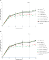
Effect of TGFβ knockdown on PRRSV copy numbers in PRRSV-2-inoculated PBMCs. Monocytes were transfected with TGFβAS1, then inoculated with either cPRRSV-2 or HP-PRRSV-2 and co-cultured with PBL (0 h), and finally stimulated with inducers i.e., (A) ConA or (B) PMAi (48 h). Monocytes inoculated with cPRRSV-2 or HP-PRRSV-2, then co-cultured with PBL and stimulated with inducer served as the PRRSV-2-inoculated control. Monocytes transfected with Scr, then inoculated with either cPRRSV-2 or HP-PRRSV-2, co-cultured with PBL, and finally stimulated with inducer served as PRRSV-2-inoculated/Scr control. Monocytes treated with transfection media (Tr.media), inoculated with cPRRSV-2 or HP-PRRSV-2, then co-cultured with PBL, and finally stimulated with inducer served as PRRSV-2-inoculated/Tr. media control. Cells receiving mock Ag plus inducer served as uninoculated control. Cell culture supernatants were collected for real-time PCR. The CT values were obtained, and PRRSV-2 ORF7 RNA copy numbers were calculated based on the CT standard curve generated from 101-108 copies of recombinant PRRSV-2 ORF7 plasmids. Data were presented in log 10 scale of copy number/ml. Error bars indicate the SD. Mean differences of PRRSV-2 ORF7 RNA copy numbers among groups at time points were tested by one-way repeated measures ANOVA, followed by Tukey HSD. Different superscript letters indicate significant difference. P<0.05 was set as a statistically significant level.
Figure 6
Figure 6
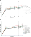
Effects of rTGFβ1 on PRRSV-2 ORF7 RNA copy numbers in PRRSV-2-inoculated monocytes co-cultured with PBL. Monocytes were treated with rTGFβ1 (10, 1 and 0.1 ng/ml final), inoculated with either cPRRSV-2 or HP-PRRSV-2, then co-cultured with PBL (0 h), and finally stimulated with inducers i.e., (A) ConA or (B) PMAi (48 h). Monocytes inoculated with cPRRSV-2 or HP-PRRSV-2, then co-cultured with PBL and stimulated with inducers served as PRRSV-2-inoculated control. Monocytes receiving mock Ag then co-cultured with PBL plus inducer served as uninoculated control. Cell culture supernatants were collected for real-time PCR. The CT values were obtained and PRRSV-2 ORF7 RNA copy numbers were calculated based on the CT standard curve generated from 101-108 copies of recombinant PRRSV-2 ORF7 plasmids. Data were presented in log 10 scale of copy number/ml. Error bars indicate the SD. Mean differences of PRRSV-2 ORF7 RNA copy numbers among groups at time points were tested by one-way repeated measures ANOVA, followed by Tukey HSD. Different superscript letters indicate significant difference. P<0.05 was set as a statistically significant level.
Figure 7
Figure 7
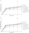
Effects of rIFNα on PRRSV-2 ORF7 RNA copy numbers in PRRSV-2-inoculated monocytes co-cultured with PBL. Monocytes were treated with rIFNα (10, 1 and 0.1 ng/ml final), inoculated with either cPRRSV-2 or HP-PRRSV-2, then co-cultured with PBL (0 h), and finally stimulated with inducers i.e., (A) ConA or (B) PMAi (48 h). Monocytes inoculated with cPRRSV-2 or HP-PRRSV-2, co-cultured with PBL, and finally stimulated with inducers served as PRRSV-2-inoculated control. Monocytes receiving mock Ag then co-cultured with PBL plus inducer served as uninoculated control. Cell culture supernatants were collected for real-time PCR. The CT values were obtained and PRRSV-2 ORF7 RNA copy numbers were calculated based on the CT standard curve generated from 101-108 copies of recombinant PRRSV-2 ORF7 plasmids. Data were presented in log 10 scale of copy number/ml. Error bars indicate the SD. Mean differences of PRRSV-2 ORF7 RNA copy numbers among groups at time points were tested by one-way repeated measures ANOVA, followed by Tukey HSD. Different superscript letters indicate significant difference. P<0.05 was set as a statistically significant level.
Figure 8
Figure 8
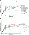
Effects of rTGFβ1 and rIFNα on PRRSV-2 ORF7 RNA copy numbers in PRRSV-2-inoculated monocytes then co-cultured with lymphocytes. Monocytes were treated with rTGFβ1 (10 ng/ml final), followed by rIFNα (10 ng/ml final), then inoculated with either cPRRSV-2 or HP-PRRSV-2, then co-cultured with PBL (0 h), and stimulated with inducers i.e., (A) ConA or (B) PMAi (48 h). Monocytes inoculated with cPRRSV-2 or HP-PRRSV-2, co-cultured with PBL, and stimulated with either ConA or PMAi served as PRRSV-2-inoculated control. Monocytes treated with rTGFβ1, co-cultured with PBL, then inoculated with either cPRRSV-2 or HP-PRRSV-2 (0 h), and stimulated with inducers (48 h) served as rTGFβ1-treated/PRRSV-2-inoculated control. Monocytes treated with rIFNα, co-cultured with PBL, then inoculated with either cPRRSV-2 or HP-PRRSV-2 (0 h), and stimulated with inducers (48 h) served as rIFNα -treated/PRRSV-2-inoculated control. Monocytes co-cultured with PBL receiving mock Ag plus inducers served as uninoculated control. Cell culture supernatants were collected for real-time PCR. The CT values were obtained and PRRSV-2 ORF7 RNA copy numbers were calculated based on the CT standard curve generated from 101-108 copies of recombinant PRRSV-2 ORF7 plasmids. Data were presented in log 10 scale of copy number/ml. Error bars indicate the SD. Mean differences of PRRSV-2 ORF7 RNA copy numbers among groups at time points were tested by one-way repeated measures ANOVA, followed by Tukey HSD. Different letters indicate significant differences. P<0.05 was set as a statistically significant level.

Similar articles

References

    1. Nathues H, Alarcon P, Rushton J, Jolie R, Fiebig K, Jimenez M, et al. . Cost of porcine reproductive and respiratory syndrome virus at individual farm level–an economic disease model. Prev veterinary Med. (2017) 142:16–29. doi: 10.1016/j.prevetmed.2017.04.006 - DOI - PubMed
    1. Zimmerman JJ, Dee SA, Holtkamp DJ, Murtaugh MP, Stadejek T, Stevenson GW, et al. . Porcine reproductive and respiratory syndrome viruses (porcine arteriviruses). Dis swine. (2019), 685–708. doi: 10.1002/9781119350927.ch41 - DOI
    1. Brinton MA, Gulyaeva AA, Balasuriya UB, Dunowska M, Faaberg KS, Goldberg T, et al. . ICTV virus taxonomy profile: Arteriviridae 2021. J Gen virology. (2021) 102:001632–3. doi: 10.1099/jgv.0.001632 - DOI - PMC - PubMed
    1. Kuhn JH, Lauck M, Bailey AL, Shchetinin AM, Vishnevskaya TV, Bào Y, et al. . Reorganization and expansion of the nidoviral family Arteriviridae. Arch virology. (2016) 161:755–68. doi: 10.1007/s00705-015-2672-z - DOI - PMC - PubMed
    1. Lalonde C, Provost C, Gagnon CA. Whole-genome sequencing of porcine reproductive and respiratory syndrome virus from field clinical samples improves the genomic surveillance of the virus. J Clin Microbiol. (2020) 58:e00097–e120. doi: 10.1128/JCM.00097-20 - DOI - PMC - PubMed

Publication types