. 2024 Jan 23;6(1):38-50.
doi: 10.1016/j.jaccao.2023.11.008. eCollection 2024 Feb.

Functional Validation of Doxorubicin-Induced Cardiotoxicity-Related Genes

Affiliations

Functional Validation of Doxorubicin-Induced Cardiotoxicity-Related Genes

Hananeh Fonoudi et al. JACC CardioOncol. .

Abstract

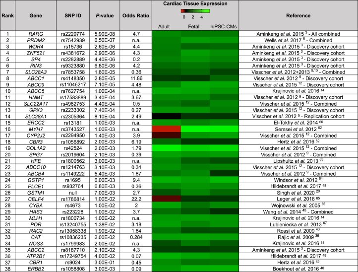
Background: Genome-wide association studies and candidate gene association studies have identified more than 180 genetic variants statistically associated with anthracycline-induced cardiotoxicity (AIC). However, the lack of functional validation has hindered the clinical translation of these findings.

Objectives: The aim of this study was to functionally validate all genes associated with AIC using human induced pluripotent stem cell-derived cardiomyocytes (hiPSC-CMs).

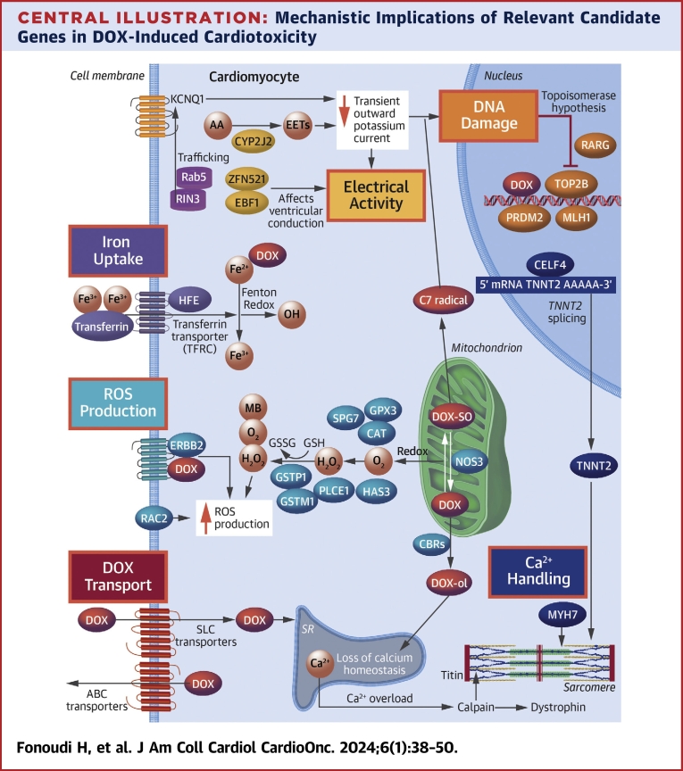
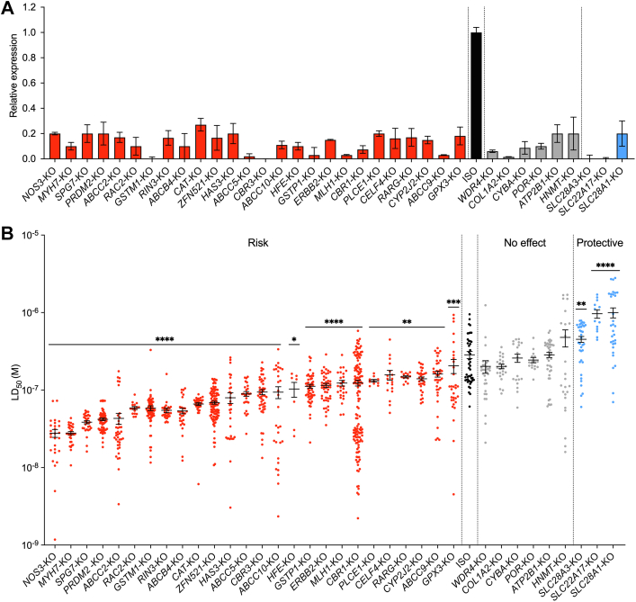
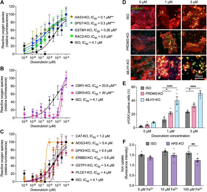
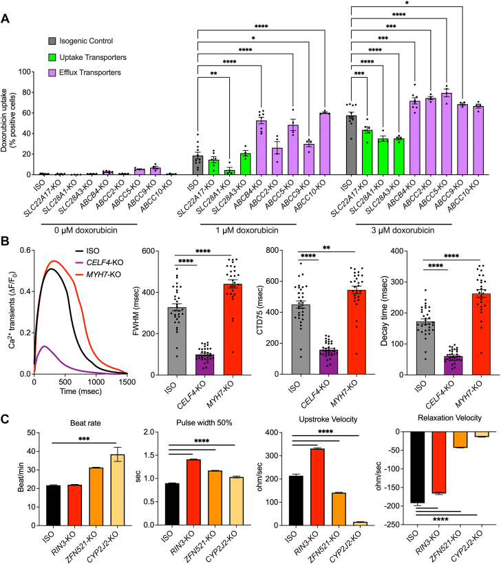
Methods: Through a systemic literature search, 80 genes containing variants significantly associated with AIC were identified. Additionally, 3 more genes with potential roles in AIC (GSTM1, CBR1, and ERBB2) were included. Of these, 38 genes exhibited expression in human fetal heart, adult heart, and hiPSC-CMs. Using clustered regularly interspaced short palindromic repeats/Cas9-based genome editing, each of these 38 genes was systematically knocked out in control hiPSC-CMs, and the resulting doxorubicin-induced cardiotoxicity (DIC) phenotype was assessed using hiPSC-CMs. Subsequently, functional assays were conducted for each gene knockout on the basis of hypothesized mechanistic implications in DIC.

Results: Knockout of 26 genes increased the susceptibility of hiPSC-CMs to DIC. Notable genes included efflux transporters (ABCC10, ABCC2, ABCB4, ABCC5, and ABCC9), well-established DIC-associated genes (CBR1, CBR3, and RAC2), and genome-wide association study-discovered genes (RARG and CELF4). Conversely, knockout of ATP2B1, HNMT, POR, CYBA, WDR4, and COL1A2 had no significant effect on the in vitro DIC phenotype of hiPSC-CMs. Furthermore, knockout of the uptake transporters (SLC28A3, SLC22A17, and SLC28A1) demonstrated a protective effect against DIC.

Conclusions: The present findings establish a comprehensive platform for the functional validation of DIC-associated genes, providing insights for future studies in DIC variant associations and potential mechanistic targets for the development of cardioprotective drugs.

Keywords: GWAS; cardiomyocytes; doxorubicin; genomics; human induced pluripotent stem cells.


Conflict of interest statement

This work was supported by National Institutes of Health grants R01 CA220002 and R01 CA261898, American Heart Association Transformational Project Award 18TPA34230105, and the Leducq Foundation (to Dr Burridge). The authors have reported that they have no relationships relevant to the contents of this paper to disclose.

Figures

None
Graphical abstract
Figure 1
Figure 1
Prioritization of Doxorubicin-Induced Cardiotoxicity–Associated Loci Table showing the 38 doxorubicin-induced cardiotoxicity–associated loci ranked on the basis of the single-nucleotide polymorphism (SNP) with highest P value from their respective publications., , , , , , , The heat map of cardiac tissue expression shows the expression of anthracycline-induced cardiotoxicity–associated genes in adult human heart (n = 2), in fetal human heart (n = 2), and in human induced pluripotent stem cell–derived cardiomyocytes (hiPSC-CMs) (n = 7) by RNA sequencing. n = number of distinct patient-specific samples.
Figure 2
Figure 2
Validation of KOs of 36 Genes Significantly Associated With Doxorubicin-Induced Cardiotoxicity (A) Validation of successful clustered regularly interspaced short palindromic repeats/Cas9–based gene knockout (KO) in human induced pluripotent stem cell–derived cardiomyocytes (hiPSC-CMs) using quantitative reverse transcriptase polymerase chain reaction. The data presented here are the relative expression of each studied gene in its own KO lines relative to isogenic control (ISO). (B) Effect of gene KOs on hiPSC-CM viability after doxorubicin treatments (72 hours). Each data point represents 1 median lethal dose (LD50) calculation on the basis of an individual experimental replicate derived from a 5-log doxorubicin dosing. Central bar represents mean. Error bars represent ±SEM. P values (∗P < 0.05, ∗∗P < 0.01, ∗∗∗P < 0.001, and ∗∗∗∗P < 0.0001) are derived from differences between gene KO and ISO (Mann-Whitney U test). Raw dose-response curves from which these LD50 values are derived are provided in Supplemental Figures 4 to 6.
Central Illustration
Mechanistic Implications of Relevant Candidate Genes in DOX-Induced Cardiotoxicity The different mechanisms of cardiotoxicity after exposure to doxorubicin (DOX) are highlighted in colored boxes. AA = arachidonic acid; ABC = adenosine triphosphate–binding cassette; EET = epoxyeicosatrienoic acid; mRNA = messenger RNA; ROS = reactive oxygen species; SLC = solute carrier; SR = sarcoplasmic reticulum.
Figure 3
Figure 3
Reactive Oxygen Species Production, DNA Damage, and Iron Uptake (A-C) Hydrogen peroxide levels measured by ROS-Glo assay (luminescence) in hiPSC-CMs after doxorubicin treatments (24 hours). (D) Representative images for γH2AX immunofluorescent staining in hiPSC-CMs after treatments with doxorubicin (24 hours, 1 and 3 μM). (E) Quantification of DNA damage on the basis of γH2AX staining in hiPSC-CMs using flow cytometry (ISO, n = 5; PRDM2-KO, n = 5; and MLH1-KO, n = 5). (F) Effect of HFE knockout on hiPSC-CM iron uptake (ISO, n = 6; and HFE-KO, n = 6) measured using calcein staining. Error bars represent ±SEM. n = full independent experimental replicates. ∗P < 0.05, ∗∗P ≤ 0.01, ∗∗∗P < 0.001, and ∗∗∗∗P < 0.0001 by Mann-Whitney U test (A-C) and 2-way analysis of variance (E and F). DAPI = 4′,6-diamidino-2-phenylindole; IC50 = half maximal inhibitory concentration; other abbreviations as in Figure 2.
Figure 4
Figure 4
Doxorubicin Uptake, Calcium Handling, and Contractility (A) Doxorubicin uptake in hiPSC-CMs with knockouts of SLC and ABC transporters (isotype, n = 11; SLC28A3-KO, n = 4; SLC22A17-KO, n = 6; SLC28A1-KO, n = 4; ABCC2-KO, n = 4; ABCB4-KO, n = 7; ABCC5-KO, n = 4; ABCC9-KO, n = 4; and ABCC10-KO, n = 4). n = full independent experimental replicates. (B) Effect of knocking out MYH7 and CELF4 on calcium transients. Left: representative calcium transients. Middle left: full width at half maximum (FWHM). Middle right: calcium transient duration 75% (CTD75). Right: decay time. (C) Contractility analysis using impedance measurement in RIN3-KO, ZFN521-KO, and CYP2J2-KO hiPSC-CMs (ISO, n = 41; RIN3-KO, n = 48; ZFN521-KO, n = 48; and CYP2J2-KO, n = 89). n = number of assessed wells of 96-well plate derived from at least 3 independent rounds of differentiation (B and C). Error bars represent ±SEM. ∗P < 0.05, ∗∗P ≤ 0.01, ∗∗∗P < 0.001, and ∗∗∗∗P < 0.0001 by 2-way analysis of variance (A) and 1-way analysis of variance (B and C). Abbreviations as in Figure 2.

References

    1. Bhatia S. Genetics of anthracycline cardiomyopathy in cancer survivors: JACC: CardioOncology state-of-the-art review. J Am Coll Cardiol CardioOnc. 2020;2(4):539–552. doi: 10.1016/j.jaccao.2020.09.006. - DOI - PMC - PubMed
    1. Cardinale D., Colombo A., Bacchiani G., et al. Early detection of anthracycline cardiotoxicity and improvement with heart failure therapy. Circulation. 2015;131(22):1981–1988. doi: 10.1161/CIRCULATIONAHA.114.013777. - DOI - PubMed
    1. Aminkeng F., Bhavsar A.P., Visscher H., et al. A coding variant in RARG confers susceptibility to anthracycline-induced cardiotoxicity in childhood cancer. Nat Genet. 2015;47(9):1079–1084. doi: 10.1038/ng.3374. - DOI - PMC - PubMed
    1. Schneider B.P., Shen F., Gardner L., et al. Genome-wide association study for anthracycline-induced congestive heart failure. Clin Cancer Res. 2017;23(1):43–51. doi: 10.1158/1078-0432.CCR-16-0908. - DOI - PMC - PubMed
    1. Wells Q.S., Veatch O.J., Fessel J.P., et al. Genome-wide association and pathway analysis of left ventricular function after anthracycline exposure in adults. Pharmacogenet Genomics. 2017;27(7):247–254. doi: 10.1097/FPC.0000000000000284. - DOI - PMC - PubMed