Review
. 2024 Feb 7:89:e70-e79.
doi: 10.5114/pjr.2024.135304. eCollection 2024.

Hand and wrist - what the hand surgeon wants to know from the radiologist

Affiliations
Review

Hand and wrist - what the hand surgeon wants to know from the radiologist

Piotr Czarnecki et al. Pol J Radiol. .

Abstract

Hand surgeons, as unique specialists, appreciate the complexity of the anatomy of the hand. A hand is not merely a group of anatomic structures but a separate organ that works by feeling, sending information to the brain, and enabling a variety of movements, from precise skills to firm tasks. Acute and chronic problems interfere with complicated hand function and potentially influence work or daily life activities for a long time. Thus, the surgeon's role is to propose appropriate treatment with predictable results. This paper attempts to specify the preoperative considerations and their influence on the choice of surgical procedure and the assessment of results potentially influencing further treatment. We have divided the manuscript by anatomical structures, which is a natural surgical assessment and planning approach. The most common problems were highlighted to introduce the method of decision-making and surgical solutions.

Keywords: hand and wrist; imaging; preoperative planning.


Conflict of interest statement

The authors report no conflicts of interest.

Figures

Figure 1
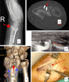
Figure 1
Neglected nonreduced lunate dislocation. A) Plain lateral radiogram (red arrow). B, C) 3D reconstruction. D) Blue arrow – scaphoid. E) After proximal row carpectomy joint is formed between the distal radius and capitate)
Figure 2
Figure 2
Ulnar screw blocking and chronically damaging the distal radioulnar joint. A) Plain radiogram. B) CT scan showing protruding screw causing ulnar impingement and erosion – red arrow
Figure 3
Figure 3
Volar plate screws conflict with extensors. A) Plain radiogram. B, C) CT scans showing screws between non-malreduced fragments (arrows). Sonographic (D) and intraoperative (E) presentation
Figure 4
Figure 4
Arthroscopic capsular repair of ulnar triangular fibrocartilage complex attachment using inside-out technique. A) Scoping from 3-4 portal, sutures are pulled out on the ulnar side. B) Inside out technique of suture placement with needles
Figure 5
Figure 5
Reinsertion of ulnar collateral ligament of the thumb with the usage of soft mini anchor. A) Anchor in the base of the proximal phalanx (blue arrow), torn ligament (red arrow), examples of soft (B) and titanium (C) implants
Figure 6
Figure 6
Staged flexor tendons reconstruction in massive scarring and no tendon continuity (A – between arrows): first stage – silicone rod (blue arrow) application with pulley reconstruction (B – pulleys reconstructed with FDS strip: red arrows)
Figure 7
Figure 7
Nerve reconstruction of the median nerve with sural grafts, the gap after neuroma resection (A – between arrows) can be assessed for planning a proper number of grafts (B – arrow pointing each graft)
Figure 8
Figure 8
Brachial plexus exploration with neurolysis of upper and middle trunks (arrows)
Figure 9
Figure 9
Lipoma (red arrows) of palmar region splitting fascicles of the median nerve (blue arrows)
Figure 10
Figure 10
Fast-growing chondroma of fourth metacarpal bone (A) in a 16-year-old patient, treated with excision and fibular graft, stabilised with Kirschner wires (B), graft united (C)

References

    1. Meals C, Meals R. Hand fractures: a review of current treatment strategies. J Hand Surg 2013; 38: 1021-1031. - PubMed
    1. Amrami KK, Frick MA, Matsumoto JM. Imaging for acute and chronic scaphoid fractures. Hand Clin 2019; 35: 241-257. - PubMed
    1. Chunara MH, McLeavy CM, Kesavanarayanan V, Paton D, Ganguly A. Current imaging practice for suspected scaphoid fracture in patients with normal initial radiographs: UK-wide national audit. Clin Radiol 2019; 74: 450-455. - PubMed
    1. Faccioli N, Santi E, Foti G, et al. . Cost-effectiveness of introducing cone-beam computed tomography (CBCT) in the management of complex phalangeal fractures: economic simulation. Musculoskelet Surg 2022; 106: 169-177. - PMC - PubMed
    1. Rhee PC, Medoff RJ, Shin AY. Complex distal radius fractures: an anatomic algorithm for surgical management. J Am Acad Orthop Surg 2017; 25: 77-88. - PubMed