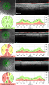
From the past to the present, optical coherence tomography in glaucoma: a practical guide to a common disease
- PMID: 38511134
- PMCID: PMC10951567
- DOI: 10.12688/f1000research.139975.2
From the past to the present, optical coherence tomography in glaucoma: a practical guide to a common disease
Abstract
Glaucoma comprises a group of disorders of the optic nerve that cause degenerative optic neuropathy, characterised by failure of neuroretinal rim tissue in the optic nerve head, retinal nerve fibre layer, and retinal ganglion cells. Glaucoma imposes a serious epidemiological threat, with an steady increase in the global number of cases. In the current ophthalmological practice, glaucoma is diagnosed via a series of examinations, including routine funduscopic examination, ocular tonometry, gonioscopy, measurement of the visual field, and assessment using the optical coherence tomography (OCT) technique. Nowadays, the OCT technique helps in systematising the diagnostic pathway and is a basic diagnostic tool for detection of early glaucomatous eye changes. It is also vital in assessing progression and monitoring treatment results of patients. The aim of this review was to present the OCT technique as a main tool in diagnosing and monitoring glaucoma.
Keywords: glaucoma; open-angle glaucoma; optic neuropathy; optical coherence tomography; retinal ganglion cells.
Copyright: © 2024 Zawadzka I and Konopińska J.
Conflict of interest statement
No competing interests were disclosed.
Figures

References
-
- Gupta D, Chen PP: Glaucoma. Am. Fam. Physician. 2016;93:668–674. - PubMed
Publication types
MeSH terms
LinkOut - more resources
Full Text Sources
Medical
Miscellaneous

