. 2024 Mar 21;134(9):e174726.
doi: 10.1172/JCI174726.

Dietary carbohydrates regulate intestinal colonization and dissemination of Klebsiella pneumoniae

Affiliations

Dietary carbohydrates regulate intestinal colonization and dissemination of Klebsiella pneumoniae

Aaron L Hecht et al. J Clin Invest. .

Abstract

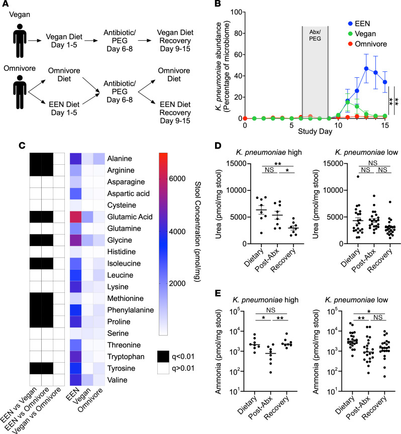
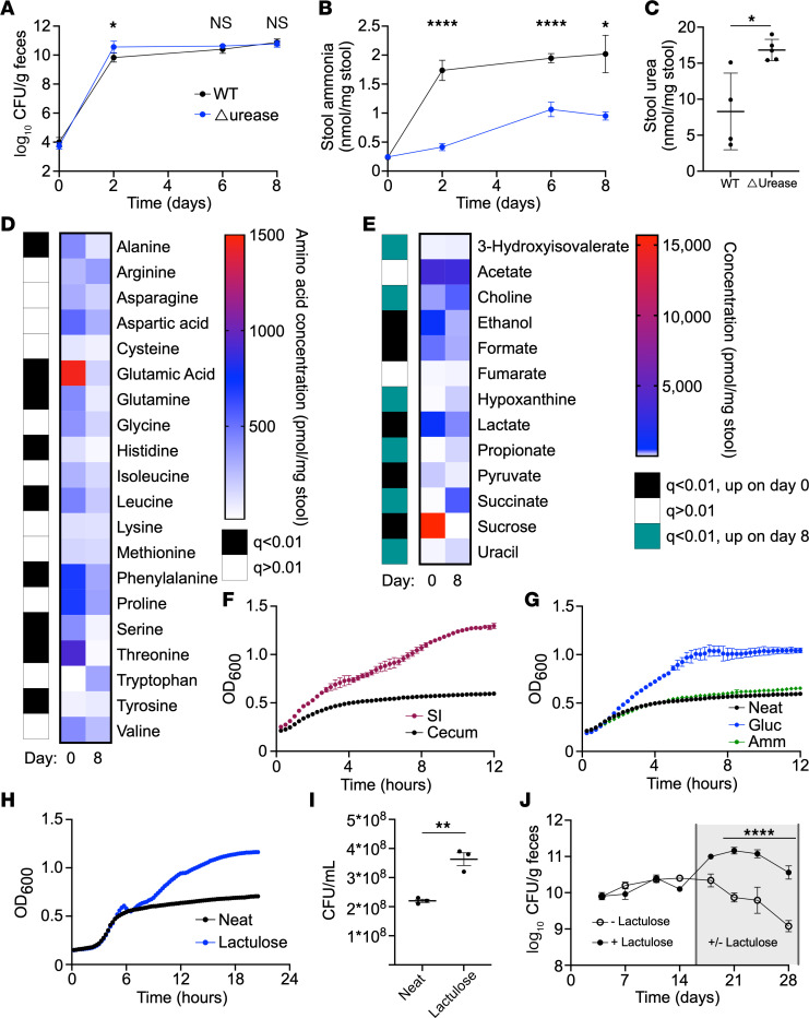
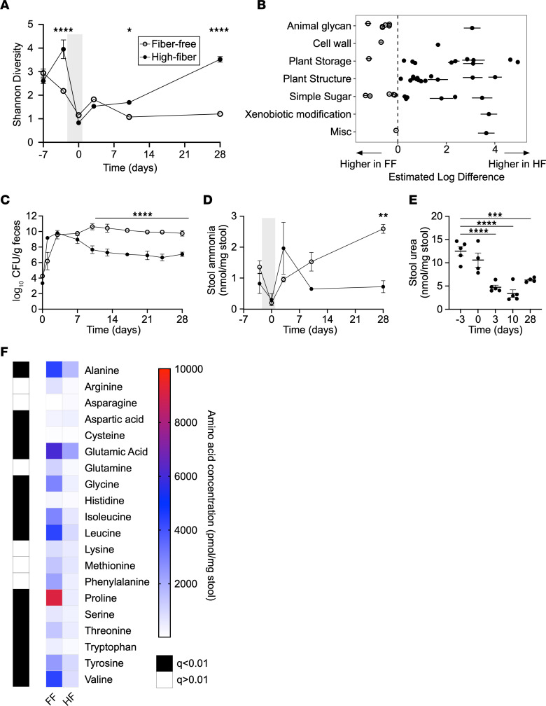
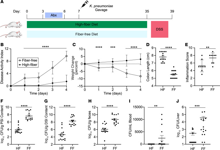
Bacterial translocation from the gut microbiota is a source of sepsis in susceptible patients. Previous work suggests that overgrowth of gut pathobionts, including Klebsiella pneumoniae, increases the risk of disseminated infection. Our data from a human dietary intervention study found that, in the absence of fiber, K. pneumoniae bloomed during microbiota recovery from antibiotic treatment. We thus hypothesized that dietary nutrients directly support or suppress colonization of this gut pathobiont in the microbiota. Consistent with our study in humans, complex carbohydrates in dietary fiber suppressed the colonization of K. pneumoniae and allowed for recovery of competing commensals in mouse models. In contrast, through ex vivo and in vivo modeling, we identified simple carbohydrates as a limiting resource for K. pneumoniae in the gut. As proof of principle, supplementation with lactulose, a nonabsorbed simple carbohydrate and an FDA-approved therapy, increased colonization of K. pneumoniae. Disruption of the intestinal epithelium led to dissemination of K. pneumoniae into the bloodstream and liver, which was prevented by dietary fiber. Our results show that dietary simple and complex carbohydrates were critical not only in the regulation of pathobiont colonization but also disseminated infection, suggesting that targeted dietary interventions may offer a preventative strategy in high-risk patients.

Keywords: Bacterial infections; Carbohydrate metabolism; Gastroenterology; Inflammatory bowel disease.


Conflict of interest statement

Conflict of interest: MG and GDW are co-inventors of the patent entitled “Compositions and methods comprising a defined microbiome and methods thereof” (US 20160243175A1).

Figures

Figure 1
Figure 1. A defined formula diet favors K. pneumoniae growth in humans.
(A) Diagram of the FARMM study design. Patients were randomized to a FF diet (EEN) or an omnivore diet; a third group remained on a vegan diet throughout the study. Recovery of the microbiome was monitored after antibiotic and PEG depletion. (B) Relative abundance of K. pneumoniae as a percentage of the microbiome determined via shotgun metagenomic sequencing, stratified by dietary group. (C) Heatmap of average stool amino acid concentrations during the microbiome recovery phase (day 14) of the FARMM study, stratified by diet. Black boxes denote a statistically significant difference of amino acid concentration between the indicated dietary groups. (D and E) Quantification of stool urea (D) and ammonia (E) at each phase of the FARMM study in samples from individuals with a high or low relative abundance of K. pneumoniae (defined as >20% K. pneumoniae by relative abundance during the recovery phase). Data are presented as the mean ± SEM. n = 10 participants per dietary group. *P < 0.05 and **P < 0.01, by 1-way ANOVA with Holm-Šidák’s correction for multiple comparisons (B), comparing the EEN versus omnivore and the EEN versus vegan groups on day 15, (D) multiple Mann-Whitney U test with the FDR method of Benjamini, Krueger, Yokutieli (C), or Kruskal-Wallis test with Dunn’s multiple-comparison test (E). Abx, antibiotics.
Figure 2
Figure 2. K. pneumoniae colonization is limited by carbon source availability and alters the nitrogen environment in the gut.
(AE) GF mice were colonized with WT or Δurease K. pneumoniae, and serial stool collections were done throughout the 1-week study. Fecal CFU (A) and stool ammonia (B) were monitored for 1 week after colonization. Fecal urea (C) was tested on day 8. Stool amino acid levels (D) and metabolites (E) were quantified from stool before (day 0) and after (day 8) WT K. pneumoniae colonization. (F) Growth of WT K. pneumoniae in small intestine (SI) or cecal extracts from mice monitored via OD600. (GI) Growth in cecal extracts supplemented with ammonia (Amm) or glucose (Gluc) (G) or lactulose (H and I) quantified by OD600 and CFU (I). Data for neat cecal extracts are presented in both F and G for reference. (J) Mice colonized with K. pneumoniae were subsequently treated with lactulose in the drinking water or water-only control. Data are presented as the mean ± SEM (A and J) or the mean ± SD (B, C, and FI). n = 4–5 mice per group (AE, J) or n = 3 wells per group (FI). Data represent combined results from 2 independent experiments (AE) or are from a single experiment representative of 3 independent experiments (FJ). *P < 0.05, **P < 0.01, and ****P < 0.001, by multiple unpaired, 2-tailed t tests with Benjamini Hochberg multiple corrections (D and E), with Bonferroni’s multiple corrections (A, B, and J) or by unpaired, 2-tailed t test (C and I).
Figure 3
Figure 3. Complex carbohydrates increase microbiome diversity and reduce K. pneumoniae colonization after antibiotic depletion.
(AF) Mice were provided a FF or a HF diet starting on day –7, treated with oral antibiotics on day –3 to day 0 (gray shading), and gavaged with K. pneumoniae (day 0). Serial stool samples were subjected to analysis as follows: (A) Shannon α diversity of stool microbiome from mice provided a FF or HF diet including the antibiotic treatment period (gray shading) and recovery, as determined by shotgun metagenomic sequencing. (B) GH genes with significantly different abundances between FF and FH diets 4 weeks after gavage with K. pneumoniae grouped by substrate type. Open circles represent genes with higher levels in mice on the FF diet, and closed circles represent genes with higher levels in mice on the HF diet. (C) K. pneumoniae fecal CFU for mice on a FF (open circles) or HF (closed circles) diet measured 4 weeks after K. pneumoniae gavage. (D) Stool ammonia was quantified before and after antibiotic treatment (gray shading) and K. pneumoniae gavage for mice subjected to a FF (open circles) or HF (closed circles) diet. (E) Stool urea levels were quantified for mice provided a FF diet. (F) Heatmap of amino acid concentrations in mice on a FF or HF diet after colonization with K. pneumoniae. Data are presented as the mean ± SD (AE). n = 5 mice per group. Data are representative of 2–3 independent experiments (CF). *P < 0.05, **P < 0.01, ***P < 0.001, and ****P < 0.0001, by multiple 2-tailed t tests with Bonferroni’s correction for multiple comparisons (A, C, and D), 1-way ANOVA with Bonferroni’s correction for multiple comparisons (E), or multiple Mann-Whitney U tests with the FDR method of Benjamini, Krueger, and Yokutieli (F).
Figure 4
Figure 4. A HF diet protects against DSS colitis and K. pneumoniae dissemination from microbiota.
(A) Diagram of experimental design. Mice were placed on a HF or a FF diet and colonized with WT K. pneumoniae after antibiotic pretreatment followed by treatment with 5% DSS in the drinking water. (B and C) Disease activity (B) and weight change (C) were monitored during DSS treatment. After 4 days of treatment, mice were euthanized. (D) Colon length was quantified. (E) Colon tissue was microscopically scored for evidence of inflammation. (FJ) CFU were quantified in the proximal small intestine (F), distal small intestine (G), feces (H), blood (I), and liver (J). Data are presented as the mean ± SD. Results are a combination of 3 independent experiments (B, C, and FJ; n = 15 mice per group) or a combination of 2 independent experiments (E; n = 8–9 mice per group). **P < 0.01, ***P < 0.001, and ****P < 0.0001, by multiple unpaired, 2-tailed t tests with Bonferroni’s multiple correction (B and C), unpaired, 2-tailed t test (DH), or Mann-Whitney U test (I and J).

Update of

References

    1. Bajaj JS, et al. The evolving challenge of infections in cirrhosis. N Engl J Med. 2021;384(24):2317–2330. doi: 10.1056/NEJMra2021808. - DOI - PubMed
    1. Wiest R, et al. Pathological bacterial translocation in liver cirrhosis. J Hepatol. 2014;60(1):197–209. doi: 10.1016/j.jhep.2013.07.044. - DOI - PubMed
    1. McMahon S, et al. Contribution of the oral and gastrointestinal microbiomes to bloodstream infections in Leukemia patients. Microbiol Spectr. 2023;11(3):e00415-23. doi: 10.1128/spectrum.00415-23. - DOI - PMC - PubMed
    1. Taur Y, et al. Intestinal domination and the risk of bacteremia in patients undergoing allogeneic hematopoietic stem cell transplantation. Clin Infect Dis. 2012;55(7):905–914. doi: 10.1093/cid/cis580. - DOI - PMC - PubMed
    1. Freedberg DE, et al. Pathogen colonization of the gastrointestinal microbiome at intensive care unit admission and risk for subsequent death or infection. Intensive Care Med. 2018;44(8):1203–1211. doi: 10.1007/s00134-018-5268-8. - DOI - PMC - PubMed

Publication types