. 2024 Mar 21;19(3):e0300718.
doi: 10.1371/journal.pone.0300718. eCollection 2024.

The role of GAPDH in the selective toxicity of CNP in melanoma cells

Affiliations

The role of GAPDH in the selective toxicity of CNP in melanoma cells

Claudia von Montfort et al. PLoS One. .

Abstract

Background: Malignant melanoma is the most aggressive form of skin cancer with a rather poor prognosis. Standard chemotherapy often results in severe side effects on normal (healthy) cells finally being difficult to tolerate for the patients. Shown by us earlier, cerium oxide nanoparticles (CNP, nanoceria) selectively killed A375 melanoma cells while not being cytotoxic at identical concentrations on non-cancerous cells. In conclusion, the redox-active CNP exhibited both prooxidative as well as antioxidative properties. In that context, CNP induced mitochondrial dysfunction in the studied melanoma cells via generation of reactive oxygene species (primarily hydrogen peroxide (H2O2)), but that does not account for 100% of the toxicity.

Aim: Cancer cells often show an increased glycolytic rate (Warburg effect), therefore we focused on CNP mediated changes of the glucose metabolism.

Results: It has been shown before that glyceraldehyde 3-phosphate dehydrogenase (GAPDH) activity is regulated via oxidation of a cysteine in the active center of the enzyme with a subsequent loss of activity. Upon CNP treatment, formation of cellular lactate and GAPDH activity were significantly lowered. The treatment of melanoma cells and melanocytes with the GAPDH inhibitor heptelidic acid (HA) decreased viability to a much higher extent in the cancer cells than in the studied normal (healthy) cells, highlighting and supporting the important role of GAPDH in cancer cells.

Conclusion: We identified glyceraldehyde 3-phosphate dehydrogenase (GAPDH) as a target protein for CNP mediated thiol oxidation.


Conflict of interest statement

The authors have declared that no competing interests exist.

Figures

Fig 1
Fig 1. A375 melanoma cells are more susceptible to increasing concentrations of hydrogen peroxide and cerium oxide nanoparticles.
(A) Subconfluent melanoma cells (A375) and normal human epidermal melanocytes (NHEM) were treated with 10 μM to 1000 μM H2O2 for 24 h. Cell viability was determined by MTT assay. The percentage of cell viability in comparison to the untreated control (set to 100%) is presented. Data are presented as means ± SEM. * p = 0.0313 A375 vs NHEM (student´s t test performed for each concentration). (B) Non-linear curve fit analysis for determination of the IC50 values of the data shown in (A) using Prism software (GraphPad, San Diego, USA) (A375: 95%CI, 55.81 to 275; NHEM: 95%CI, 223.6 to 1298). Data are presented as means ± SD. (C) Subconfluent A375 melanoma cells and normal human epidermal melanocytes (NHEM) were treated with 150 μM, 300 μM and 450 μM CNP for 96 h. Thereafter, cell viability was determined by MTT assay. The percentage of cell viability in comparison to the mock-treated control (set to 100%) is presented. Three independent experiments were performed (n = 3). Data are presented as means ± SEM. **p<0.01 and *p<0.05 A375 vs NHEM (student´s t test performed for each concentration).
Fig 2
Fig 2. Incubation with CNP induced thiol oxidation of GAPDH in A375 melanoma cells.
Subconfluent A375 melanoma cells were treated with either 2 mM H2O2 for 15 minutes or 300 μM CNP for 4 h and 24 h, respectively. Thiol oxidation was assessed via Sulfo Biotics Redox State Monitoring Kit with subsequent Western Blotting and probing for GAPDH. SHx0 to SHx3 bands refer to the amount of linker which was bound to thiol groups of the protein. If all thiol groups were oxidized prior to the incubation with the protein linker/SHifter, no SHifter was able to bind to the oxidized protein (SHx0). Experiments were performed in triplicates (n = 3).
Fig 3
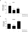
Fig 3. CNP lowered GAPDH activity in melanoma cells.
Subconfluent A375 melanoma cells (A) or melanocytes (B) were treated with either 500 μM H2O2 for 2 h or 300 μM CNP for 24 h. The GAPDH activity was determined with the GAPDH activity Kit. Three independent experiments were performed (n = 3). The results represent means ± SEM and were normalized to cell numbers. *p<0.05 vs control (ANOVA and Dunett´s test).
Fig 4
Fig 4. CNP lowered GAPDH activity and the intracellular lactate amount in melanoma cells.
Subconfluent A375 melanoma cells were treated with 300 μM CNP for 24 h, 48 h and 72 h. As positive control, cells were treated with 500 μM H2O2 for 2 h and 1 mM 2-DG for 24 h. The L-Lactate level in melanoma cells was determined with the L-Lactate Assay Kit. Three independent experiments were performed (n = 3). The results represent means ± SEM and were normalized to the number of the cells in each sample. ***p<0.001 vs control (ANOVA and Dunett´s test).
Fig 5
Fig 5. A375 melanoma cells show a higher sensitivity to GAPDH inhibition than normal melanocytes.
Subconfluent A375 melanoma cells (A) or melanocytes (B) were either mock treated or treated with the indicated concentrations of heptelidic acid (HA) for 96 h. Cell viability was determined by MTT assay. The percentage of viable cells was calculated compared to the mock-treated control which was set to 100%. Three independent experiments were performed (n = 3). Data are presented as means ± SEM. **p<0.01 and *p<0.05 vs control (ANOVA and Dunett´s test). # p<0.05 vs control and HA treatment.
Fig 6
Fig 6. Lactate administration partially rescues CNP induced toxicity in melanoma cells.
Subconfluent A375 melanoma cells were either untreated (ct) or treated for 96 h with 300 μM CNP alone, lactate (10 mM, 20 mM) alone or a combination. Cell viability was determined by MTT assay. The percentage of viable cells was calculated compared to the mock-treated control which was set to 100%. Three independent experiments were performed (n = 3). Data are presented as means ± SEM. (p value was determined by ANOVA and Dunett´s test).

Similar articles

References

    1. Lo JA, Fisher DE. The melanoma revolution: from UV carcinogenesis to a new era in therapeutics. Science. 2014;346: 945–949. doi: 10.1126/science.1253735 - DOI - PMC - PubMed
    1. Del Fiore P, Russo I, Ferrazzi B, Monico AD, Cavallin F, Filoni A, et al.. Melanoma in Adolescents and Young Adults: Evaluation of the Characteristics, Treatment Strategies, and Prognostic Factors in a Monocentric Retrospective Study. Front Oncol. 2021;11: 725523. doi: 10.3389/fonc.2021.725523 - DOI - PMC - PubMed
    1. Patra JK, Das G, Fraceto LF, Campos EVR, Rodriguez-Torres M del P, Acosta-Torres LS, et al.. Nano based drug delivery systems: recent developments and future prospects. J Nanobiotechnol. 2018;16: 71. doi: 10.1186/s12951-018-0392-8 - DOI - PMC - PubMed
    1. Brenneisen P, Reichert A. Nanotherapy and Reactive Oxygen Species (ROS) in Cancer: A Novel Perspective. Antioxidants. 2018;7: 31. doi: 10.3390/antiox7020031 - DOI - PMC - PubMed
    1. Gagnon J, Fromm KM. Toxicity and Protective Effects of Cerium Oxide Nanoparticles (Nanoceria) Depending on Their Preparation Method, Particle Size, Cell Type, and Exposure Route. Eur J Inorg Chem. 2015;2015: 4510–4517. doi: 10.1002/ejic.201500643 - DOI

Substances