. 2024 Mar 26;121(13):e2319856121.
doi: 10.1073/pnas.2319856121. Epub 2024 Mar 21.

IL7 increases targeted lipid nanoparticle-mediated mRNA expression in T cells in vitro and in vivo by enhancing T cell protein translation

Affiliations

IL7 increases targeted lipid nanoparticle-mediated mRNA expression in T cells in vitro and in vivo by enhancing T cell protein translation

Caitlin M Tilsed et al. Proc Natl Acad Sci U S A. .

Abstract

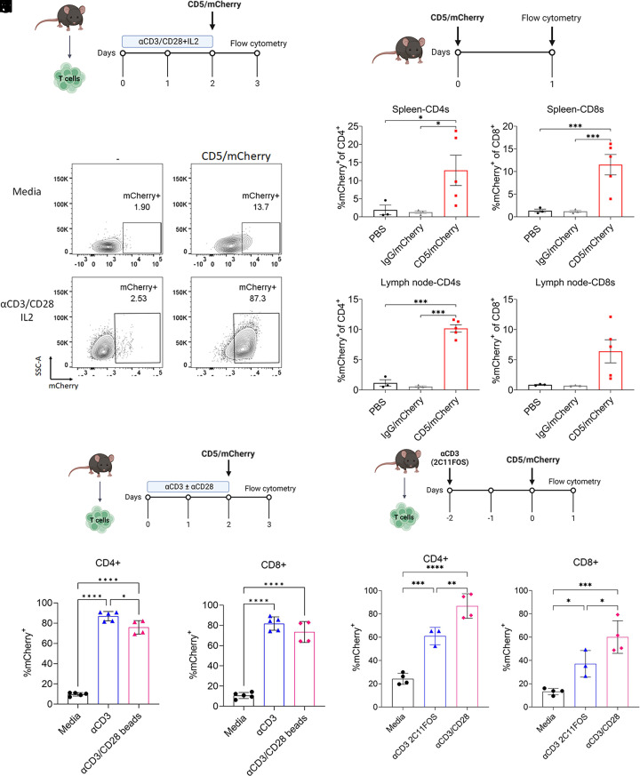
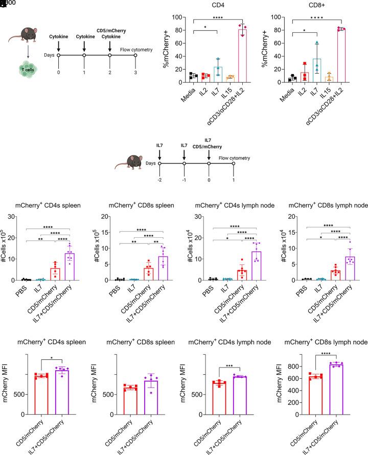
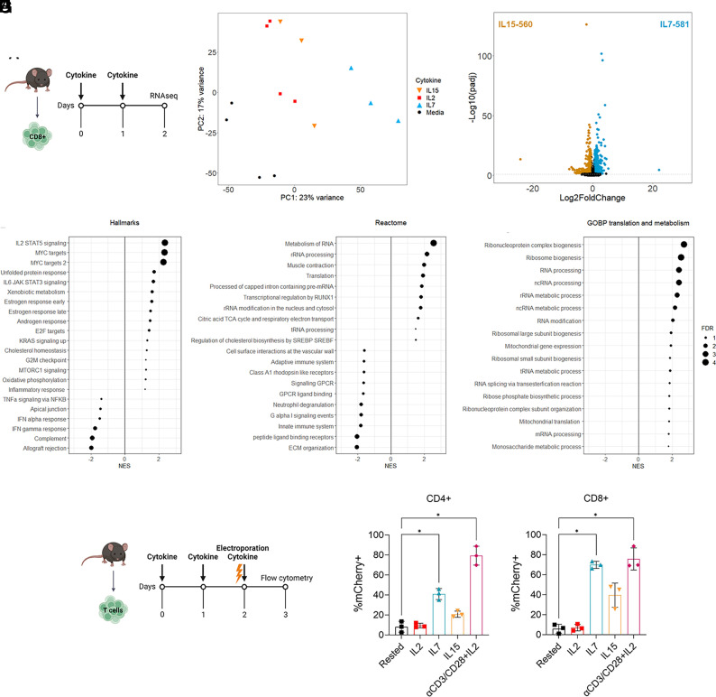
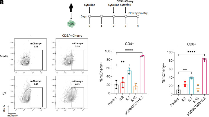
The use of lipid nanoparticles (LNP) to encapsulate and deliver mRNA has become an important therapeutic advance. In addition to vaccines, LNP-mRNA can be used in many other applications. For example, targeting the LNP with anti-CD5 antibodies (CD5/tLNP) can allow for efficient delivery of mRNA payloads to T cells to express protein. As the percentage of protein expressing T cells induced by an intravenous injection of CD5/tLNP is relatively low (4-20%), our goal was to find ways to increase mRNA-induced translation efficiency. We showed that T cell activation using an anti-CD3 antibody improved protein expression after CD5/tLNP transfection in vitro but not in vivo. T cell health and activation can be increased with cytokines, therefore, using mCherry mRNA as a reporter, we found that culturing either mouse or human T cells with the cytokine IL7 significantly improved protein expression of delivered mRNA in both CD4+ and CD8+ T cells in vitro. By pre-treating mice with systemic IL7 followed by tLNP administration, we observed significantly increased mCherry protein expression by T cells in vivo. Transcriptomic analysis of mouse T cells treated with IL7 in vitro revealed enhanced genomic pathways associated with protein translation. Improved translational ability was demonstrated by showing increased levels of protein expression after electroporation with mCherry mRNA in T cells cultured in the presence of IL7, but not with IL2 or IL15. These data show that IL7 selectively increases protein translation in T cells, and this property can be used to improve expression of tLNP-delivered mRNA in vivo.

Keywords: IL7; lipid nanoparticles; translation.


Conflict of interest statement

Competing interests statement:B.A.S. and A.B. are employees of Capstan Therapeutics. C.H.J., H.A., D.W., S.M.A., and H.P. are scientific founders and hold equity in Capstan Therapeutics. Authors have patent filings in the field of RNA therapeutics.

Figures

Fig. 1.
Fig. 1.
Activating T cells using anti-CD3 antibodies improves tLNP-induced mCherry expression rates in vitro but not in vivo. (A) Experimental design for in vitro T cell activation. T cells were magnetically isolated from the spleens of C57BL/6 mice, activated with αCD3/CD28 beads, and supplemented with IL2. Forty-eight hours later, beads were removed, 1 µg of tLNP was added per million cells, and flow cytometry was performed 24 h later. (B) Representative flow plots of nonactivated (media) and activated T cells treated with mCherry tLNP. (C) Experimental design for the in vivo experiment. Mice were given 10 µg of IgG or anti-CD5-targeted LNP i.v, with spleen and lymph nodes collected 24 h later. (D and E) Percent mCherry+ CD4+ (D) or CD8+ (E) T cells in the spleen. (F and G) Percent mCherry+ CD4+ (F) or CD8+ (G) T cells in the lymph node. (H) Experimental design. T cells were isolated and cultured with 1 µg/mL αCD3 or αCD3/CD28 beads for 48 h. One microgram of CD5/mCherry tLNP was added per million cells and incubated for an additional 24 h before flow cytometry was performed (I and J) Percentage of mCherry+ CD4+ (I) or CD8+ (J) T cells. (K) Experimental design. T cells were isolated and cultured with or without 10 µg/mL αCD3 2C11FOS for 48 h while bead-activated T cells were used as a positive control. One microgram of CD5-mCherry tLNP was added per million cells and incubated for an additional 24 h before flow cytometry was performed (L and M) Percentage of mCherry+ CD4+ (L) or CD8+ (M) T cells after tLNP transfection. One-way ANOVA with Sidak’s test was used for multiple comparisons. *P < 0.05, **P < 0.01, ***P < 0.001, and ****P < 0.0001.
Fig. 2.
Fig. 2.
IL7 enhances CD5/mCherry tLNP-induced protein expression in vitro and in vivo. (A) Experimental design for in vitro cytokine treatment. T cells were isolated and cultured with either IL2, IL7, or IL15. Cytokines were refreshed daily, and tLNPs were added on day 2. (B and C) Proportion of mCherry+ CD4+ (B) and CD8+ (C) T cells in vitro in the presence of IL2, IL7, or IL15. Media-treated and activated T cells were used as controls. (D) Experimental design for in vivo experiments. C57BL/5 mice were injected i.p with 5 µg of recombinant murine IL7 daily for 3 d. On the third day, mice received 10 µg CD5-mCherry tLNPs i.v. Twenty-four hours after tLNP treatment, spleens and lymph nodes were collected for flow cytometry. (E and F) Total number of CD4+ (E) and CD8+ (F) T cells in the spleen expressing mCherry. (G and H) Total number of CD4+ (G) and CD8+ (H) T cells in the lymph node expressing mCherry. (I and J) Median fluorescent intensity (MFI) of mCherry of the mCherry+ CD4+ (I) and CD8+ (J) T cells in the spleen. (K and L) MFI of mCherry of the mCherry+ CD4+ (K) and CD8+ (L) T cells in the spleen. One-way ANOVA with Sidak’s test was used for multiple comparisons. *P < 0.05, **P < 0.01, ***P < 0.001, and ****P < 0.0001.
Fig. 3.
Fig. 3.
IL7 increases specific changes in genes related to protein translation in T cells and supports increased protein production after mRNA electroporation. (A) Experimental design. CD8+ T cells were isolated from the spleens of C57BL/6 mice and cultured with T cell media alone or supplemented with IL2, IL7, or IL15. After 48 h, T cells were collected and sent for bulk RNA sequencing. (B) PCA of cytokine-treated T cells. Counts were VST normalized. (C) Volcano plot showing the differentially expressed genes between IL7- and IL15-treated CD8 T cells. Genes with a positive log2fold change are up-regulated with IL7 treatment compared to IL15, while a negative log2fold change indicates upregulation with IL15 treatment compared to IL7. (DF) Gene set enrichment analysis using the list of differentially expressed genes between IL7- and IL15-treated cells using the Hallmarks (D), Reactome (E), or Gene Ontology Biological Processes (GOBP) (F) databases. Gene sets associated with translation and metabolism are shown from the GOBP analysis. Size of point indicates the FDR (−log10Padj), with a positive NES indicating enrichment in IL7-treated cells and a negative NES indicating enrichment in IL15-treated cells. (G) Experimental design for electroporation experiment. T cells were isolated from the spleen C57BL/6 mice and either activated using CD3/CD28 beads or cultured in T cell media supplemented with IL2, IL7, or IL15. After 48 h T cells were electroporated with 2 µg of mCherry mRNA per 1 million cells. mCherry expression was measured 24 h later. (H and I) Proportion of mCherry+ CD4+ (H) or CD8+ (I) T cells after electroporation with mCherry mRNA. One-way ANOVA with Sidak’s test was used for multiple comparisons. *P < 0.05, **P < 0.01, ***P < 0.001, and ****P < 0.0001.
Fig. 4.
Fig. 4.
IL7 pretreatment of human T cells improves CD5-mCherry tLNP-induced protein expression rates in vitro. (A) Experimental design for tLNP transfection. Human T cells were isolated from PBMC and either activated using anti-CD3/CD28 beads with IL2 or cultured in T cell media supplemented with IL2, IL7, or IL15 and replenished after 48 h. After 72 h, T cells were transfected with 0.6 µg of CD5-LNP-mCherry per 2 × 10⁵ cells. mCherry expression was measured 24 h later. (B) Representative flow plots of rested (Media) and IL7-cultured T cells transfected with CD5-LNP mCherry. Data are from two donors across three experiments. (C and D) Proportion of mCherry+ CD4+ (B) or CD8+ (C) T cells after transfection with mCherry mRNA. One-way ANOVA with Sidak’s test was used for multiple comparisons. *P < 0.05, **P < 0.01, ***P < 0.001, and ****P < 0.0001.

References

    1. Agrawal S., Garg A., Varshney V., Recent updates on applications of lipid-based nanoparticles for site-specific drug delivery. Pharm. Nanotechnol. 10, 24–41 (2022). - PubMed
    1. Godbout K., Tremblay J. P., Delivery of RNAs to specific organs by lipid nanoparticles for gene therapy. Pharmaceutics 14, 2129 (2022). - PMC - PubMed
    1. Rurik J. G., et al. , CAR T cells produced in vivo to treat cardiac injury. Science 375, 91–96 (2022). - PMC - PubMed
    1. Tombácz I., et al. , Highly efficient CD4+ T cell targeting and genetic recombination using engineered CD4+ cell-homing mRNA-LNPs. Mol. Ther. J. Am. Soc. Gene Ther. 29, 3293–3304 (2021). - PMC - PubMed
    1. Breda L., et al. , In vivo hematopoietic stem cell modification by mRNA delivery. Science 381, 436–443 (2023). - PMC - PubMed