. 2024 Jun 25;8(12):3109-3119.
doi: 10.1182/bloodadvances.2023012125.

Genomic profiling of mycosis fungoides identifies patients at high risk of disease progression

Affiliations

Genomic profiling of mycosis fungoides identifies patients at high risk of disease progression

Léa Fléchon et al. Blood Adv. .

Abstract

Mycosis fungoides (MF) is the most prevalent primary cutaneous T-cell lymphoma, with an indolent or aggressive course and poor survival. The pathogenesis of MF remains unclear, and prognostic factors in the early stages are not well established. Here, we characterized the most recurrent genomic alterations using whole-exome sequencing of 67 samples from 48 patients from Lille University Hospital (France), including 18 sequential samples drawn across stages of the malignancy. Genomic data were analyzed on the Broad Institute's Terra bioinformatics platform. We found that gain7q, gain10p15.1 (IL2RA and IL15RA), del10p11.22 (ZEB1), or mutations in JUNB and TET2 are associated with high-risk disease stages. Furthermore, gain7q, gain10p15.1 (IL2RA and IL15RA), del10p11.22 (ZEB1), and del6q16.3 (TNFAIP3) are coupled with shorter survival. Del6q16.3 (TNFAIP3) was a risk factor for progression in patients at low risk. By analyzing the clonal heterogeneity and the clonal evolution of the cohort, we defined different phylogenetic pathways of the disease with acquisition of JUNB, gain10p15.1 (IL2RA and IL15RA), or del12p13.1 (CDKN1B) at progression. These results establish the genomics and clonality of MF and identify potential patients at risk of progression, independent of their clinical stage.


Conflict of interest statement

Conflict-of-interest disclosure: The authors declare no competing financial interests.

Figures

None
Graphical abstract
Figure 1.
Figure 1.
Landscape of most recurrent genomic alterations in MF. (A) Landscape of genomic alterations in 67 tumor samples of MF divided into LR and HR disease based on the TNMB stages. Alterations are divided into SNVs and SCNAs. (B) Driver oncogene maps of JUNB and MAPK1. (C) Relative enrichment of signature activities per samples divided into LR, LR progressors, and HR.
Figure 2.
Figure 2.
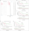
Correlation of genomic events to the disease stage. (A) Forest plot showing the association between individual genes alteration and clinical stage of MF divided into LR and HR, as depicted by OR. (B-E) Kaplan-Meier plots of individual genetic factors predictive of OS in univariate and multivariate models of 48 patients with a newly diagnosed MF: del10p11.22 (B); gain of 10p15.1 (C); gain of 7q (D); and del6q16.3 (E). (F) Kaplan-Meier curves for analysis of time to progression in patients with LR disease. P values were derived from log-rank test. (G) CD25 immunohistochemistry at diagnosis of MF skin biopsies in patients with gain of 10p15.1 (top panels, MF sample of patient 18 and patient 14 with presence of transformed MF cells) or without gain of 10p15.1 (bottom panels, MF sample of patient 23 and patient 16). Scale bars indicate 150 μm.
Figure 2.
Figure 2.
Correlation of genomic events to the disease stage. (A) Forest plot showing the association between individual genes alteration and clinical stage of MF divided into LR and HR, as depicted by OR. (B-E) Kaplan-Meier plots of individual genetic factors predictive of OS in univariate and multivariate models of 48 patients with a newly diagnosed MF: del10p11.22 (B); gain of 10p15.1 (C); gain of 7q (D); and del6q16.3 (E). (F) Kaplan-Meier curves for analysis of time to progression in patients with LR disease. P values were derived from log-rank test. (G) CD25 immunohistochemistry at diagnosis of MF skin biopsies in patients with gain of 10p15.1 (top panels, MF sample of patient 18 and patient 14 with presence of transformed MF cells) or without gain of 10p15.1 (bottom panels, MF sample of patient 23 and patient 16). Scale bars indicate 150 μm.
Figure 3.
Figure 3.
Clonal heterogeneity and inferred timing of genetic drivers. (A) Proportion in which recurrent drivers are found as clonal or subclonal across the 67 samples (top), along with the individual CCF values for each sample affected by a driver (bottom). Median CCF values are shown (bottom, bars represent the median and interquartile range for each driver). (B) Correlation matrix for the most recurrent genomic alterations for the 48 patients with MF, with Fisher exact test to detect such significant pair of mutations. (C) Timing of genomic alterations with early events at top and late events at bottom. Color indicates alteration types. Arrows between 2 alterations were drawn when 2 drivers were found in 1 sample with an excess of clonal to subclonal events. Dashed arrows indicate 1 clonal-subclonal pair and solid arrows indicate ≥2 clonal-subclonal pairs.
Figure 4.
Figure 4.
Clonal evolution of sequential samples. (A-E) Fish plots of 5 serial cases of MF with samples at LR stages who progressed to HR stages. (F) One serial case with both time points at LR stage because the patient did not progress. TNMB classification has been indicated (green for LR and dark red for HR samples). tMF, transformed MF.
Figure 5.
Figure 5.
Recurrent genetic alterations affecting key signaling pathways involved in MF. In this diagram, frequently mutated genes with well-established roles in these signaling pathways have been depicted representing the proteins they encode. TCR, T-Cell Receptor.

References

    1. Willemze R. Cutaneous T-cell lymphoma: epidemiology, etiology, and classification. Leuk Lymphoma. 2003;44(Suppl 3):S49–S54. - PubMed
    1. Willemze R, Jaffe ES, Diaz-Perez JL, et al. WHO-EORTC classification for cutaneous lymphomas. Blood. 2005;105(10):3768–3785. - PubMed
    1. Olsen E, Vonderheid E, Pimpinelli N, et al. Revisions to the staging and classification of mycosis fungoides and Sézary syndrome: a proposal of the International Society for Cutaneous Lymphomas (ISCL) and the cutaneous lymphoma task force of the European Organization of Research and Treatment of Cancer (EORTC) Blood. 2007;110(6):1713–1722. - PubMed
    1. Diamandidou E, Colome-Grimmer M, Fayad L, Duvic M, Kurzrock R. Transformation of mycosis fungoides/sezary syndrome: clinical characteristics and prognosis. Blood. 1998;92(4):1150–1159. - PubMed
    1. Vergier B, de Muret A, Beylot-Barry M, et al. Transformation of mycosis fungoides: clinicopathological and prognostic features of 45 cases. Blood. 2000;95(7):2212–2218. - PubMed

Publication types