. 2024 May 28;8(10):2410-2423.
doi: 10.1182/bloodadvances.2023011484.

RUNX1 C-terminal mutations impair blood cell differentiation by perturbing specific enhancer-promoter networks

Affiliations

RUNX1 C-terminal mutations impair blood cell differentiation by perturbing specific enhancer-promoter networks

Nathan D Jayne et al. Blood Adv. .

Abstract

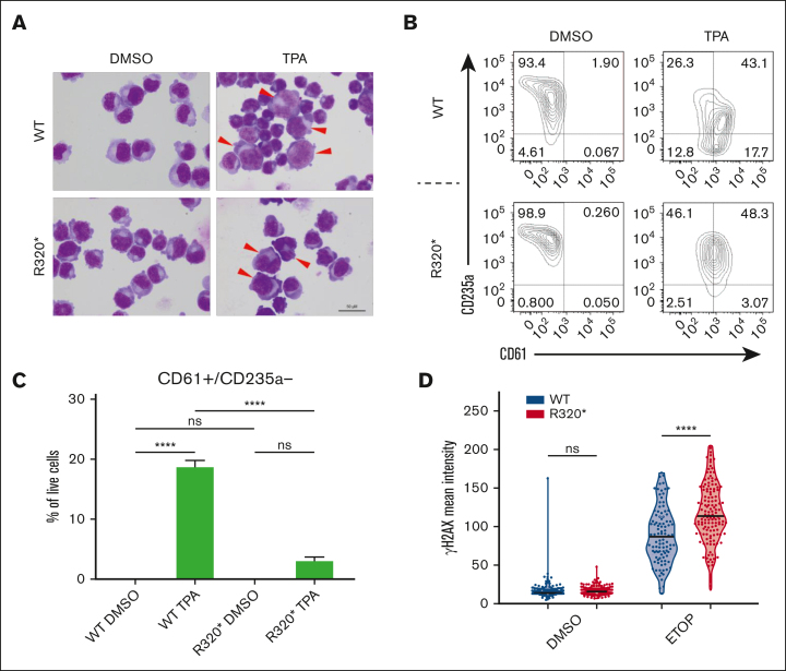
The transcription factor RUNX1 is a master regulator of hematopoiesis and is frequently mutated in myeloid malignancies. Mutations in its runt homology domain (RHD) frequently disrupt DNA binding and result in loss of RUNX1 function. However, it is not clearly understood how other RUNX1 mutations contribute to disease development. Here, we characterized RUNX1 mutations outside of the RHD. Our analysis of the patient data sets revealed that mutations within the C-terminus frequently occur in hematopoietic disorders. Remarkably, most of these mutations were nonsense or frameshift mutations and were predicted to be exempt from nonsense-mediated messenger RNA decay. Therefore, this class of mutation is projected to produce DNA-binding proteins that contribute to the pathogenesis in a distinct manner. To model this, we introduced the RUNX1R320∗ mutation into the endogenous gene locus and demonstrated the production of RUNX1R320∗ protein. Expression of RUNX1R320∗ resulted in the disruption of RUNX1 regulated processes such as megakaryocytic differentiation, through a transcriptional signature different from RUNX1 depletion. To understand the underlying mechanisms, we used Global RNA Interactions with DNA by deep sequencing (GRID-seq) to examine enhancer-promoter connections. We identified widespread alterations in the enhancer-promoter networks within RUNX1 mutant cells. Additionally, we uncovered enrichment of RUNX1R320∗ and FOXK2 binding at the MYC super enhancer locus, significantly upregulating MYC transcription and signaling pathways. Together, our study demonstrated that most RUNX1 mutations outside the DNA-binding domain are not subject to nonsense-mediated decay, producing protein products that act in concert with additional cofactors to dysregulate hematopoiesis through mechanisms distinct from those induced by RUNX1 depletion.


Conflict of interest statement

Conflict-of-interest disclosure: B.R. is a cofounder of Epigenome Technologies, Inc and has equity in Arima Genomics, Inc. The remaining authors declare no competing financial interests.

The current affiliation for Z.L. is School of Life Sciences, Southern University of Science and Technology, Shenzhen, Guangdong, China.

The current affiliation for D.-H.L. is School of Systems Biomedical Science, Soongsil University, Seoul, Republic of Korea.

The current affiliation for P.B.C. is Genome Institute of Singapore, Agency for Science, Technology and Research (A∗STAR), Singapore.

Figures

None
Graphical abstract
Figure 1.
Figure 1.
C-terminal RUNX1 mutations are frequently frameshift and nonsense, resulting in transcripts that are exempt from nonsense-mediated decay. (A) Lollipop plot of hematopoietic mutations in RUNX1 (isoform 1c NP_001754.2) in the COSMIC database with accompanying transcript exons displayed (top). Truncating mutations include nonsense, nonstop, frameshift deletion, frameshift insertion, and splice site. In-frame deletions and in-frame insertions are considered in-frame mutations, and all other nonmissense mutations are labeled as “Other.” Enlarged region of exons 7 and 8 of RUNX1 denoting NMD exempt mutations (bottom). Mutations that result in a premature stop codon in the final exon (exon 8) or within 50 nucleotides upstream of the last exon-exon junction (exon 7-8) are predicted to be exempt from NMD. (B) Analysis of C-terminal RUNX1 mutations beyond the RHD. Frameshift and nonsense mutations represented 304 of 387 mutations (78.55%), whereas all other in frame mutations consisting of missense, in-frame insertions and deletions, coding silent substitutions, and compound substitution combined account for 83 of 387 mutations (21.45%). NMD analysis was performed on 304 frameshift and nonsense mutations, examining premature stop codons within the region defined in panel (A). A total of 76.3% (232/304) of C-terminal frameshift and nonsense mutations were predicted to be exempt from NMD. (C) Schematic of RUNX1 protein domains and knock-in R320 mutation using CRISPR-Cas9. (D) Sanger sequencing of the RUNX1R320∗ homozygous knock-in mutation compared with the wild-type RUNX1 sequence. K562 cells were nucleofected with Cas9, RUNX1 targeting gRNA, and R320 donor template. The gRNA (black underline) targeted exon 7 (isoform 1c NM_001754.5) and the donor oligo template results in a TAA codon from TCG at R320. Single-cell clones were screened for homozygous mutations, confirmed by sequencing the targeted region, and analyzed using the ICE tool by Synthego. (E) Western blot of wild-type (WT) RUNX1 and RUNX1R320∗ K562 cells along with β-actin loading control. Both lines were subjected to the same nucleofection process as the +/− CRISPR-Cas9 editing components. Whole cell lysate was extracted and used to confirm the presence of both wild-type and RUNX1R320∗ proteins. Densitometry calculations were performed using β-actin normalization. Arrow indicates a possible nonspecific signal. (F) RUNX1 transcript levels in RUNX1 wild-type and RUNX1R320∗ cells and RUNX family members as measured using the DESeq2 analysis software package. Each line was subjected to RNA-seq and sampled in triplicate (n = 3). Student t test was used, significance: ∗ P ≤ .05; ∗∗ P ≤ .01; ∗∗∗ P ≤ .001; ∗∗∗∗ P ≤ .0001.
Figure 2.
Figure 2.
RUNX1R320∗ results in differentiation block and increased DNA damage sensitivity. (A) Representative images of RUNX1 wild-type and RUNX1R320∗ cells treated with DMSO or 10 nM TPA for 48 hours. Differentiating cells are denoted with arrows. (B-C) Representative flow cytometry analysis of RUNX1 wild-type and RUNX1R320∗ K562 cells which were treated with DMSO and 5 nM TPA for 48 hours. The MK marker CD61 (integrin β3 chain) was analyzed, along with the erythroid marker CD235a (glycophorin A). Live cells were divided into 4 groups using the FACS diva software based on the presence (+/−) of CD61 and CD235a. DMSO-treated control cells were compared with TPA-treated cells for both the RUNX1 wild-type and RUNX1R320∗ genotypes (n = 3). Significance was determined using 2-way ANOVA. (D) DNA damage levels in RUNX1 wild-type and RUNX1R320∗ cells upon treatment with ETOP and CPT relative to the DMSO control. The cells were treated with 25 μM ETOP or CPT for 1 hour at 37°C before fixation and staining. DAPI was used to identify the nuclei of cells and the γH2AX mean signal intensity was measured per cell within the nucleus. Student t test was used to determine significance. ∗ P ≤ .05; ∗∗ P ≤ .01; ∗∗∗ P ≤ .001; ∗∗∗∗ P ≤ .0001. DMSO, dimethyl sulfoxide; FACS, fluorescence-activated cell sorting; DAPI, 4′,6-diamidino-2-phenylindole.
Figure 3.
Figure 3.
RUNX1R320∗ results in significant transcriptional dysregulation of megakaryocytic differentiation pathways and MYC targets. (A) Principal component analysis of RUNX1 wild-type (n = 3) and RUNX1R320∗ (n = 3) RNA-seq samples following analysis using DESeq2. (B) Volcano plot showing differentially expressed genes between RUNX1 wild-type and RUNX1R320∗ cells. Genes were considered significantly differentially expressed (red) with FDR ≤ 0.05 and fold-change ≥ ±1.5). (C) Comparison of differentially expressed genes between RUNX1R320∗ and RUNX1 knockdown experiments. RUNX1R320∗ cells were compared with RUNX1 wild-type controls and RUNX1 shRNA knockdown cells to shRNA control cells in triplicate. Both data sets were analyzed with DESeq2, with significance determined by FDR ≤ 0.05, and fold-change ≥ ±1.5. (D) Reactome pathway analysis of differentially expressed genes in RUNX1R320∗ and RUNX1 knockdown cells, as described in panels (A-C). Pathways were considered significant with P-value < .05. (E-F) GSEA enrichment results between wild-type and RUNX1R320∗ RNA-seq data sets, NES = normalized enrichment score. (G) MYC expression in RUNX1 and RUNX1R320∗ cells via RNA-seq. Student t test was used to determine significance: ∗ P ≤ .05; ∗∗ P ≤ .01; ∗∗∗ P ≤ .001; ∗∗∗∗ P ≤ .0001.
Figure 4.
Figure 4.
RUNX1R320∗ differential binding is most enriched at enhancer regions. (A-B) Annotation of RUNX1 and RUNX1R320∗ binding sites using the ChIPSeeker annotation of the hg38 genome for all peaks. Wild-type peaks = 40,679; RUNX1R320∗ peaks = 38,233. (C) Differential binding volcano plot between RUNX1 wild-type and RUNX1R320∗ ChIP-seq data sets, significantly upregulated binding shown in (red) and downregulated binding (blue) compared with R320/WT. (D) Analysis of gene expression in RUNX1R320∗ cells relative to RUNX1 WT for genes with RUNX1 promoter binding. (E) Enrichment of RUNX1R320∗ peaks genome-wide using ENCODE K562 annotation data across up, nc (no change), and downregulated binding relative to RUNX1 WT. H3K27ac, H3K4me1, H3K4me3, H3K27me3, and H3K9me3 were used to annotate the enhancers, promoters, transcribed regions, repressed regions, and heterochromatin, respectively. (F) RUNX1 motif presence across enhancers and promoters with up- or downregulated binding of RUNX1R320∗ relative to RUNX1.
Figure 5.
Figure 5.
GRID-seq reveals extensive remodeling of enhancer-promoter connections in RUNX1R320∗ cells. (A) Representative heat map of the GRID-seq data set detecting RNA association with DNA regions across chromosome 21, only interactions within chromosome 21 are shown. (B) Z-score of detected RNA-DNA interactions classified as local, cis, and trans. Local interactions represent nascent RNA interactions with the gene body, cis interactions are within the same chromosome and outside the gene body region, and trans interactions are interchromosomal. (C) RNA-DNA interaction density across distance after log transformation, demonstrating the power law model of DNA looping and interaction described by Lieberman-Aiden et al (D) Enhancer-promoter interactions identified solely in either RUNX1 wild-type (WT) or RUNX1R320∗ (R320∗) cells or present in both (shared), as detected by GRID-seq. (E) Motif analysis of RUNX1R320∗ regulated enhancer and promoter regions in (D) selected significantly enriched motifs shown. (F) Normalized read counts of forkhead box gene expression in K562 RUNX1 wild-type and RUNX1R320∗ cells via RNA-seq. Each bar represents the mean of 3 replicates and standard deviation. FOXK1 and FOXK2 subfamilies were measured against the remaining FOX subfamilies using 1-way ANOVA. ∗ P ≤ .05; ∗∗ P ≤ .01; ∗∗∗ P ≤ .001; ∗∗∗∗ P ≤ .0001.
Figure 6.
Figure 6.
FOXK2 cooperates with RUNX1R320∗ to regulate MYC. (A) Analysis of K562 H3K27ac (ENCODE ENCFF465GBD), FOXK2 (ENCODE ENCFF286IOU), RUNX1 wild-type, and RUNX1R320∗ binding at the MYC and MYC enhancer regions upstream and downstream of MYC (top). GRID-seq long-range interaction map of chromatin-associated RNAs at MYC locus (bottom). Interaction strength with a greater score between RUNX1 and RUNX1R320∗ is denoted in blue and red, respectively. (B-C) CUT&RUN qPCR analysis of FOXK2, RUNX1, and RUNX1R320∗ at MYC BENC enhancer element 3 ‘E3’. (D-E) Western blots examining FOXK2 and MYC protein levels in wild-type (WT) RUNX1 and RUNX1R320∗ cells transduced with nontargeting shCtl or FOXK2 shRNAs with a β-actin loading control. (F) Model describing the roles of RUNX1R320 and FOXK2 in MYC enhancer regulation. Student t test was used to determine significance: ∗ P ≤ .05; ∗∗ P ≤ .01; ∗∗∗ P ≤ .001; ∗∗∗∗ P ≤ .0001.

References

    1. Sood R, Kamikubo Y, Liu P. Role of RUNX1 in hematological malignancies. Blood. 2017;129(15):2070–2082. - PMC - PubMed
    1. Chen MJ, Yokomizo T, Zeigler BM, Dzierzak E, Speck NA. Runx1 is required for the endothelial to haematopoietic cell transition but not thereafter. Nature. 2009;457(7231):887–891. - PMC - PubMed
    1. Wang Q, Stacy T, Binder M, Marín-Padilla M, Sharpe AH, Speck NA. Disruption of the Cbfa2 gene causes necrosis and hemorrhaging in the central nervous system and blocks definitive hematopoiesis. Proc Natl Acad Sci U S A. 1996;93(8):3444–3449. - PMC - PubMed
    1. Okuda T, Van Deursen J, Hiebert SW, Grosveld G, Downing JR. AML1, the target of multiple chromosomal translocations in human leukemia, is essential for normal fetal liver hematopoiesis. Cell. 1996;84(2):321–330. - PubMed
    1. Dowdy CR, Xie R, Frederick D, et al. Definitive hematopoiesis requires Runx1 C-terminal-mediated subnuclear targeting and transactivation. Hum Mol Genet. 2010;19(6):1048–1057. - PMC - PubMed

Publication types

MeSH terms

Substances