. 2024 May 2;63(5):2301232.
doi: 10.1183/13993003.01232-2023. Print 2024 May.

Gasdermin B, an asthma-susceptibility gene, promotes MAVS-TBK1 signalling and airway inflammation

Affiliations

Gasdermin B, an asthma-susceptibility gene, promotes MAVS-TBK1 signalling and airway inflammation

Tao Liu et al. Eur Respir J. .

Abstract

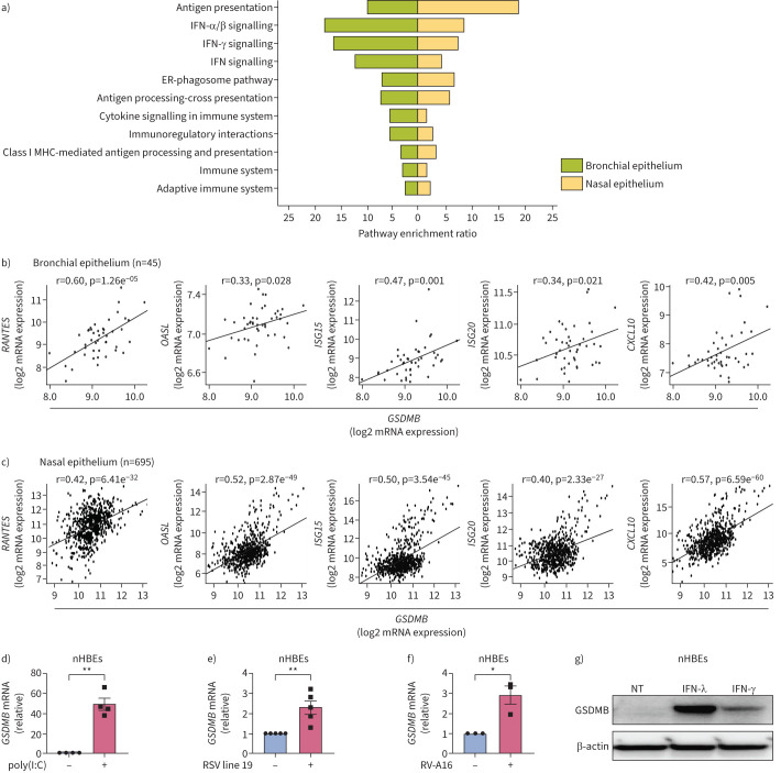
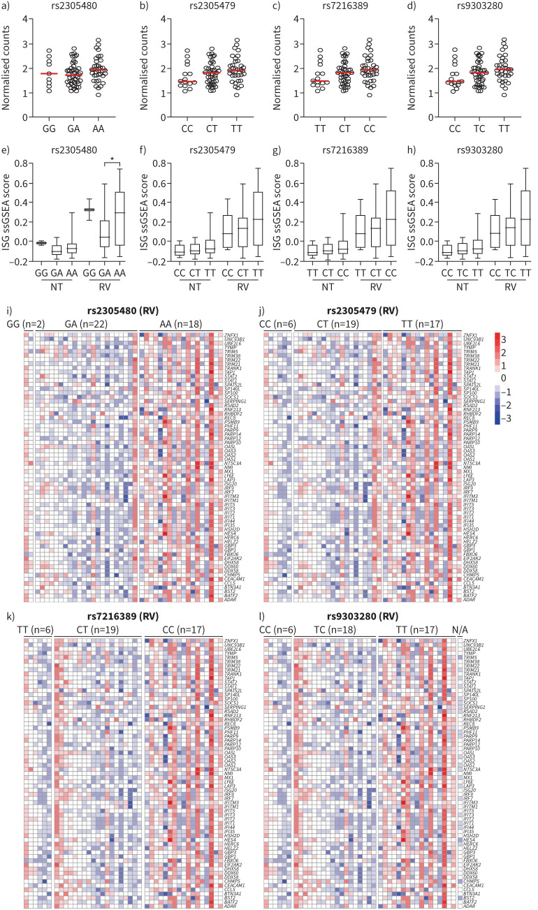
Rationale: Respiratory virus-induced inflammation is the leading cause of asthma exacerbation, frequently accompanied by induction of interferon-stimulated genes (ISGs). How asthma-susceptibility genes modulate cellular response upon viral infection by fine-tuning ISG induction and subsequent airway inflammation in genetically susceptible asthma patients remains largely unknown.

Objectives: To decipher the functions of gasdermin B (encoded by GSDMB) in respiratory virus-induced lung inflammation.

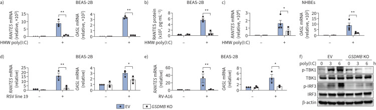
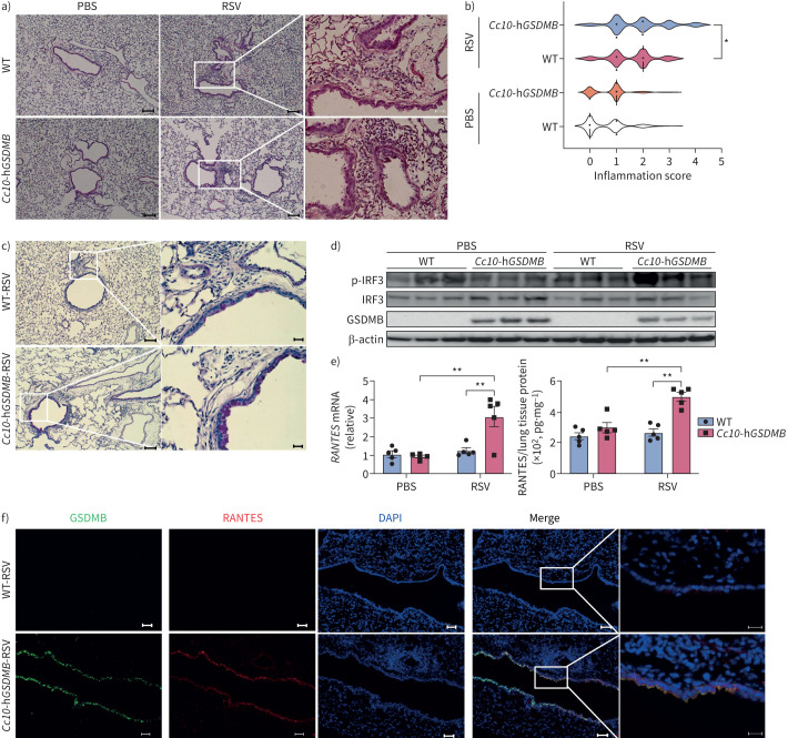
Methods: In two independent cohorts, we analysed expression correlation between GSDMB and ISG s. In human bronchial epithelial cell line or primary bronchial epithelial cells, we generated GSDMB-overexpressing and GSDMB-deficient cells. A series of quantitative PCR, ELISA and co-immunoprecipitation assays were performed to determine the function and mechanism of GSDMB for ISG induction. We also generated a novel transgenic mouse line with inducible expression of human unique GSDMB gene in airway epithelial cells and infected the mice with respiratory syncytial virus to determine the role of GSDMB in respiratory syncytial virus-induced lung inflammation in vivo.

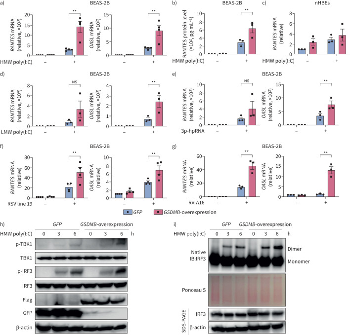
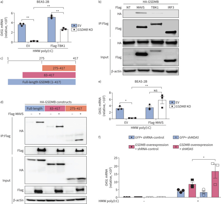
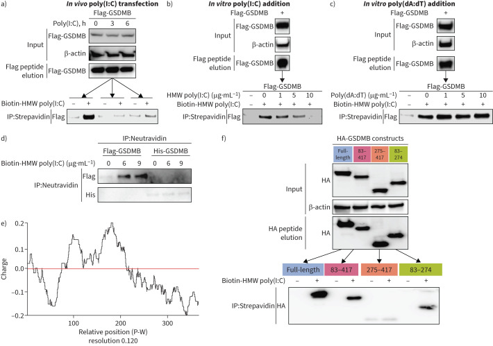
Results: GSDMB is one of the most significant asthma-susceptibility genes at 17q21 and acts as a novel RNA sensor, promoting mitochondrial antiviral-signalling protein (MAVS)-TANK binding kinase 1 (TBK1) signalling and subsequent inflammation. In airway epithelium, GSDMB is induced by respiratory viral infections. Expression of GSDMB and ISGs significantly correlated in respiratory epithelium from two independent asthma cohorts. Notably, inducible expression of human GSDMB in mouse airway epithelium led to enhanced ISGs induction and increased airway inflammation with mucus hypersecretion upon respiratory syncytial virus infection.

Conclusions: GSDMB promotes ISGs expression and airway inflammation upon respiratory virus infection, thereby conferring asthma risk in risk allele carriers.


Conflict of interest statement

Conflict of interest: The authors have no potential conflicts of interest to disclose.

Figures

FIGURE 1
FIGURE 1
Gasdermin B (GSDMB) is closely related to interferon-stimulated gene (ISG) activation in respiratory epithelial cells. a) The Gene Ontology enrichment analysis of these genes significantly correlated with GSDMB expression in both Asthma BRIDGE (BioRepository for Integrative Genomic Exploration) samples (bronchial, green) and Genes–Environments & Admixture in Latino Americans (GALA) II study (nasal, yellow). b, c) Expression of RANTES, OASL, ISG15, ISG20 and CXCL10 significantly correlated with expression of GSDMB in Asthma BRIDGE samples (b) and GALA II study samples (c). d–f) Expression of GSDMB in normal human bronchial epithelial cells (nHBEs) transfected with polyinosinic-polycytidylic acid (poly(I:C)) for 6 h (d) or infected with respiratory syncytial virus (RSV) line 19 (multiplicity of infection=1) for 24 h (e) or rhinovirus 16-A (RV-A16) (multiplicity of infection=1) for 72 h (f). g) Protein levels of GSDMB were measured by Western blotting in nHBEs treated with interferon (IFN)-λ (500 ng·mL−1) or IFN-γ (500 ng·mL−1) for 24 h. For b and c, expression levels of each gene were log2-transformed and quantile-normalised. Then, the linear correlation between the expression levels of two genes was determined using a t-test for Pearson's correlation coefficient. For b–f, mean±sem shown from at least three independent biological replicates. ER: endoplasmic reticulum; MHC: major histocompatibility complex; NT: no treatment. *: p<0.05; **: p<0.01 (t-test).
FIGURE 2
FIGURE 2
Gasdermin B (GSDMB) promotes RNA mimic or RNA virus-induced interferon-stimulated gene (ISG) induction. a, b) mRNA levels of RANTES and OASL (a) or extracellular RANTES protein levels (b) were measured in BEAS-2B cells with overexpression of GSDMB and transfected with high molecular weight (HMW) polyinosinic-polycytidylic acid (poly(I:C)) for 6 h. c) Expression of RANTES in normal human bronchial epithelial cells (nHBEs) with overexpression of GSDMB after transfection of HMW poly(I:C) for 6 h. d, e) RNA level of RANTES and OASL in BEAS-2B cells with overexpression of GSDMB and transfected with low molecular weight (LMW) poly(I:C) (d) or 5′-triphosphate hairpin RNA (3p-hpRNA) for 6 h (e), respectively. f, g) Expression of RANTES and OASL in BEAS-2B cells with overexpression of GSDMB and infected with respiratory syncytial virus (RSV) line 19 (multiplicity of infection=1, 24 h) (f) or rhinovirus A16 (RV-A16) (multiplicity of infection=1, 72 h) (g). h) Measurements of interferon regulatory factor 3 (IRF3) and TANK binding kinase 1 (TBK1) phosphorylation by immunoblotting in green fluorescent protein (GFP)-overexpressing versus GSDMB-overexpressing cells. i) IRF3 dimerisation in BEAS-2B cells with overexpression of GSDMB after transfection of HMW poly(I:C) for 0, 3 and 6 h. Cells expressing GFP were used as controls. For a–g, mean±sem shown from at least three independent biological replicates. For all immunoblot data, representative results were shown from two independent biological experiments. ns: nonsignificant. **: p<0.01 (two-way ANOVA).
FIGURE 3
FIGURE 3
Deficiency of gasdermin B (GSDMB) impairs induction of interferon-stimulated genes (ISGs) by RNA mimic or RNA virus. a) Expression of RANTES and OASL in BEAS-2B cells with or without knockout (KO) of GSDMB after transfection of high molecular weight (HMW) polyinosinic-polycytidylic acid (poly(I:C)) for 6 h. b) RANTES protein levels in supernatants were measured in BEAS-2B cells transfected with HMW poly(I:C) for 6 h. c) Expression of RANTES and OASL in normal human bronchial epithelial cells (nHBEs) with or without GSDMB KO after transfection of HMW poly(I:C) for 6 h. d, e) Expression of RANTES in empty vector (EV) or GSDMB KO BEAS-2B cells infected with respiratory syncytial virus (RSV) line 19 (multiplicity of infection=1, 24 h) (d) or rhinovirus 16-A (RV-A16) (multiplicity of infection=1, 72 h) (e). f) Measurements of interferon regulatory factor 3 (IRF3) and TANK binding kinase 1 (TBK1) phosphorylation by immunoblotting in EV versus GSDMB-overexpressing cells. EV-transfected cells were used as controls. Mean±sems shown from at least three independent biological replicates. *: p<0.05; **: p<0.01 (two-way ANOVA).
FIGURE 4
FIGURE 4
Gasdermin B (GSDMB) binds to mitochondrial antiviral-signalling protein (MAVS). a) Expression of OASL in control or GSDMB knockout (KO) BEAS-2B cells transfected with either empty vector (EV) or Flag-tagged TANK binding kinase 1 (TBK1), followed by high molecular weight (HMW) polyinosinic-polycytidylic acid (poly(I:C)) transfection for 6 h. b) Co-immunoprecipitation (IP) studies: anti-haemagglutinin (HA) immunoblots of either input or immunoprecipitated (IP:Flag) protein complex with anti-Flag from HEK293T cells transfected with HA-GSDMB and either Flag-MAVS, Flag-TBK1 or Flag-interferon regulatory factor 3 (IRF3). c) Schematic illustration of HA-GSDMB constructs used in co-IP assays in d. d) Flag-MAVS co-immunoprecipitated with various domains of HA-GSDMB in HEK 293T cells. e) Expression of OASL in control or GSDMB KO BEAS-2B cells transfected with EV or Flag-MAVS, followed by HMW poly(I:C) transfection for 6 h. f) Expression of OASL in GFP-overexpressing or GSDMB-overexpressing cells with MDA5 depletion, followed by transfection of HMW poly(I:C) for 6 h. For a, e, f, mean±sems shown from at least three independent biological replicates. For all immunoblot data, representative results are shown from two independent biological experiments. NT: no treatment; ns: nonsignificant; sh: small hairpin. *: p<0.05; **: p<0.01 (two-way ANOVA).
FIGURE 5
FIGURE 5
Gasdermin B (GSDMB) binds RNA as a potential RNA sensor. a) In vitro binding of biotin-high molecular weight (HMW) polyinosinic-polycytidylic acid (poly(I:C)) (3 µg·mL−1) and immunoprecipitated Flag-GSDMB from BEAS-2B cells with stable expression of Flag-GSDMB detected by immunoprecipitation (IP) with streptavidin beads and immunoblot analysis with anti-Flag antibody. b, c) Dose-dependent competitive binding of unlabelled HMW poly(I:C) (b), but not unlabelled poly(deoxyadenylic-deoxythymidylic) acid (poly(dA:dT) (c) with Flag-GSDMB against biotin-labelled poly(I:C) (3 µg·mL−1). d) Pure Flag-GSDMB instead of Flag-GSDMD protein binds to biotin-HMW poly(I:C) as shown by IP with neutravidin beads and immunoblotting analysis with anti-Flag antibody. e) Distribution of GSDMB electric charge was predicted based on each amino acid in the protein sequence. f) Haemagglutinin (HA)-GSDMB and its deletion mutants underwent IP and were eluted from BEAS-2B cells after transfection, followed by subsequent IP for their binding with biotin-labelled HMW poly(I:C). For all immunoblot data, representative results are shown from two independent biological experiments. His: histidine; P-W: position-window.
FIGURE 6
FIGURE 6
Human gasdermin B (hGSDMB) promotes respiratory syncytial virus (RSV)-induced mitochondrial antiviral-signalling protein (MAVS)-TANK binding kinase 1 (TBK1) signalling, inflammation and mucus production in mouse models. a) Representative haematoxylin and eosin (H&E) staining images of lung specimens from wild-type (WT), hGSDMB mice intranasally infected with 2×10⁴ pfu RSV line 19 (n=5) or PBS (n=5), respectively. Scale bars: 100 μm or 20 μm (inset). b) Peri-airway inflammation was scored based on the number of immune cells infiltrating surrounding airways as shown by H&E staining. c) Periodic acid–Schiff staining in WT and hGSDMB mice. Scale bars: 100 μm or 20 μm (inset). d) Western blotting of GSDMB and activated interferon regulatory factor 3 (IRF3) in lungs from mice in a. e) mRNA and protein levels of RANTES in murine lungs were measured by quantitative PCR and ELISA. f) Immunofluorescence staining of GSDMB (green) and RANTES (red) in murine lung slides. Nuclei were counterstained with DAPI (blue). Scale bars: 50 μm or 20 μm (inset). *: p<0.05; **: p<0.01 (two-way ANOVA).
FIGURE 7
FIGURE 7
The genotypes of gasdermin B (GSDMB) correlate with the expression of induced interferon-stimulated genes (ISGs) in airway epithelial cells of individuals with asthma after rhinovirus 16-A (RV) infection from a public dataset (GSE172368). a–d) Expression of GSDMB in healthy and asthmatic airway epithelial cells with different 17q21 genotypes: rs2305480 (a), rs2305479 (b), rs7216389 (c) and rs9303280 (d) without RV infection. e–h) Box plots showing genotypes of GSDMB and single-sample gene set enrichment analysis (ssGSEA) score for ISGs gene signature in asthmatic airway epithelial cells before and after RV infection by different genotypes of rs2305480 (e), rs2305479 (f), rs7216389 (g) and rs9303280 (h). i–l) Heatmap showing the expression of ISGs in RV-infected asthmatic airway epithelial cells with different genotypes in the GSDMB GWAS locus rs2305480 (i), rs2305479 (j), rs7216389 (k) and rs9303280 (l), scaled by each gene. NT: no treatment. *: p<0.05.

Comment in

  • doi: 10.1183/13993003.02223-2023

References

    1. Vos T, Flaxman AD, Naghavi M, et al. . Years lived with disability (YLDs) for 1160 sequelae of 289 diseases and injuries 1990–2010: a systematic analysis for the Global Burden of Disease Study 2010. Lancet 2012; 380: 2163–2196. doi:10.1016/S0140-6736(12)61729-2 - DOI - PMC - PubMed
    1. Galanter J, Choudhry S, Eng C, et al. . ORMDL3 gene is associated with asthma in three ethnically diverse populations. Am J Respir Crit Care Med 2008; 177: 1194–1200. doi:10.1164/rccm.200711-1644OC - DOI - PMC - PubMed
    1. Durbin RK, Kotenko SV, Durbin JE. Interferon induction and function at the mucosal surface. Immunol Rev 2013; 255: 25–39. doi:10.1111/imr.12101 - DOI - PMC - PubMed
    1. Pervolaraki K, Rastgou Talemi S, Albrecht D, et al. . Differential induction of interferon stimulated genes between type I and type III interferons is independent of interferon receptor abundance. PLoS Pathog 2018; 14: e1007420. doi:10.1371/journal.ppat.1007420 - DOI - PMC - PubMed
    1. Bhakta NR, Christenson SA, Nerella S, et al. . IFN-stimulated gene expression, type 2 inflammation, and endoplasmic reticulum stress in asthma. Am J Respir Crit Care Med 2018; 197: 313–324. doi:10.1164/rccm.201706-1070OC - DOI - PMC - PubMed

Publication types

MeSH terms