Review
. 2024 Mar 22;9(1):61.
doi: 10.1038/s41392-024-01764-w.

TGF-β signaling in health, disease, and therapeutics

Affiliations
Review

TGF-β signaling in health, disease, and therapeutics

Ziqin Deng et al. Signal Transduct Target Ther. .

Abstract

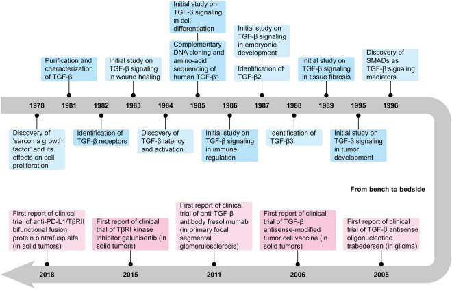
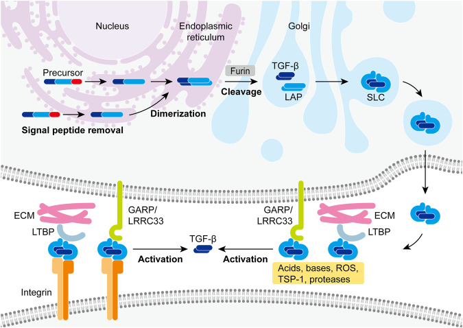
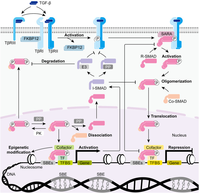
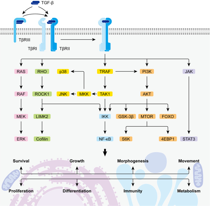
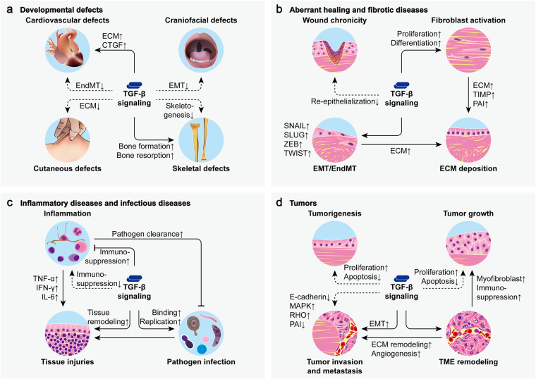
Transforming growth factor (TGF)-β is a multifunctional cytokine expressed by almost every tissue and cell type. The signal transduction of TGF-β can stimulate diverse cellular responses and is particularly critical to embryonic development, wound healing, tissue homeostasis, and immune homeostasis in health. The dysfunction of TGF-β can play key roles in many diseases, and numerous targeted therapies have been developed to rectify its pathogenic activity. In the past decades, a large number of studies on TGF-β signaling have been carried out, covering a broad spectrum of topics in health, disease, and therapeutics. Thus, a comprehensive overview of TGF-β signaling is required for a general picture of the studies in this field. In this review, we retrace the research history of TGF-β and introduce the molecular mechanisms regarding its biosynthesis, activation, and signal transduction. We also provide deep insights into the functions of TGF-β signaling in physiological conditions as well as in pathological processes. TGF-β-targeting therapies which have brought fresh hope to the treatment of relevant diseases are highlighted. Through the summary of previous knowledge and recent updates, this review aims to provide a systematic understanding of TGF-β signaling and to attract more attention and interest to this research area.


Conflict of interest statement

The authors declare no competing interests.

Figures

Fig. 1
Fig. 1
History of research on TGF-β signaling
Fig. 2
Fig. 2
Biosynthesis and activation of TGF-β. Each TGF-β monomer is initially synthesized as a precursor polypeptide. In the endoplasmic reticulum, TGF-β precursors lose their signal peptides and dimerize through disulfide bonds. The dimers then transit into the Golgi where they are cleaved by protease furin into mature cytokine segments and latency-associated peptides (LAPs) to form small latent complexes (SLCs). The secreted SLCs can further link to latent TGF-β-binding proteins (LTBPs) which target them into the extracellular matrix (ECM) for storage, or they can link to glycoprotein-A repetitions predominant protein (GARP) or leucine-rich repeat-containing protein 33 (LRRC33) which tethers them to the cell surface. Numerous factors such as acids, bases, reactive oxygen species (ROS), thrombospondin-1 (TSP-1), certain proteases, and integrins can release the mature cytokines from the latent complexes and thus are known as TGF-β activators
Fig. 3
Fig. 3
Canonical TGF-β signaling. TGF-β can initially bind to its co-receptor TGF-β receptor III (TβRIII) or directly bind to its receptor TβRII which subsequently recruits TβRI to form a TGF-β-TβRI-TβRII complex. TβRII then actives TβRI through phosphorylation, leading to its dissociation with signaling inhibitor FK506-binding protein 1A (FKBP12) and interaction with signaling effectors receptor-activated SMADs (R-SMADs). R-SMADs which are presented to TβRI by adaptor protein SMAD anchor for receptor activation (SARA) get activated through phosphorylation and undergo oligomerization with common-partner SMAD (co-SMAD). The SMAD oligomers then translocate into the nucleus where they function as transcription factors (TFs), mediating the transcriptional activation or repression of target genes by binding to specific DNA sequences known as SMAD-binding elements (SBEs) and generally in cooperation with other TFs as well as transcriptional cofactors. In this way, TGF-β signaling can activate the expression of inhibitory SMADs (I-SMADs) which in turn function to attenuate the transcriptional regulation mediated by TGF-β signaling through several mechanisms. Moreover, many protein kinases (PKs), protein phosphatases (PPs), and (E3) ubiquitin ligases can also modulate canonical TGF-β signaling through various post-translational modifications of SMADs. (TFBS, TF-binding site)
Fig. 4
Fig. 4
Non-canonical TGF-β signaling. TGF-β can signal through non-canonical pathways to activate extracellular signal-regulated kinase (ERK) signaling, rat sarcoma (RAS) homolog (Rho)-guanosine triphosphatase (GTPase) signaling, p38 mitogen-activated protein kinase (MAPK) signaling, c-Jun N-terminal kinase (JNK) signaling, nuclear factor-κB (NF-κB) signaling, phosphatidylinositol 3-kinase (PI3K)/AKR mouse thymoma proto-oncogene (AKT) signaling, as well as Janus kinase (JAK)/signal transducer and activator of transcription (STAT) signaling. These non-canonical TGF-β signaling pathways are actively involved in an extensive range of cellular events. (RAF, RAS-associated factor; MEK, MAPK/ERK kinase; ROCK1, Rho-associated coiled-coil-containing protein kinase 1; LIMK2, LIM domain kinase 2; TRAF, tumor necrosis factor (TNF) receptor-associated factor; TAK1, TGF-β-activated kinase 1; MKK, MAPK kinase; IKK, NF-κB inhibitor (IκB) kinase; GSK-3β, glycogen synthase kinase-3β; MTOR, mechanistic target of rapamycin; FOXO, forkhead box O; S6K, S6 kinase; 4EBP1, 4E-binding protein 1)
Fig. 5
Fig. 5
TGF-β signaling in health. TGF-β signaling plays a critical role in physiological conditions. a During embryonic development, TGF-β regulates cell differentiation, epithelial/endothelial-mesenchymal transition (EMT/EndMT), and apoptosis to ensure proper histogenesis and organogenesis. b TGF-β promotes wound healing by participating in inflammation, re-epithelialization, angiogenesis, and fibroblast activation. c TGF-β is indispensable for tissue homeostasis as it generally suppresses cell proliferation and induces cell apoptosis through various mechanisms. d TGF-β functions to suppress the activity of multiple immunocompetent cells while inducing the phenotypes of several immune immunosuppressive cells to maintain immune homeostasis. (SMC, smooth muscle cell; VEGF, vascular endothelial growth factor; MMP, matrix metalloproteinase; TIMP tissue inhibitor of MMP, PAI plasminogen activator inhibitor, CDK cyclin-dependent kinase, CKI CDK inhibitor, ID inhibitor of DNA binding, MYC cellular-myelocytomatosis viral oncogene, CDC25A cell division cycle 25A, BCL-2 B-cell lymphoma-2, BAX BCL-2-associated X protein, BIM BCL-2-interacting mediator of cell death, BCL-XL BCL-extra-large, GADD45β growth arrest and DNA damage-inducible β, SHIP sarcoma (SRC) homology 2 (SH2) domain-containing inositol 5’-phosphatase, TIEG TGF-β-inducible early gene, CTL cytotoxic T lymphocyte, Th T helper, Treg regulatory T cell, Breg regulatory B cell, NK natural killer, DC dendritic cell)
Fig. 6
Fig. 6
TGF-β signaling in disease. Dysfunctional TGF-β signaling is involved in numerous pathological processes. a Mutations that lead to decreased or increased TGF-β signaling can cause various developmental defects. b Deficient TGF-β signaling contributes to wound chronicity while excess TGF-β signaling leads to wound scarring and tissue fibrosis by stimulating ECM deposition through fibroblast activation and EMT/EndMT. c Dysfunctional TGF-β signaling exacerbates tissue injuries in inflammatory diseases and infectious diseases by promoting inflammation, pathogen infection, and tissue remodeling. d Aberrant TGF-β signaling is implicated in all aspects of tumor development including tumorigenesis, tumor growth, tumor invasion, tumor metastasis, as well as tumor microenvironment (TME) remodeling. (CTGF, connective tissue growth factor; IFN-γ, interferon-γ; IL-6, interleukin-6; solid arrows from TGF-β indicate excessive TGF-β signaling, dashed arrows from TGF-β indicate deficient TGF-β signaling)

Similar articles

Cited by

References

    1. de Larco, J. E. & Todaro, G. J. Growth factors from murine sarcoma virus-transformed cells. Proc. Natl Acad. Sci. USA75, 4001–4005 (1978). 10.1073/pnas.75.8.4001 - DOI - PMC - PubMed
    1. Derynck, R. et al. Human transforming growth factor-beta complementary DNA sequence and expression in normal and transformed cells. Nature316, 701–705 (1985). 10.1038/316701a0 - DOI - PubMed
    1. Roberts, A. B., Anzano, M. A., Lamb, L. C., Smith, J. M. & Sporn, M. B. New class of transforming growth factors potentiated by epidermal growth factor: isolation from non-neoplastic tissues. Proc. Natl Acad. Sci. USA78, 5339–5343 (1981). 10.1073/pnas.78.9.5339 - DOI - PMC - PubMed
    1. Proper, J. A., Bjornson, C. L. & Moses, H. L. Mouse embryos contain polypeptide growth factor(s) capable of inducing a reversible neoplastic phenotype in nontransformed cells in culture. J. Cell Physiol.110, 169–174 (1982). 10.1002/jcp.1041100210 - DOI - PubMed
    1. Childs, C. B., Proper, J. A., Tucker, R. F. & Moses, H. L. Serum contains a platelet-derived transforming growth factor. Proc. Natl Acad. Sci. USA79, 5312–5316 (1982). 10.1073/pnas.79.17.5312 - DOI - PMC - PubMed

Publication types

MeSH terms