. 2024 Mar 21;9(1):66.
doi: 10.1038/s41541-024-00856-6.

Immunization-induced antigen archiving enhances local memory CD8+ T cell responses following an unrelated viral infection

Affiliations

Immunization-induced antigen archiving enhances local memory CD8+ T cell responses following an unrelated viral infection

Thu A Doan et al. NPJ Vaccines. .

Abstract

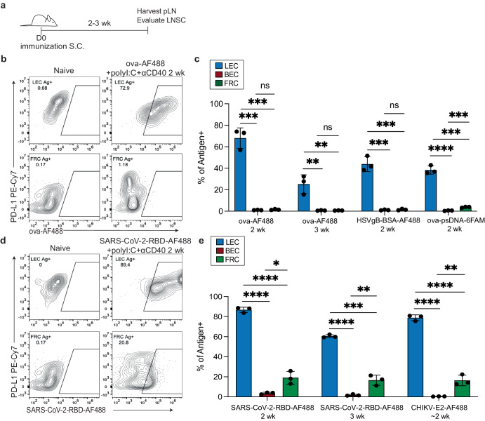
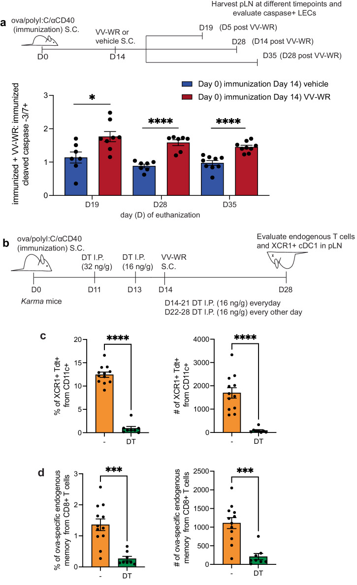
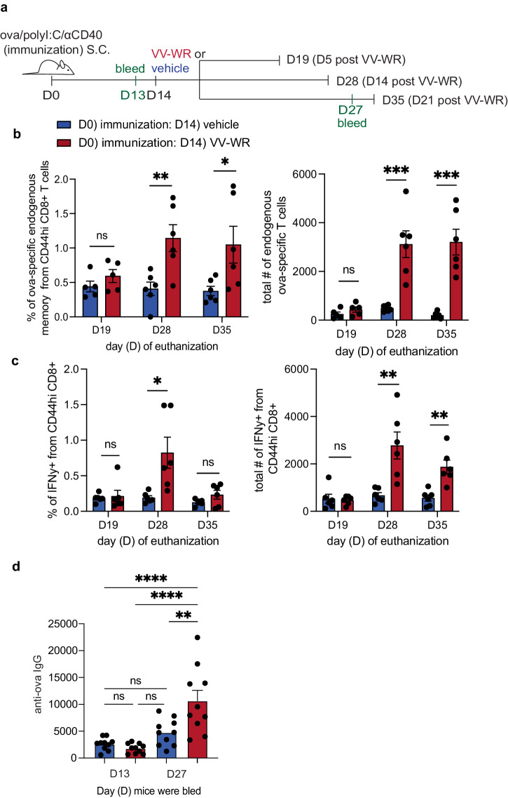
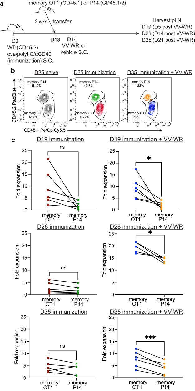
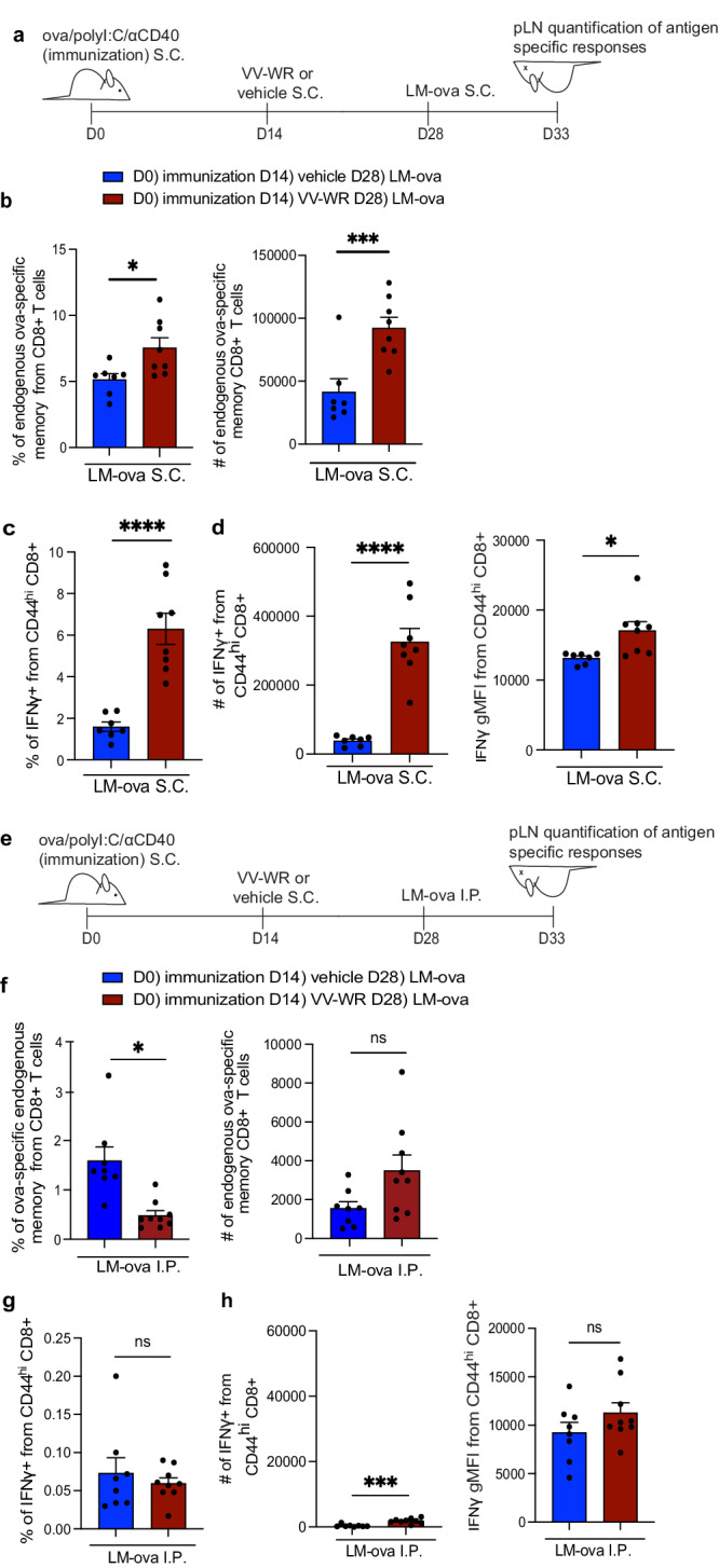
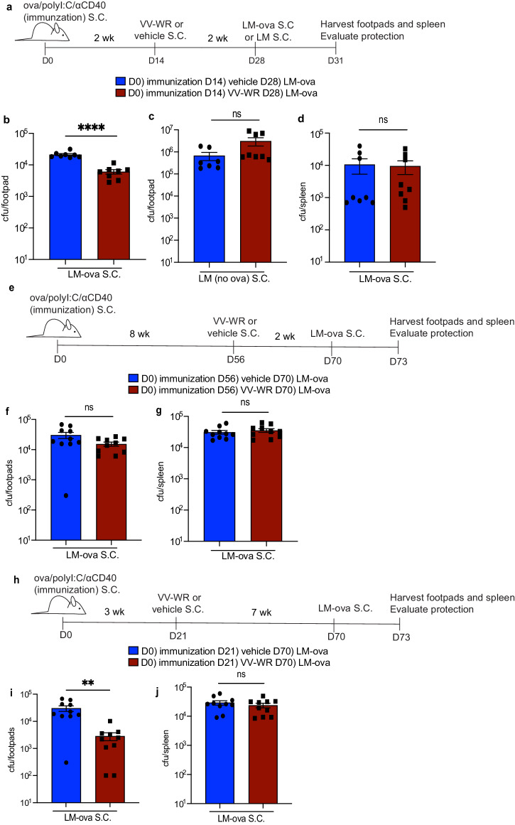
Antigens from viruses or immunizations can persist or are archived in lymph node stromal cells such as lymphatic endothelial cells (LEC) and fibroblastic reticular cells (FRC). Here, we find that, during the time frame of antigen archiving, LEC apoptosis caused by a second, but unrelated, innate immune stimulus such as vaccina viral infection or CpG DNA administration resulted in cross-presentation of archived antigens and boosted memory CD8 + T cells specific to the archived antigen. In contrast to "bystander" activation associated with unrelated infections, the memory CD8 + T cells specific to the archived antigen from the immunization were significantly higher than memory CD8 + T cells of a different antigen specificity. Finally, the boosted memory CD8 + T cells resulted in increased protection against Listeria monocytogenes expressing the antigen from the immunization, but only for the duration that the antigen was archived. These findings outline an important mechanism by which lymph node stromal cell archived antigens, in addition to bystander activation, can augment memory CD8 + T cell responses during repeated inflammatory insults.


Conflict of interest statement

The authors declare no competing interest.

Figures

Fig. 1
Fig. 1. Antigen levels in lymph node stromal cells after immunization.
a Experimental schematic for be. C57/BL6 mice were immunized subcutaneously in the footpad and/or flank with the indicated antigens and adjuvants. b Cells were stained with CD45, PDPN, CD31 and PD-L1. Cells were gated on CD45-PDPN + CD31- for FRC and CD45-PDPN + CD31+ for LECs. Shown are examples of LEC and FRC antigen-positive cells based on PD-L1 expression (floor, MARCO LEC) and ova-AF488+ from mice 2–3 weeks after immunization with ova conjugated to Alexa-Fluor 488 (AF488) and polyI:C and αCD40. c Quantification of the frequency of LEC, BEC, and FRC in the popliteal lymph node (pLN) that are positive for the indicated antigens administered with polyI:C and αCD40 at indicated time. d Same as (b) except for mice were immunized with SARS-CoV-2-RBD-AF488, polyI:C, and αCD40. e Same as in (c) except for SARS-CoV-2-RBD and CHIKV-E2 with polyI:C and αCD40. CHIKV-E2 was repeated for 9–14 days post-vaccine (~2 weeks). Statistical analysis was done using an unpaired t-test where the p-value between naïve and indicated antigen is <0.0001. In each experiment, at least n = 2–3 mice per group were evaluated and the experiment was repeated n = 2–5 times for ce. Shown is the representative data from one of the experiments. Error bars are mean ± standard error of the mean. ns not significant, *p < 0.05, **p < 0.01, ***p < 0.001, ****p < 0.0001.
Fig. 2
Fig. 2. Increased LEC apoptosis following VV-WR coincides with cDC1 presentation of archived antigens.
a Experimental schematic. Mice were immunized and infected with VV-WR at indicated time points. Popliteal lymph node (pLN) were harvested at respective time points and cleaved caspase 3/7+ LECs were evaluated. Quantification of the fold change of each treatment at the respective timepoint over vaccine only. The fold change was calculated by taking the percentage of cleaved caspase-3/7 + LEC and dividing by the percentage of cleaved caspase-3/7 + LEC for immunization only at each time point. In each experiment, n = 2–5 mice per group were evaluated and the experiment was repeated n = 2–3 times depending on the time-point. Shown is the combined data from all experiments. b Experimental schematic for (c, d) Karma mice were immunized and infected with VV-WR at indicated timepoints as in a. Except, Karma mice were treated at indicated timepoints with DT as described in figure and methods. c Quantification of flow cytometric analysis, performed on day 28 after euthanization (CO2 followed by cervical dislocation), of cDC1s (CD11chiMHCIIhi XCR1 + CD11b-) in vehicle (orange) or DT depleted (green) mice. Shown are percent and numbers from draining popliteal lymph node. d As in (c) except cells were gated as CD44hi and SIINFEKL tetramer + (ova specific) from (B220 - CD8 + lymphocytes). Shown are percent and number from draining popliteal lymph node. In each experiment n = 3–5 mice per group were evaluated and the experiment was repeated (n = 3). Shown is combined data from three independent experiments. Error bars are mean ± standard error of the mean. Statistical analysis was done using an unpaired t-test where the p-value is indicated by an asterisk. ns = not significant, *p < 0.05, ***p < 0.001, ****p < 0.0001.
Fig. 3
Fig. 3. Increased immunization-specific memory CD8 + T cell quantity and function following vaccinia infection.
a Experimental schematic for (bd). Mice were immunized subcutaneously in the footpad with a subunit immunization containing ova, polyI:C, and αCD40. Two weeks later, mice were infected with VV-WR or vehicle (PBS). At time of euthanasia (CO2 followed by cervical dislocation) popliteal lymph nodes (pLN) were harvested at respective time points post-VV-WR infection. Half the cells were used to evaluate endogenous CD8 + T cells and the other half were used for ex vivo stimulation with SIINFEKL peptide. b Quantification of frequency and number of ova-specific endogenous memory CD8 + T cells in the draining popliteal LN. In each experiment, n = 2–5 mice per group were evaluated and each experiment was repeated 3–7 times depending on the time point with similar results. Shown is the representative data from two of the experiments. c Quantification of frequency and number of IFNγ -producing B220-CD44hi CD8 + T cells from the draining popliteal lymph node. In each experiment, n = 2–5 mice per group were evaluated and each experiment was repeated 3–7 times depending on the time point with similar results. Shown is the representative data from two experiments. (d) Quantification of IgG specific antibodies to ovalbumin in the serum using ELISA after the indicated times/treatments from (a). Shown are combined data from two independent experiments with n = 5 mice per group. Statistical analysis was done using an unpaired t-test where the p-value between vaccine + vehicle (blue bar) and vaccine + VV-WR (red bar) is <0.0001. Error bars are mean ± standard error of the mean. ns not significant, *p < 0.05, **p < 0.01, ***p < 0.001, ****p < 0.0001.
Fig. 4
Fig. 4. Archived antigen-specific memory CD8 + T cells derived from immunization expand preferentially following vaccinia infection.
a Experimental schematic for (b, c). Mice were immunized and infected with VV-WR as in Fig. 2. One day prior to VV-WR infection congenically different memory OT1 and memory P14 CD8 + T cells were isolated following euthanasia (CO2 followed by cervical dislocation) and transferred intravenously into WT mice. To establish memory, naïve OT1 or P14 cells were transferred into naïve WT mice and immunized with their cognate antigen (ovalbumin or gp33 peptide) and isolated by CD8 negative selection 2–6 weeks later as described in materials and methods. Memory OT1 and memory P14 were also transferred into naïve WT host to calculate fold expansion over OT1/P14 “take”. Popliteal LNs (pLN) were harvested at time of euthanasia (CO2 followed by cervical dislocation) and processed at indicated time points. b Representative flow cytometric plots of co-transferred memory P14 and memory OT1 fold expansion transferred at 1:1 ratio. c Memory OT1 (CD45.1/1) and P14 (CD45.1/2) were co-transferred into immunized mice (CD45.2/2) 1 day before VV-WR as in (a). c The fold expansion from plots shown in (b) was calculated as the total number of memory OT1 or memory P14 in antigen-bearing mice over the total number of memory OT1 or memory P14 in the naïve WT host (to accommodate for differences in ratio and “take”) at each respective time point. Red squares represent memory OT1s and green circles represent memory P14s transferred into the same mouse that received ova/polyI:C/αCD40 immunization 13 days prior and PBS (vehicle) 1 day after (D14). Blue squares represent memory OT1s and orange circles represent memory P14s transferred into the same mouse that received ova/polyI:C/αCD40 immunization 13 days prior and VV-WR 1 day after (D14). Each line is an individual mouse where both cell types were transferred into the same mouse. In each experiment, at least n = 3 mice per group were evaluated and the experiment was repeated n = 2 times. In each case, a different congenic marker was used for transferred cells (e.g. OT1 was CD45.1/1 and P14 was CD45.1/2 and hosts were CD45.2/2 or OT1 was CD45.1/2 and P14 was CD45.1/1 and host was CD45.2/2). Results were similar across congenic marker combinations used. In Fig. 4b, representative flow plots from one experiment are shown as an example (in the other experiment, OT1 were CD45.1/2 and P14 were CD45.1/1). Shown in Fig. 4c is the combined data from both experiments. A third replicate was not performed as our experiments were adequately powered to provide statistical significance in accordance with our IACUC policies regarding animal experiments with consistent data points. Statistical analysis was done using a paired t-test. Error bars are mean ± standard error of the mean. ns not significant, *p < 0.05, ***p < 0.001.
Fig. 5
Fig. 5. Archived antigen-specific memory CD8 + T cells derived from immunization are recalled during antigenic challenge.
(a) Experimental schematic for (bd). Mice were immunized with ova/polyI:C/αCD40 and infected with VV-WR 2 weeks later. Two weeks after VV-WR, mice were challenged with LM-ova subcutaneously (S.C.). Five days post-LM-ova mice were euthanized (CO2 followed by cervical dislocation) and popliteal lymph nodes (pLN) were harvested to assess endogenous archived-antigen (ova)-specific memory CD8 + T cells in the draining pLN. b Quantification of frequency and the total number of ova-specific endogenous memory CD8 + T cells in the popliteal LN as assessed by a SIINFEKL specific MHC class I Kb tetramer. c Quantification of frequency of IFNγ -produced from CD44hi CD8 + T cells in the draining popliteal lymph node. d Quantification of the total number and geometric mean fluorescence intensity (gMFI) of IFNγ from CD44hi CD8 + T cells in the draining lymph node. e Experimental schematic for (fh). Mice were challenged with LM-ova intraperitoneally (I.P.) f Same as (b). except for the mice were challenged with LM-ova I.P. g Same as (c) except for the mice were challenged with LM-ova I.P. h Same as (d) except for the mice were challenged with LM-ova I.P. Statistical analysis was done using an unpaired t-test where the p-value between vaccine + vehicle + LM-ova (blue bar) and vaccine + VV-WR + LM-ova (red bar) is <0.0001. Errors bars are mean ± standard error of the mean. In (ad) and (eh) n = 3–5 mice per group were evaluated and the experiment was repeated n = 2 times. Shown is the combined data from both experiments. A third replicate was not performed as our experiments were adequately powered to provide statistical significance in accordance with our IACUC policies regarding animal experiments with consistent data points. ns not significant, *p < 0.05, ***p < 0.001, ****p < 0.0001.
Fig. 6
Fig. 6. Archived antigen-specific memory CD8 + T cells derived from immunization improve protective immunity during antigenic rechallenge over the time-frame of antigen archiving.
a Experimental schematic for (bd). Mice were immunized with ova/polyI:C/αCD40, infected with VV-WR, and challenged with LM-ova or LM at indicated time points. Foot and ankle skin or spleen were harvested after mice were euthanized (CO2 followed by cervical dislocation). bd Respective tissues were processed as described in the methods section. Homogenized tissues were plated on BHI + erythromycin (LM-ova) or streptomycin (LM) plates and colonies were counted after 3 days of growth. e Experimental schematic for (f, g). Mice were immunized, infected with VV-WR, and challenged with LM-ova at indicated time points. f, g Same as (bd). h Experimental schematic for (i, j). Mice were immunized, infected with VV-WR, and rechallenged with LM-ova at indicated time points. i, j Same as (bd). Statistical analysis was done using unpaired t-test where the p-value between vaccine + vehicle (blue bar) and vaccine + VV-WR (red bar) is <0.0001. Errors bars are mean ± standard error of the mean. In each experiment, at least n = 3–5 mice per group were evaluated and the experiment was repeated n = 2 times. Shown are all data points from both experiments. A third replicate was not performed as our experiments were adequately powered to provide statistical significance in accordance with our IACUC policies regarding animal experiments with consistent data points. ns not significant, **p < 0.01, ****p < 0.0001.

Update of

References

    1. Sprenger KG, Louveau JE, Murugan PM, Chakraborty AK. Optimizing immunization protocols to elicit broadly neutralizing antibodies. Proc. Natl Acad. Sci. USA. 2020;117:20077–20087. doi: 10.1073/pnas.1919329117. - DOI - PMC - PubMed
    1. Hvidt AK, et al. Comparison of vaccine-induced antibody neutralization against SARS-CoV-2 variants of concern following primary and booster doses of COVID-19 vaccines. Front. Med. (Lausanne) 2022;9:994160. doi: 10.3389/fmed.2022.994160. - DOI - PMC - PubMed
    1. Sogaard OS, et al. Characteristics associated with serological COVID-19 vaccine response and durability in an older population with significant comorbidity: the danish nationwide ENFORCE Study. Clin. Microbiol. Infect. 2022;28:1126–1133. doi: 10.1016/j.cmi.2022.03.003. - DOI - PMC - PubMed
    1. Klein JS, Bjorkman PJ. Few and far between: how HIV may be evading antibody avidity. PLoS Pathog. 2010;6:e1000908. doi: 10.1371/journal.ppat.1000908. - DOI - PMC - PubMed
    1. van de Sandt CE, Kreijtz JH, Rimmelzwaan GF. Evasion of influenza a viruses from innate and adaptive immune responses. Viruses. 2012;4:1438–1476. doi: 10.3390/v4091438. - DOI - PMC - PubMed