. 2024 May;31(5):544-557.
doi: 10.1038/s41418-024-01278-6. Epub 2024 Mar 21.

Identification of FasL as a crucial host factor driving COVID-19 pathology and lethality

Affiliations

Identification of FasL as a crucial host factor driving COVID-19 pathology and lethality

Marie-Christine Albert et al. Cell Death Differ. 2024 May.

Abstract

The dysregulated immune response and inflammation resulting in severe COVID-19 are still incompletely understood. Having recently determined that aberrant death-ligand-induced cell death can cause lethal inflammation, we hypothesized that this process might also cause or contribute to inflammatory disease and lung failure following SARS-CoV-2 infection. To test this hypothesis, we developed a novel mouse-adapted SARS-CoV-2 model (MA20) that recapitulates key pathological features of COVID-19. Concomitantly with occurrence of cell death and inflammation, FasL expression was significantly increased on inflammatory monocytic macrophages and NK cells in the lungs of MA20-infected mice. Importantly, therapeutic FasL inhibition markedly increased survival of both, young and old MA20-infected mice coincident with substantially reduced cell death and inflammation in their lungs. Intriguingly, FasL was also increased in the bronchoalveolar lavage fluid of critically-ill COVID-19 patients. Together, these results identify FasL as a crucial host factor driving the immuno-pathology that underlies COVID-19 severity and lethality, and imply that patients with severe COVID-19 may significantly benefit from therapeutic inhibition of FasL.


Conflict of interest statement

HW is co-founder of Apogenix, a biotech company that develops asunercept. The authors declare no other competing interests.

Figures

Fig. 1
Fig. 1. Mouse-adapted SARS-CoV-2 induces lethal disease.
a Illustration of mouse adaptation process with SARS-CoV-2 Alpha variant by serial passages (pass.) to obtain SARS-CoV-2 MA20. Severity score (b), weight loss curves (c, left) and survival curves (c, right) of 2 m BALB/c, 2 m C57BL/6 and 8 m C57BL/6 mice infected with indicated viral titres of MA20(n = 4 or 6). d Persistent (light orange) and acquired (dark orange) mutations in SARS-CoV-2 isolates during mouse adaptation as compared to SARS-CoV-2 Wuhan strain. e Location of mutations within genes of SARS-CoV-2 MA20, acquired mutations depicted in bold. f (a–d) Depiction of predicted interactions with H-bonds (black) and CH-π interactions (magenta) of Alpha Spike RBD (cyan) in complex with hACE2 (green) (a), MA20 Spike RBD (cyan) in complex with hACE2 (green) (b), Alpha Spike RBD (cyan) in complex with mACE2 (green) (c), MA20 Spike RBD (cyan) in complex with mACE2 (green) (d). Only interactions up to 3.5 Å are shown. For H-bonds indicated distances correspond to distance between the corresponding hydrogen atom and acceptor oxygen atoms of either D38 or E35. g ELISA-style binding assay of the RBD of Spike proteins from Alpha (blue) and MA20 (orange) with mACE2 (n = 3). h Experimental design for sample collection after infection with MA20. Viral titres of infected 2 m BALB/c (i), 2 m C57BL/6 and 8 m C57BL/6 (j) mice (n = 4 to 6). p values were determined by One-way ANOVA with post-hoc Tukey.*p < 0.0332, **p < 0.0021, ***p < 0.0002, ****p < 0.0001. IHC staining of lung sections of SARS-CoV-2 Spike protein of infected 2 m BALB/c (k), 2 m C57BL/6 and 8 m C57BL/6 (l) mice. Values represent mean ± SEM. Scale bars indicate 500 µm. 2 m:2-month-old. 8 m: 8-month-old.
Fig. 2
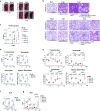
Fig. 2. MA20-induced pathology resembles COVID-19.
a Macroscopic pictures of left lung lobes at 4 dpi of infected 2 m BALB/c, 2 m C57BL/6 and 8 m C57BL/6 (right). b Histological Pathology of H&E-stained lung sections of infected 2 m BALB/c (upper panel), 2 m C57BL/6 (middle panel) and 8 m C57BL/6 (lower panel) mice at 2 dpi and 4 dpi with indicated prominent pathological features. c ALI score at 4 dpi of infected mice as indicated (n = 4–6). p values were determined by unpaired t-test with Welch’s correction. *p < 0.0332, **p < 0.0021, ***p < 0.0002, ****p < 0.0001. Flow cytometry analysis of immune cell populations in lungs of infected 2 m BALB/c, (d) 2 m C57BL/6 and 8 m C57BL/6 (e) mice as indicated (n = 3 to 6). p values were determined by One-way ANOVA with post-hoc Tukey. *p < 0.0332, **p < 0.0021, ***p < 0.0002, ****p < 0.0001. NLR of MA20-infected 2 m BALB/c (f), 2 m C57BL/6 and 8 m C57BL/6 (g) mice. p values were determined by One-way ANOVA with post-hoc Tukey (n = 3–6). *p < 0.0332, **p < 0.0021, ***p < 0.0002, ****p < 0.0001. Values represent mean ± SEM. Scale bars indicate 500 µm. ALI acute lung injury. Inflamm. Mono-Macs inflammatory monocytic-macrophages. NLR neutrophil/lymphocyte ratio.2 m 2-month-old. 8 m 8-month-old.
Fig. 3
Fig. 3. MA20 induces cell death in infected lungs.
a Pathway ontology analysis of RNAseq results of lungs of infected 2 m BALB/c mice at 2 dpi. Percentage indicates number of genes dysregulated in respective pathway. Representative pictures (b) and quantification (c) of TUNEL-stained and cleaved Caspase 3 staining of lung sections of infected 2 m BALB/c mice at 2 and 4 dpi (n = 5 or 10). p values were determined by One-way ANOVA with post-hoc Dunnett. *p < 0.0332, **p < 0.0021, ***p < 0.0002, ****p < 0.0001. d RNAseq expression analysis of TNFSF members of lungs of infected 2 m BALB/c mice (n = 4 to 5). Data are represented as log2 fold change relative to mock. e Individual expression values of Fasl (left panel) and Tnf (right panel) (n = 5 to 6). p values were determined by One-way ANOVA with post-hoc Dunnett. *p < 0.0332, **p < 0.0021, ***p < 0.0002, ****p < 0.0001. f Flow cytometry analysis of FasL expression on immune cell populations as indicated in infected 2 m BALB/c mice at 2 dpi (n = 5). p values were determined by unpaired t-test with Welch’s correction. *p < 0.0332, **p < 0.0021, ***p < 0.0002, ****p < 0.0001. Scale bars indicate 50 µm in overview and 10 µm in enlarged frames. Values represent mean ± SEM. NK cells: Natural Killer cells. Inflammatory Mono-Macs inflammatory monocytic-macrophages, 2 m 2-month-old.
Fig. 4
Fig. 4. Therapeutic FasL inhibition prevents lethality in MA20-infected mice.
a Experimental design for i.p. treatment after infection of 2 mBALB/c mice. Survival curves (b) and single weight loss curves (c) of MA20-infected 2m BALB/c mice with i.p. injections at 2 dpi with indicated treatments (n = 30). Coloured dots indicate when mice reached humane endpoint. Survival of mice with either treatment was compared by log-rank test. *p < 0.0332, **p < 0.0021, ***p < 0.0002, ****p < 0.0001. d Experimental design for i.p. treatment after infection of 8 m C57BL/6 mice. Survival curves (e) and single weight loss curves (f) of MA20-infected 8 m C57BL/6 mice with i.p. injections at 2 dpi with each indicated treatments (n = 10). Coloured dots indicate when mice reached humane endpoint. Survival of mice with either treatment was compared by log-rank test. *p < 0.0332, **p < 0.0021, ***p < 0.0002, ****p < 0.0001. D Day. i.p. intraperitoneal, 2 m 2-month-old. 8 m 8-month-old.
Fig. 5
Fig. 5. Inhibition of FasL decreases overall cell death and inflammation in infected lungs.
a Experimental design for i.p. treatment and sample collection after infection. b Viral titres of lungs of infected 2 m BALB/c mice with indicated treatments (n = 9 or 5). p values were determined by One-way ANOVA with post-hoc Tukey. *p < 0.0332, **p < 0.0021, ***p < 0.0002, ****p < 0.0001. Representative pictures (c) and quantification (d) of TUNEL and cleaved caspase 3 staining of lung sections of infected 2 m BALB/c mice with indicated treatments (n = 9 or 5). p values were determined by One-way ANOVA with post-hoc Dunnett. *p < 0.0332, **p < 0.0021, ***p < 0.0002, ****p < 0.0001. e Inflammation score depicting accumulation of 39 cytokines and chemokines quantified by Luminex Multiplex Assay normalized to protein amount in lung homogenates of infected 2 m BALB/c mice with indicated treatments (n = 9 or 5). p values were determined by Two-way ANOVA with post-hoc Tukey. *p < 0.0332, **p < 0.0021, ***p < 0.0002, ****p < 0.0001. f Protein levels of FasL in lung samples of infected 2 m BALB/c mice with indicated treatments (n = 9 or 5). p values were determined by One-way ANOVA with post-hoc Dunnett. *p < 0.0332, **p < 0.0021, ***p < 0.0002, ****p < 0.0001. Values represent mean ± SEM. Scale bars indicate 50 µm in overview and 10 µm in enlarged frames. D: Day. i.p.: intraperitoneal.2 m:2-month-old.
Fig. 6
Fig. 6. FasL upregulation in the bronchoalveolar lavage fluid of COVID-19 patients.
Human BALF scRNAseq data for UMAP projection (a) and Violin plots (b) depicting Fasl mRNA expression in immune cell populations as indicated (NK cells, healthy n = 97; COVID-19 n = 984; T cells, healthy n = 1225; COVID-19 n = 6491; macrophages, healthy n = 18539; COVID-19 n = 30878). p values in Violin plots (B) were determined by Wilcoxon Rank Sum test. *p < 0.0332, **p < 0.0021, ***p < 0.0002, ****p < 0.0001. c Cytokine protein analysis of FasL in BALF of 24 COVID-19 ICU patients with serial sample collection during hospitalization (n = 109) and IAV patients (n = 28) and healthy donors (n = 36) quantified by Luminex Multiplex Assay. p values were determined by Mann-Whitney test. *p < 0.0332, **p < 0.0021, ***p < 0.0002, ****p < 0.0001. Values represent mean ± SEM. NK cells: Natural Killer cells. BALF bronchoalveolar lavage fluid.

References

    1. Kumari M, Lu R-M, Li M-C, Huang J-L, Hsu F-F, Ko S-H, et al. A critical overview of current progress for COVID-19: development of vaccines, antiviral drugs, and therapeutic antibodies. J Biomed Sci. 2022;29:68. doi: 10.1186/s12929-022-00852-9. - DOI - PMC - PubMed
    1. Tregoning JS, Flight KE, Higham SL, Wang Z, Pierce BF. Progress of the COVID-19 vaccine effort: viruses, vaccines and variants versus efficacy, effectiveness and escape. Nat Rev Immunol. 2021;21:626–36. doi: 10.1038/s41577-021-00592-1. - DOI - PMC - PubMed
    1. Subissi L, von Gottberg A, Thukral L, Worp N, Oude Munnink BB, Rathore S, et al. An early warning system for emerging SARS-CoV-2 variants. Nat Med. 2022;28:1110–5. doi: 10.1038/s41591-022-01836-w. - DOI - PMC - PubMed
    1. Bhatraju PK, Ghassemieh BJ, Nichols M, Kim R, Jerome KR, Nalla AK, et al. Covid-19 in critically ill patients in the Seattle region—case series. N Engl J Med. 2020;382:2012–22. doi: 10.1056/NEJMoa2004500. - DOI - PMC - PubMed
    1. Huang C, Wang Y, Li X, Ren L, Zhao J, Hu Y, et al. Clinical features of patients infected with 2019 novel coronavirus in Wuhan, China. Lancet. 2020;395:497–506. doi: 10.1016/S0140-6736(20)30183-5. - DOI - PMC - PubMed

MeSH terms

Substances