. 2024 May;11(18):e2308276.
doi: 10.1002/advs.202308276. Epub 2024 Mar 21.

Bioprinting Soft 3D Models of Hematopoiesis using Natural Silk Fibroin-Based Bioink Efficiently Supports Platelet Differentiation

Affiliations

Bioprinting Soft 3D Models of Hematopoiesis using Natural Silk Fibroin-Based Bioink Efficiently Supports Platelet Differentiation

Christian Andrea Di Buduo et al. Adv Sci (Weinh). 2024 May.

Abstract

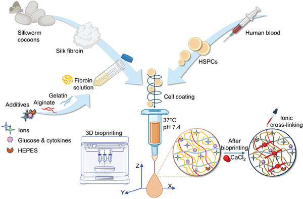
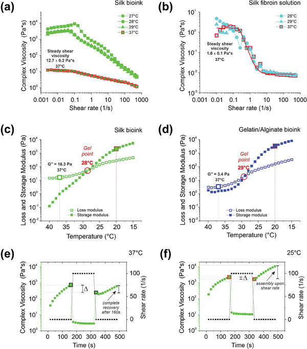
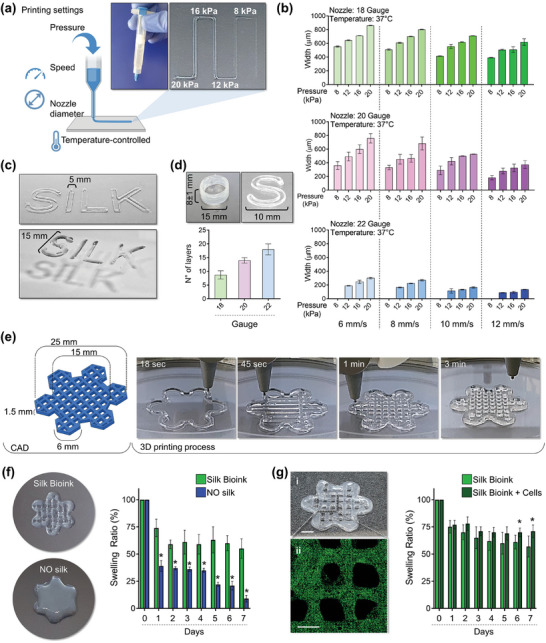
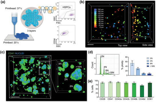
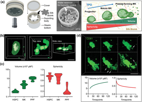
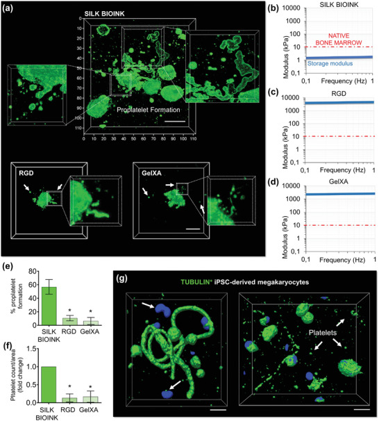
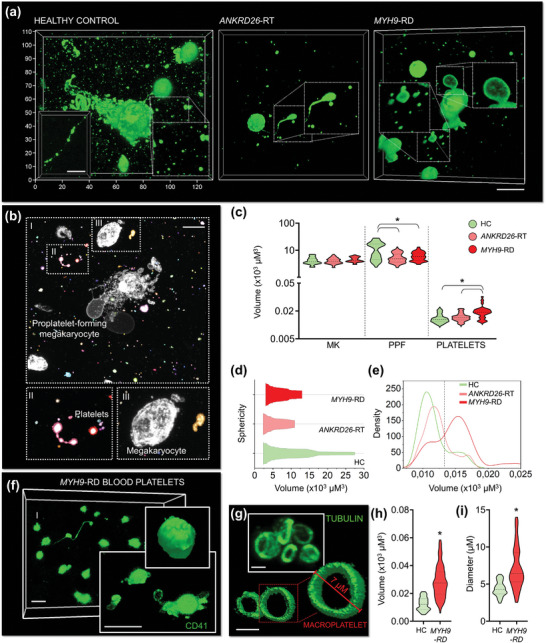
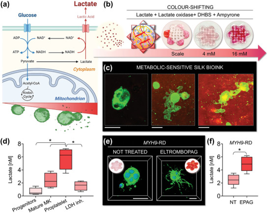
Hematopoietic stem and progenitor cells (HSPCs) continuously generate platelets throughout one's life. Inherited Platelet Disorders affect ≈ 3 million individuals worldwide and are characterized by defects in platelet formation or function. A critical challenge in the identification of these diseases lies in the absence of models that facilitate the study of hematopoiesis ex vivo. Here, a silk fibroin-based bioink is developed and designed for 3D bioprinting. This bioink replicates a soft and biomimetic environment, enabling the controlled differentiation of HSPCs into platelets. The formulation consisting of silk fibroin, gelatin, and alginate is fine-tuned to obtain a viscoelastic, shear-thinning, thixotropic bioink with the remarkable ability to rapidly recover after bioprinting and provide structural integrity and mechanical stability over long-term culture. Optical transparency allowed for high-resolution imaging of platelet generation, while the incorporation of enzymatic sensors allowed quantitative analysis of glycolytic metabolism during differentiation that is represented through measurable color changes. Bioprinting patient samples revealed a decrease in metabolic activity and platelet production in Inherited Platelet Disorders. These discoveries are instrumental in establishing reference ranges for classification and automating the assessment of treatment responses. This model has far-reaching implications for application in the research of blood-related diseases, prioritizing drug development strategies, and tailoring personalized therapies.

Keywords: bioprinting; bone marrow; eltrombopag; hematopoiesis; megakaryocyte; platelet; silk.


Conflict of interest statement

C.A.D.B., V.K., P‐A.L., I.N.R., and A.B. have submitted a patent application associated with this work. The other authors declare no conflict of interest.

Figures

Figure 1
Figure 1
Workflow of silk bioink production and usage. Schematic representation of silk bioink preparation. Regenerated silk fibroin from natural Bombyx mori silkworm cocoons is first solubilized and then blended with gelatin and alginate. Electrolyte balance is guaranteed by the addition of ions. Glucose and cytokine feeding is used to provide fuel to cell metabolism and differentiation, both during and after the bioprinting process. Cells are mixed directly inside the bioink before the bioprinting process that takes place at 37 °C using a heated printhead. After the bioprinting, ionic cross‐linking stabilizes the 3D structure‐supporting pattern. Made using BioRender.com.
Figure 2
Figure 2
Rheological characterization. a,b), Flow sweep was performed at different temperatures to evaluate the complex viscosity as a function of the shear rate of a), silk bioink, and b), silk fibroin solution. Red lines are the values of complex viscosity at 37 °C fitted with the Carreau–Yasuda model. Representative of three independent experiments. c,d), Temperature ramp tests showed the behavior of loss (G’’) and storage (G’) modulus as a function of temperature decrease from 40 to 15 °C of (c), silk bioink and (d), gelatin/alginate bioink. The crossover point (G’ = G’’) indicates the gel point (red circle). e,f), Thixotropic test was performed to monitor silk bioink complex viscosity during a deformation and a recovery phase at (e), 37 °C and (f), 25  C. The analysis was performed by applying an initial shear rate of 0.01 1 s−1 for 160 s, which was ramped up to 100 1 s−1 for 160 s (deformation phase) and finally ramped down to 0.01 1 s−1 (recovery phase). The Δ indicates the recovery step after the deformation process. Representative of three independent experiments.
Figure 3
Figure 3
Silk bioink printability. a), Printing pressure, speed, nozzle diameter, and temperature determine the resolution of a filament extrusion‐based 3D printer. Cartoon was made using BioRender.com b), The silk bioink can be 3D printed using needles of different diameters 18G (top, green), 20G (middle, pink), and 22G (bottom, blue), at various printing speeds (6,8,10,12 mm −1s) and pressures (8,12,16,20 kPa) (n = 3). c), The silk bioink can be extruded to obtain constructs of tailored widths and heights. d), 3D printing with a 20 G nozzle at 8–10 mm −1 s speed, with 12–16 kPa pneumatic pressure, ensure a good balance of print fidelity and accurate deposition to obtain 3D constructs having a height with stable junctions between the layers (n = 3). e), Model (left) of a layered grid hexagon intersecting smaller hexagons at its borders designed to model a “flower”‐like shape. The 3D printing process into a petri dish is shown (right). f), The presence of silk in the bioink formulation is crucial for ensuring printing fidelity when compared to the same formulation without silk (left). The analysis of silk bioink swelling ratio compared to bioink formulations without silk (NO silk) is shown (right) (n = 3, * p<0.01). g), Confocal microscopy analysis of HSPCs after bioprinting into the silk bioink (i: scale bar = 4 mm; ii: green = CD34; scale bar = 2 mm) (left). The analysis of silk bioink swelling ratio in the presence or absence of HSPCs is shown (n = 3, * p<0.05).
Figure 4
Figure 4
HSPC bioprinting and differentiation into the 3D silk‐based construct. a), Primary human blood progenitor cells are mixed with the silk bioink and 3D bioprinted into a 3‐layer “flower” construct. The flow cytometry analysis of CD34+ cells before 3D bioprinting is shown. Cartoon was made using BioRender.com b), Confocal microscopy analysis of cells after bioprinting (pseudo colors are used to indicate cell distribution; scale bar = 60 µm). c), 3D confocal reconstruction of polyploid megakaryocytes cultured into the silk bioink (green = CD41; blue = nuclei; scale bar = 40 µm, representative of three independent experiments). d), Flow cytometry analysis of the ploidy of megakaryocyte retrieved from the silk bioink using the dissolution buffer (n = 3). e), Percentage of megakaryocytes expressing lineage‐surface markers as assessed by flow cytometry analysis of samples retrieved from the 3D construct (n = 3).
Figure 5
Figure 5
3D bioprinted silk bioink supports functional platelet production. a), A custom‐made “flowerholder” was used for performing live‐image analysis (MK = megakaryocyte). b), 3D confocal reconstruction of polyploid megakaryocytes differentiated into the silk bioink (green = CD41; white = nuclei; scale bar = 20 µm) and c), Evaluation of single cell volume and sphericity during differentiation (HSPC = hematopoietic stem and progenitor cell, MK = megakaryocyte; PPF = proplatelet‐forming megakaryocyte, n = 100). d), Spatiotemporal volumetric imaging of samples in the last phase of differentiation shows the process of proplatelet formation. Image segmentation analysis demonstrates that megakaryocytes increase their volume while decreasing sphericity during proplatelet formation. Arrows indicate platelets released from branching filaments (green = CD61; scale bar = 30 µm).
Figure 6
Figure 6
The unique softness of 3D bioprinted silk‐bioink is crucial to support thrombopoiesis. a), The silk bioink recreates a favorable environment for supporting increased proplatelet branching (top) compared to other conventional bioinks (bottom) (green = CD61; scale bar = 20 µm, representative of three independent experiments). b–d), Storage modulus of silk bioink compared to other conventional crosslinked bioinks b), silk bioink, c), RGD; d), GelXA). The red line identifies the maximal stiffness measured for the native bone marrow tissue. e), Percentage of proplatelet formation in the different tested conditions (n = 10; * p<0.001). f), Fold increase of platelet count/area in the different tested conditions (n = 10; * p<0.001). g), Human iPSC‐derived megakaryocytes (imMKCL) engineered to express fluorescent β1‐tubulin (green) show that proplatelet formation and platelet release are sustained by cytoskeleton remodeling (scale bar = 10 µm).
Figure 7
Figure 7
Silk bioink for disease modeling and diagnosis in Inherited Thrombocytopenia. a), Confocal microscopy analysis of proplatelet formation by 3D bioprinted megakaryocytes from healthy controls (HC) and patients affected by ANKRD26‐RT and MYH9‐RD (green = CD61; scale bar = 20 µm. Representative of n = 5 HC; n = 5 ANKRD26‐RT patients; n = 5 MYH9‐RD patients). b), Image segmentation analysis allows the discrete classification of mature round megakaryocytes (MK), proplatelet‐forming megakaryocytes (PPF), and released platelets (scale bar = 30 µm). c), Volumetric analysis of the different cell subpopulations (MK: n = 100; PPF: n = 100; platelets: n = 300; * p<0.001). d), Kite diagram of megakaryocyte volume and sphericity. e, Density plot of platelet volume. f), Representative confocal microscopy and rendering of platelets from the peripheral blood of patients affected by MYH9‐RD (green = CD41; scale bar = 8 µm. Representative of n = 3 MYH9‐RD patients). g), Representative confocal microscopy and rendering of β1‐tubulin staining of peripheral blood platelets of patients affected by MYH9‐RD (green = β1‐tubulin; scale bar = 5 µm. Representative of n = 3 MYH9‐RD patients). h), Volumetric analysis of 3D‐bioprinted platelets from the peripheral blood of patients affected by MYH9‐RD with respect to healthy controls (HC) (n = 150; * p<0.001). i), Analysis of peripheral blood platelet diameter of patients affected by MYH9‐RD and healthy controls (HC) 3D‐printed into the silk bioink (n = 100; * p<0.05).
Figure 8
Figure 8
Silk bioink for easy‐readable detection of platelet production. a), Schematic of megakaryocyte metabolism. During differentiation and platelet formation, megakaryocytes undergo an increase of glycolytic activity and consequent release of lactate. Cartoon made using BioRender.com b), The silk bioink functionalized with horseradish peroxidase (HRP), lactate oxidase, and colorimetric sensors sense lactate concentrations. When lactate oxidase converts lactate into pyruvate and hydrogen peroxide, this latter is used as a cofactor by HRP that leads to a colorimetric shift of the silk bioink formulation in the presence of DHBS, and 4‐aminoantipyrine (4‐AAP). c), Imaging of silk bioink after the colorimetric reaction (green = CD61; scale bar = 20 µm). Colorimetric variations can be detected. The most intense color‐shift is detected at the stage of platelet production. d), A significantly increased release of lactate can be quantified during proplatelet formation by healthy megakaryocytes. A lactate dehydrogenase inhibitor (LDH‐inh.) has been used as control to assess the specificity of the colorimetric reaction (n = 5; * p<0.01). e), Silk bioink supports the 3D bioprinting of MYH9‐RD megakaryocytes. Treatment with the TPO‐RA Eltrombopag promotes increased proplatelet branching of MYH9‐RD megakaryocytes with respect to the untreated condition (green = CD42b; blue = nuclei; scale bar = 30 µm). f), Lactate levels are low in thrombocytopenic states (NT = not treated). Treatment with Eltrombopag increases the production of lactate (n = 5 MYH9‐RD patients; * p<0.05).

Similar articles

Cited by

References

    1. a) Gurkan U. A., Akkus O., Ann. Biomed. Eng. 2008, 36, 1978; - PubMed
    2. b) Morrison S. J., Scadden D. T., Nature 2014, 505, 327; - PMC - PubMed
    3. c) Pinho S., Frenette P. S., Nat. Rev. Mol. Cell Biol. 2019, 20, 303. - PMC - PubMed
    1. a) Abbonante V., Di Buduo C. A., Gruppi C., De Maria C., Spedden E., De Acutis A., Staii C., Raspanti M., Vozzi G., Kaplan D. L., Moccia F., Ravid K., Balduini A., Haematologica 2017, 102, 1150; - PMC - PubMed
    2. b) Chen X., Hughes R., Mullin N., Hawkins R. J., Holen I., Brown N. J., Hobbs J. K., Biophys. J. 2020, 119, 502; - PMC - PubMed
    3. c) Vining K. H., Marneth A. E., Adu‐Berchie K., Grolman J. M., Tringides C. M., Liu Y., Wong W. J., Pozdnyakova O., Severgnini M., Stafford A., Duda G. N., Hodi F. S., Mullally A., Wucherpfennig K. W., Mooney D. J., Nat. Mater. 2022, 21, 939; - PMC - PubMed
    4. d) Ivanovska I. L., Shin J. W., Swift J., Discher D. E., Trends Cell Biol. 2015, 25, 523. - PMC - PubMed
    1. a) Adamo L., Naveiras O., Wenzel P. L., McKinney‐Freeman S., Mack P. J., Gracia‐Sancho J., Suchy‐Dicey A., Yoshimoto M., Lensch M. W., Yoder M. C., García‐Cardeña G., Daley G. Q., Nature 2009, 459, 1131; - PMC - PubMed
    2. b) Zhang P., Zhang C., Li J., Han J., Liu X., Yang H., Stem Cell Res. Ther. 2019, 10, 327; - PMC - PubMed
    3. c) Li H., Luo Q., Shan W., Cai S., Tie R., Xu Y., Lin Y., Qian P., Huang H., Cell. Mol. Life Sci. 2021, 78, 5881. - PMC - PubMed
    1. a) Abbonante V., Karkempetzaki A. I., Leon C., Krishnan A., Huang N., Di Buduo C. A., Cattaneo D., Ward C. M., Matsuura S., Guinard I., Weber J., De Acutis A., Vozzi G., Iurlo A., Ravid K., Balduini A., Am J. Hematol. 2024, 99, 339; - PMC - PubMed
    2. b) De Belly H., Paluch E. K., Chalut K. J., Nat. Rev. Mol. Cell Biol. 2022, 23, 465. - PubMed
    1. a) Di Buduo C. A., Aguilar A., Soprano P. M., Bocconi A., Miguel C. P., Mantica G., Balduini A., Haematologica 2021, 106, 947; - PMC - PubMed
    2. b) Currao M., Malara A., Di Buduo C. A., Abbonante V., Tozzi L., Balduini A., Exp. Cell Res. 2016, 346, 1; - PMC - PubMed
    3. c) Housler G. J., Miki T., Schmelzer E., Pekor C., Zhang X., Kang L., Voskinarian‐Berse V., Abbot S., Zeilinger K., Gerlach J. C., Tissue Eng Part C Methods 2012, 18, 133; - PMC - PubMed
    4. d) Aguilar A., Pertuy F., Eckly A., Strassel C., Collin D., Gachet C., Lanza F., Léon C., Blood 2016, 16, 2022; - PubMed
    5. e) Chou D. B., Frismantas V., Milton Y., David R., Pop‐Damkov P., Ferguson D., MacDonald A., Vargel Bölükbaşı Ö., Joyce C. E., Moreira Teixeira L. S., Rech A., Jiang A., Calamari E., Jalili‐Firoozinezhad S., Furlong B. A., O'Sullivan L. R., Ng C. F., Choe Y., Marquez S., Myers K. C., Weinberg O. K., Hasserjian R. P., Novak R., Levy O., Prantil‐Baun R., Novina C. D., Shimamura A., Ewart L., Ingber D. E., Nat. Biomed. Eng. 2020, 4, 394. - PMC - PubMed

LinkOut - more resources