. 2024 Mar 21;13(1):33.
doi: 10.1186/s40164-024-00500-y.

Heat-killed Prevotella intermedia promotes the progression of oral squamous cell carcinoma by inhibiting the expression of tumor suppressors and affecting the tumor microenvironment

Affiliations

Heat-killed Prevotella intermedia promotes the progression of oral squamous cell carcinoma by inhibiting the expression of tumor suppressors and affecting the tumor microenvironment

Yifan Zhou et al. Exp Hematol Oncol. .

Abstract

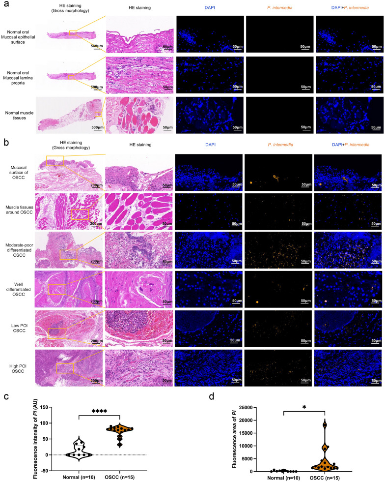
Background: Oral microbial dysbiosis contributes to the development of oral squamous cell carcinoma (OSCC). Our previous study showed that Prevotella intermedia (P. intermedia) were enriched in the oral mucosal surface, plaque, and saliva of patients with OSCC. Intratumoral microbiome could reshape the immune system and influence the development of various tumors. However, the invasion status of human OSCC tissues by P. intermedia and the pathway through which intratumoral P. intermedia potentiates tumor progression remain unexplored.

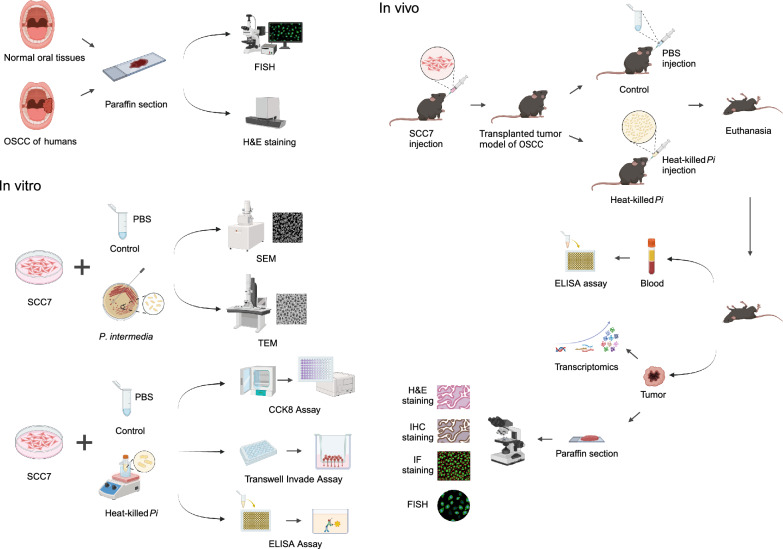
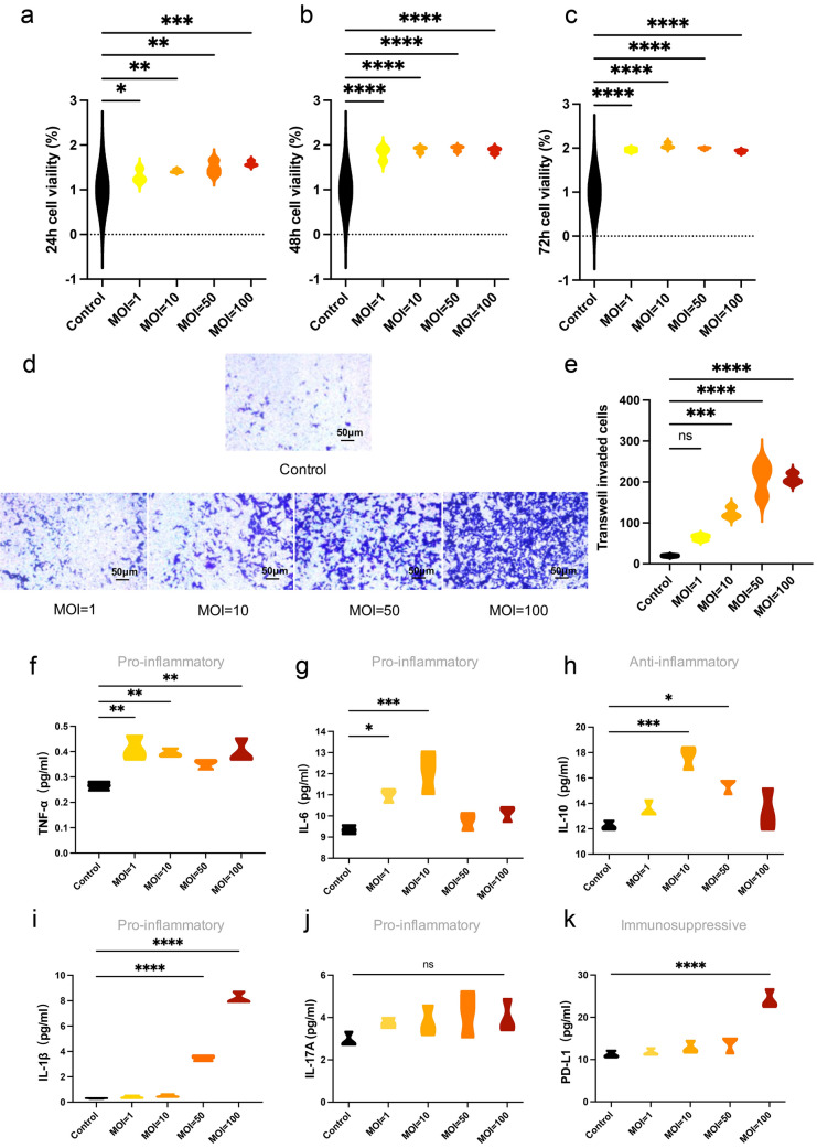
Methods: P. intermedia in human OSCC or normal tissues was detected by FISH. A mouse OSCC cell line SCC7 was adopted to investigate the effects of heat-killed P. intermedia treatment on cell proliferation, invasion, and cytokine release by using CCK-8 assay, transwell invasion assay and ELISA. Moreover, we established a mouse transplanted tumor model by using SCC7 cells, injected heat-killed P. intermedia into tumor tissues, and investigated the effects of heat-killed P. intermedia on tumor growth, invasion, cytokine levels, immune cell infiltrations, and expression levels by using gross observation, H&E staining, ELISA, immunohistochemistry, mRNA sequencing, and transcriptomic analysis.

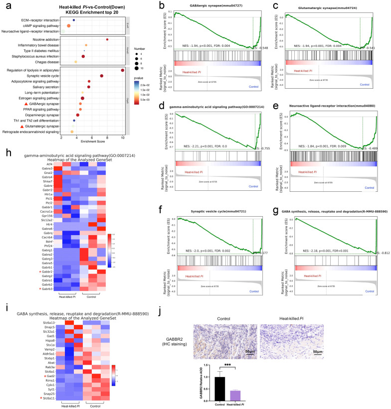
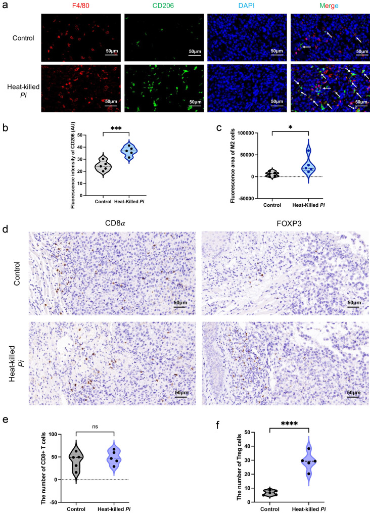
Results: Our results indicated that P. intermedia were abundant in OSCC and surrounding muscle tissues. Heat-killed P. intermedia promoted SCC7 cell proliferation, invasion and proinflammatory cytokine secretions, accelerated transplanted tumor growth in mice, exacerbate muscle and perineural invasion of OSCC, elevated the serum levels of IL-17A, IL-6, TNF-α, IFN-γ, and PD-L1, induced Treg cells M2 type macrophages in mouse transplanted tumors. The data of transcriptomic analysis revealed that heat-killed P. intermedia increased the expression levels of inflammatory cytokines and chemokines while reduced the expression levels of some tumor suppressor genes in mouse transplanted tumors. Additionally, IL-17 signaling pathway was upregulated whereas GABAergic system was downregulated by heat-killed P. intermedia treatment.

Conclusions: Taken together, our results suggest that P. intermedia could inhibit the expression of tumor suppressors, alter the tumor microenvironment, and promote the progression of OSCC.

Keywords: Prevotella intermedia; GABAergic system; IL-17 signal pathway; Oral squamous cell carcinoma; Tumor suppressor genes.


Conflict of interest statement

The authors declare that they have no competing interests.

Figures

Fig. 1
Fig. 1
Flow chart of the experiments. P. intermedia detection in OSCC and normal tissues, in vitro study of SCC7 cells, and in vivo study in a murine transplanted tumor model of OSCC
Fig. 2
Fig. 2
Representative images of H&E and FISH staining of human normal or OSCC tissues. DAPI (blue). P. intermedia labeled with specific probes (yellow). a Normal oral mucosal epithelial surface, mucosal lamina propria, and muscle tissues. b Mucosal surface of OSCC, muscle tissues around OSCC, various differentiated OSCC tissues, low or high pattern of invasion (POI) OSCC tissues. c Fluorescence intensity difference of P. intermedia between normal and OSCC tissues. d Fluorescence area difference of P. intermedia between normal and OSCC tissues. Data were analyzed by t test. *P < 0.05, ****P < 0.0001
Fig. 3
Fig. 3
Measurement of P. intermedia in mouse transplanted tumors and observation of P. intermedia-SCC7 cell cocultures. a FISH images of P. intermedia in OSCC tissues of mouse transplanted tumors using P. intermedia-specific probes. b SEM images of P. intermedia-SCC7 coculture for 40 min. c TEM images of P. intermedia-SCC7 coculture for 40 min. d TEM images of P. intermedia-SCC7 coculture for 3 h. e TEM images of P. intermedia in SCC7 cells following P. intermedia-SCC7 coculture for 3 h. White arrows indicate P. intermedia in OSCC tissues of mice. Red arrows indicate P. intermedia that is adhering to SCC7 cells. A green arrow indicates mitotic structures of P. intermedia within the SCC7 cytosol
Fig. 4
Fig. 4
Effects of heat-killed P. intermedia on SCC7 cell proliferation and cytokine release at different MOIs. ac Viability of SCC7 cells treated with/without heat-killed P. intermedia. a Cell viability at 24 h. b Cell viability at 48 h. c Cell viability at 72 h. d, e Cell invasion ability at 24 h detected by using transwell assays. fk Secretory levels of TNF-α, IL-6, IL-10, IL-1β, IL-17A, and PD-L1 by SCC7 cells treated with/without heat-killed P. intermedia. Data are the means of values from three independent experiments, shown as the mean ± SEM, and were analyzed using one-way ANOVA. *P < 0.05, **P < 0.01, ***P < 0.001, ****P < 0.0001. ns, not significant.
Fig. 5
Fig. 5
Heat-killed P. intermedia accelerated transplanted tumor growth in mice. a Flow chart of transplanted tumor experiments. b Tumor volume changes in mice. c Body weight changes in mice. d Gross morphology of dissected tumors in each group. e Differences in tumor weight between the heat-killed P. intermedia and control groups. f Differences in tumor volume between the heat-killed P. intermedia and control groups. g Invasive fronts (black arrows), muscle (M) and perineural (closed arrowheads) invasion of OSCC tissues (T) are shown according to H&E staining, KI-67 and MMP-9 expressions were determined by IHC staining. h Invasive mode comparison between the heat-killed P. intermedia and control groups. i, j The relative AOD of KI-67 and MMP-9 IHC staining was compared between the heat-killed P. intermedia and control groups. Data were analyzed by t test. #P < 0.05, ***P < 0.001, ****P < 0.0001
Fig. 6
Fig. 6
Altered serum cytokine levels by heat-killed P. intermedia in mice with transplanted tumors. The data are expressed as the mean ± SD and were analyzed using a t test. **P < 0.01, ***P < 0.001, ****P < 0.0001
Fig. 7
Fig. 7
Effects of heat-killed P. intermedia on the transcriptomics of proinflammatory cytokines and chemokines in transplanted tumor model mice. a Top 20 upregulated pathways in the heat-killed P. intermedia group by using KEGG pathway enrichment analysis. The red triangle indicates OSCC-related top 1 proinflammatory signaling pathways. b Chord diagrams of upregulated genes and pathways. Red asterisks indicate important genes in the upregulated signaling pathway. c Heatmap of the chemokine activity pathway gene set in both groups. d Representative images of IL-17A, IL-6, and IL-23 IHC staining. The relative AOD of IHC staining was compared between the control and heat-killed P. intermedia groups. The data are shown as the mean ± SD and were analyzed by t test. **P < 0.01, ****P < 0.0001
Fig. 8
Fig. 8
Effects of heat-killed P. intermedia on the transcriptomics of GABA signaling and GABAergic synapses in transplanted tumor model mice. a Top 20 downregulated pathways between the heat-killed P. intermedia and control groups in KEGG pathway enrichment analysis. Red triangles indicate the GABA-related signaling pathway. bg GSEA showing the downregulation in the heat-killed P. intermedia group. b GABAergic synapse downregulation. c Glutamatergic synapse downregulation. d GABA signaling pathway downregulation. e Neuroactive ligand‒receptor interaction downregulation. f Synaptic vesicle cycle downregulation. g GABA synthesis, release, reuptake and degradation downregulation. h Heatmap of the GABA synthesis, release, reuptake and degradation gene set. i Heatmap of the GABA signaling pathway gene set. Red asterisks indicate important genes in the GABA-related signaling pathway. j Representative images of GABBR2 IHC staining. The relative AOD of GABBR2 IHC staining was compared between the control and heat-killed P. intermedia groups. The data are shown as the mean ± SD and were analyzed by t test. ***P < 0.001
Fig. 9
Fig. 9
Effects of heat-killed P. intermedia on the transcriptomics of tumor suppressors in transplanted tumor model mice. a Chord diagrams of downregulated genes and pathways. Red asterisks indicate classic tumor suppressor genes. b Volcano plot demonstrating the differentially expressed genes in the heat-killed P. intermedia group compared with the control group. The genes with significantly increased or decreased levels are colored in red or blue, respectively (Wald test, p value < 0.05, and fold change > 2). c Representative images of FASL and P63 IHC staining. The data are shown as the mean ± SD and were analyzed by t test. *P < 0.05, ****P < 0.0001
Fig. 10
Fig. 10
Heat-killed P. intermedia significantly increased the proportions of M2 macrophages and FOXP3 + Treg cells in mouse transplanted tumors. a Representative images of F4/80 (red) and CD206 (green) immunofluorescence staining. White arrows indicate the colocalization of F4/80 and CD206 in the same cell. b Fluorescence intensity difference of CD206 between the control and heat-killed P. intermedia groups. c Fluorescence area difference of CD206 between both groups. d Representative images of CD8α and FOXP3 IHC staining. e The number difference of CD8 + T cells between both groups. f The number difference of FOXP3 + Treg cells between both groups. The data are shown as the mean ± SD and were analyzed by t test. *P < 0.05, ***P < 0.001, ****P < 0.0001. ns, not significant
Fig. 11
Fig. 11
Heat-killed P. intermedia increased tumor nerve density and perineural invasion. a Nerve density in OSCC tissues of mouse transplanted tumors. b Representative images of nerve density in normal and OSCC human tissues. c Innervation score difference between the control and heat-killed P. intermedia groups. d Innervation score difference between normal and OSCC tissues of humans. e FOXP3 + Tregs in perineural areas around the invasive fronts of OSCC can be seen in IHC staining images. Black circles and closed arrowheads show the neural structures. Black asterisks and arrows represent the main tumors and Tregs at tumor invasive fronts, respectively. The data are shown as the mean ± SD and were analyzed by t test. *P < 0.05, ****P < 0.0001
Fig. 12
Fig. 12
a Correlation analysis between the gene expression of three tumor suppressors and tumor-associated factors. Pairwise comparisons of various factors are shown, with a color gradient denoting Spearman’s correlation coefficients. Red and blue nodes denote positive and negative correlations, respectively. Line width corresponds to Mantel’s r statistic for the corresponding distance correlations, and line color denotes statistical significance. b Schematic of P. intermedia-induced OSCC progression showing the effects of heat-killed P. intermedia on the TME and tumor suppressor genes

Similar articles

Cited by

References

    1. Irfan M, Delgado RZR, Frias-Lopez J. The oral microbiome and cancer. Front Immunol. 2020;11:591088. doi: 10.3389/fimmu.2020.591088. - DOI - PMC - PubMed
    1. Xie Y, et al. Microbiota in tumors: from understanding to application. Adv Sci. 2022;9(21):e2200470. doi: 10.1002/advs.202200470. - DOI - PMC - PubMed
    1. Heng W, et al. Oral bacteriome and mycobiome across stages of oral carcinogenesis. Microbiol Spectr. 2022;10(6):e0273722. doi: 10.1128/spectrum.02737-22. - DOI - PMC - PubMed
    1. Zhang L, et al. The oral microbiota may have influence on oral cancer. Front Cell Infect Microbiol. 2019;9:476. doi: 10.3389/fcimb.2019.00476. - DOI - PMC - PubMed
    1. Okuyama K, Yanamoto S. Oral bacterial contributions to gingival carcinogenesis and progression. Cancer Prev Res. 2023 doi: 10.1158/1940-6207.CAPR-22-0511. - DOI - PubMed

LinkOut - more resources