. 2024 Mar;14(3):e1632.
doi: 10.1002/ctm2.1632.

PROM2 overexpression induces metastatic potential through epithelial-to-mesenchymal transition and ferroptosis resistance in human cancers

Affiliations

PROM2 overexpression induces metastatic potential through epithelial-to-mesenchymal transition and ferroptosis resistance in human cancers

Justine Paris et al. Clin Transl Med. 2024 Mar.

Abstract

Introduction: Despite considerable therapeutic advances in the last 20 years, metastatic cancers remain a major cause of death. We previously identified prominin-2 (PROM2) as a biomarker predictive of distant metastases and decreased survival, thus providing a promising bio-target. In this translational study, we set out to decipher the biological roles of PROM2 during the metastatic process and resistance to cell death, in particular for metastatic melanoma.

Methods and results: Methods and results: We demonstrated that PROM2 overexpression was closely linked to an increased metastatic potential through the increase of epithelial-to-mesenchymal transition (EMT) marker expression and ferroptosis resistance. This was also found in renal cell carcinoma and triple negative breast cancer patient-derived xenograft models. Using an oligonucleotide anti-sense anti-PROM2, we efficaciously decreased PROM2 expression and prevented metastases in melanoma xenografts. We also demonstrated that PROM2 was implicated in an aggravation loop, contributing to increase the metastatic burden both in murine metastatic models and in patients with metastatic melanoma. The metastatic burden is closely linked to PROM2 expression through the expression of EMT markers and ferroptosis cell death resistance in a deterioration loop.

Conclusion: Our results open the way for further studies using PROM2 as a bio-target in resort situations in human metastatic melanoma and also in other cancer types.

Keywords: breast cancer; epithelial‐to‐mesenchymal transition; ferroptosis resistance; melanoma; metastases; prominin‐2; renal cancer.


Conflict of interest statement

The authors declare that they have no competing interests.

Figures
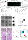
FIGURE 1
FIGURE 1
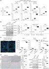
PROM2 expression level has an impact on melanoma invasion and migration. (A) mRNA expression level of PROM2 in A375 KO (Cq > 40), A375 and A375 PROM2 cell lines (***p < .001). (B) PROM2 immunofluorescence staining (in green) with nuclei stained with DAPI (in blue), and protein normalised expression using Western blot (**p < .01). (C and D) Count of invading (C) or migrating (D) cells at 24, 48 and 72 h in Boyden chamber (***p < .001). (E) The left panel displays images illustrating the formation and expansion of spheroids derived from A375 KO, A375 and A375 PROM2 cell lines. On the right, the outcomes of the thresholding process are presented, conducted on the images acquired at day 4 after 24 h of invasion in collagen. Additionally, thresholded images for all analysed spheroids are included in Figure S4. Scale bars = 100 µm (**p < .01). (F) Left panel shows histological sections of lungs from mice injected with 5 × 106 A375 KO, A375 or A375 PROM2 cell lines, using standard haematoxylin and eosin coloration (A, pulmonary alveoli; B, bronchus; M, metastasis). Right panel shows the mean surface area in percentage of lung metastases/total lung surface area quantified 8 weeks after injection (***p < .001). (G) Mean number of metastases in the five mouse models of patient‐derived melanoma xenografts (**p < .01).
FIGURE 2
FIGURE 2
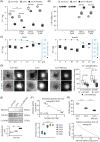
PROM2 overexpression in cells is associated with an EMT phenotype. (A) mRNA expression level of epithelial marker (CDH1), EMT markers (ZEB1, ZEB2, SNAI1, SNAI2, TWIST1, TWIST2) and mesenchymal marker (VIM) of A375 KO, A375 and A375 PROM2 cell lines (**p < .01). (B) Immunoblotting for ZEB1 and SNAI1, with normalised expression in A375 KO, A375 and A375 PROM2 cell lines (**p < .01). (C) Left panel illustrates the laser‐microdissection of a melanoma lung metastasis tagged with PRAME (green fluorescence, red arrow) from a mice injected with A375 cell line. Right panel shows mRNA quantification of ZEB1 (***p < .0001), SNAI1 (***p < .001) and TWIST1 (***p < .001) in laser‐microdissected lung metastases or tumour xenografts using digital‐droplet PCR. (D) ZEB1 and SNAIL/SLUG immunostaining of tumour xenografts expressing low (PROM2‐) or high (PROM2+) PROM2 (***p < .0001). (E) PROM2, ZEB1 and SNAIL/SLUG immunoblotting of tumour xenografts expressing low (PROM2−) or high (PROM2+) PROM2 (**p < .01).
FIGURE 3
FIGURE 3
PROM2 overexpression is associated with ferroptosis resistance. (A) Quantification of ferrous iron (Fe2+) in A375 KO, A375 and A375 PROM2 cell lines without treatment, or after 24 h of treatment with deferoxamine (DFO, ferroptosis inhibitor), or with RSL3 or Erastin (ferroptosis activators) (***p < .0001). (B) Cell viability of A375 KO, A375 and A375 PROM2 cell lines after the addition of DFO, RSL3 or Erastin during 24 h (***p < .0001). (C) Quantification of Fe2+ (bar histograms) and cell viability (dash‐dotted line) of A375 PROM2 cell line treated for 24 h with increasing concentrations of RSL3 or Erastin. (D) Left panel shows spheroids obtained from A375 cell lines and cultured with or without Erastin [using white light (left) or FerroOrange fluorescence (right)]. Right panel shows Fe2+ quantification through FerroOrange fluorescence quantification (**p < .01). (E) Immunoblotting and quantification of TSG101 (multivesicular bodies) in A375 KO (white), A375 (grey) and A375 PROM2 (black) cell lines (**p < .01). (F) Quantification of Fe2+ in patient‐derived melanoma xenografts, with the correlation with PROM2 mRNA expression in tumours (***p < .0001). (G) Quantification of 4HNE (lipid peroxidation) in A375 KO, A375 and A375 PROM2 cell lines (top panel, ***p < .0001), and the correlation with PROM2 mRNA expression in patient‐derived melanoma xenografts (bottom panel, ***p < .001).
FIGURE 4
FIGURE 4
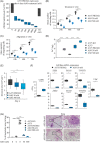
Targeting PROM2 with an antisense oligonucleotide (ASO) prevents metastases in human melanoma xenografts. (A) PROM2 mRNA expression of A375 PROM2 cells treated during 4 days with different concentrations of an anti‐PROM2. (B) Counts of invading A375 PROM2 cells treated for 72 h with two concentrations of the ASO (***p < .0001). (C) Counts of migrating A375 PROM2 cells treated for 72 h with two concentrations of the ASO (***p < .0001). (D) Quantification of Fe2+ of A375 PROM2 cell line treated for 4 days with two concentrations of the ASO (***p < .0001). (E) mRNA expression level of epithelial marker (CDH1), EMT markers (ZEB1, ZEB2, SNAI1, SNAI2, TWIST1, TWIST2) and mesenchymal marker (VIM) of A375 PROM2 cell line treated for 4 days with 0 or 50 nM of the ASO (*p = .04). (F) Mean surface area of lung metastases determined 8 weeks after injection of 5 × 106 A375 PROM2 cell line, untreated or treated in vitro for 4 days with 50 or 500 nM of the ASO before injection (***p < .001).
FIGURE 5
FIGURE 5
PROM2 is implicated in a pathophysiological loop of increased metastatic potential. (A) PROM2 mRNA expression after detachment of A375 cell line for 0, 30, 60 and 120 min (***p < .0001). (B) PROM2 mRNA expression in A375 and A375 PROM2 cell lines, and in subcutaneous xenografts and lung metastases derived from A375 PROM2 cell line and obtained 8 weeks after grafting (***p < .0001). (C) Schematic of experimental enrichment loop (BioRender and SMART Servier Medical Art). (D) Mean number of metastases between round 1 and round 2 (***p < .001). (E) PROM2, CAV1 and EMT marker mRNA expression in cultured cells after successive rounds (**p < .01 from round 0 to 3).
FIGURE 6
FIGURE 6
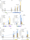
PROM2 pathophysiological loop is linked to metastatic progression in patients. (A) PROM2 and EMT marker mRNA expression in sequential biopsies of three patients with metastatic melanoma (Patient 1: Mel1, Patient 2: Mel2, Patient 3: Mel3). (B) PROM2 and EMT marker mRNA expression in sequential biopsies of two patients with renal cell carcinoma (Patient 1: RCC1, Patient 2: RCC2). Ipi, ipilimumab; nivo, nivolumab; pembro, pembrolizumab.
FIGURE 7
FIGURE 7
Pathophysiological loop by which high PROM2 expression promotes metastases and induces resistance to ferroptotic cell death. At the first step of the metastatic process, when a melanoma cell detaches from the tumour burden, the ferroptotic stress induces a ROS‐dependent cell death if PROM2 expression is low. In a melanoma cell with high PROM2 expression, PROM2 activates caveolin‐1 with an increase of EMT marker expression. This increase of EMT markers is associated with cell detachment and invasion. As caveolin‐1 is known to modify cdc‐42 activity and cell polarity through redistribution of actin fibres, we represent here PROM2 interplay with EMT markers and invadopode formation during invasion process. During migration in blood vessel, PROM2 overexpression overcomes ferroptotic cell death through the formation of microvesicular bodies and expulsion of Fe2+ from the cell. In addition, the ferroptotic stress increases itself PROM2 overexpression in an aggravating loop favouring the metastatic process.

References

    1. Wolchok JD, Chiarion‐Sileni V, Gonzalez R, et al. Long‐term outcomes with nivolumab plus ipilimumab or nivolumab alone versus ipilimumab in patients with advanced melanoma. J Clin Oncol. 2022;40(2):127‐137. - PMC - PubMed
    1. Robert C, Grob JJ, Stroyakovskiy D, et al. Five‐year outcomes with dabrafenib plus trametinib in metastatic melanoma. N Engl J Med. 2019;381(7):626‐636. - PubMed
    1. Serra‐Bellver P, Versluis JM, Oberoi HK, et al. Real‐world outcomes with ipilimumab and nivolumab in advanced melanoma: a multicentre retrospective study. Eur J Cancer. 2022;176:121‐132. - PubMed
    1. Nguyen TT, Gapihan G, Tetu P, et al. Increased risk of brain metastases among patients with melanoma and PROM2 expression in metastatic lymph nodes. Clin Transl Med. 2020;10(8):e198. - PMC - PubMed
    1. Fargeas CA, Florek M, Huttner WB, Corbeil D. Characterization of prominin‐2, a new member of the prominin family of pentaspan membrane glycoproteins. J Biol Chem. 2003;278(10):8586‐8596. - PubMed

Publication types

Substances

LinkOut - more resources