

Case Reports
doi: 10.1093/ehjcr/ytae123.
eCollection 2024 Mar.
Exercise-induced pseudo-ischaemic electrocardiographic changes in a female with concave-shaped chest wall
Affiliations
- PMID: 38515510
- PMCID: PMC10957157
- DOI: 10.1093/ehjcr/ytae123
Item in Clipboard
Case Reports
Exercise-induced pseudo-ischaemic electrocardiographic changes in a female with concave-shaped chest wall
Eur Heart J Case Rep.
.
No abstract available
Conflict of interest statement
Conflict of interest: None declared.
Figures
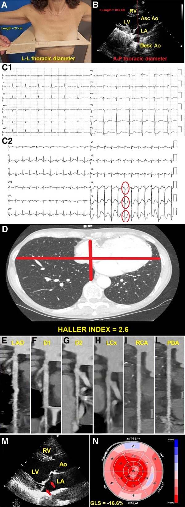
(A) L-L thoracic diameter, measured at end inspiration with the individual in the standing position and with open arms, by using a rigid ruler in centimetres coupled to a level (the measuring device), placed at the distal third of the sternum, in the point of maximum depression of the sternum. L-L, latero-lateral. (B) A-P thoracic diameter, obtained with the individual in the left-lateral decubitus position, during conventional transthoracic echocardiography, by placing a 2.5 mHz transducer near the sternum in the left third or fourth intercostal space, to obtain a parasternal long-axis view, and measuring at end inspiration the distance between the true apex of the sector and the anterior surface of the vertebral body. The vertebral body is identified by using, as a reference point, the posterior wall of the descending thoracic aorta, visualized behind the left atrium. Ao, aorta; A-P, antero-posterior; Asc, ascending; Desc, descending; LA; left atrium; LV, left ventricle; MHI, modified Haller index; RV, right ventricle. (C1) Resting electrocardiogram showing sinus rhythm with mild right ventricular conduction delay and non-specific ST-T repolarization abnormalities in the anterolateral leads. (C2) Peak-exercise electrocardiogram showing marked downsloping ST-segment depression in the anterolateral leads V4–V6 (circles) (maximum 2.5 mm in V5). (D) Axial computed tomography scan assessing conventional Haller index, the ratio of chest transverse diameter over the distance between the sternum and spine. (E–L) Computed tomography coronary angiography: curved planar reformation images of left anterior descending artery (E), D1 (F), D2 (G), left circumflex artery (H), right coronary artery (I), and posterior descending artery (L), with no evidence of obstructive coronary artery disease. D1, first diagonal branch; D2, second diagonal branch; LAD, left anterior descending artery; LCx, left circumflex artery; PDA, posterior descending artery; RCA, right coronary artery. (M) Parasternal long-axis view demonstrating bileaflet mitral valve thickening and prolapse (arrows). Ao, aorta; LA, left atrium; LA, left ventricle; RV, right ventricle. (N) Global longitudinal strain bull's eye plot assessed by strain echocardiographic imaging. This example shows a moderate impairment in global longitudinal strain magnitude (−16.6%), secondary to a significant reduction in left ventricular basal strain values, represented as light red, light pink, and pale and/or light blue. GLS, global longitudinal strain; LV, left ventricular.
Similar articles
-
False-positive electrocardiographic changes during exercise test in a patient with pectus excavatum.J Clin Ultrasound. 2020 Nov;48(9):579-584. doi: 10.1002/jcu.22831. Epub 2020 Apr 6. J Clin Ultrasound. 2020. PMID: 32249937
-
Both Bilateral Breast Volume Discrepancy and Asymmetric Anterior Chest Wall Shape Contribute to the Unsightly Breast Contour in Female Right Thoracic Idiopathic Scoliosis.Clin Spine Surg. 2017 May;30(4):E344-E350. doi: 10.1097/BSD.0000000000000128. Clin Spine Surg. 2017. PMID: 28437336
-
Prognostic Value of Modified Haller Index in Patients with Suspected Coronary Artery Disease Referred for Exercise Stress Echocardiography.J Cardiovasc Echogr. 2021 Apr-Jun;31(2):85-95. doi: 10.4103/jcecho.jcecho_141_20. Epub 2021 Jul 28. J Cardiovasc Echogr. 2021. PMID: 34485034 Free PMC article.
-
Exercise echocardiography.Aust N Z J Med. 1992 Oct;22(5 Suppl):610-2. doi: 10.1111/j.1445-5994.1992.tb00487.x. Aust N Z J Med. 1992. PMID: 1449449 Review.
-
Radiation therapy for breast cancer: Literature review.Med Dosim. 2016 Autumn;41(3):253-7. doi: 10.1016/j.meddos.2016.06.005. Med Dosim. 2016. PMID: 27545009 Review.
References
-
- Sonaglioni A, Baravelli M, Vincenti A, Trevisan R, Zompatori M, Nicolosi GL, et al. . A new modified anthropometric Haller index obtained without radiological exposure. Int J Cardiovasc Imaging 2018;34:1505–1509. - PubMed
-
- Udoshi MB, Shah A, Fisher VJ, Dolgin M. Incidence of mitral valve prolapse in subjects with thoracic skeletal abnormalities—a prospective study. Am Heart J 1979;97:303–311. - PubMed
-
- Boudoulas KD, Pitsis AA, Mazzaferri EL, Gumina RJ, Triposkiadis F, Boudoulas H. Floppy mitral valve/mitral valve prolapse: a complex entity with multiple genotypes and phenotypes. Prog Cardiovasc Dis 2020;63:308–326. - PubMed
Publication types
LinkOut - more resources
Full Text Sources