. 2024 Mar 7:14:1333640.
doi: 10.3389/fonc.2024.1333640. eCollection 2024.

Treatment response of advanced HNSCC towards immune checkpoint inhibition is associated with an activated effector memory T cell phenotype

Affiliations

Treatment response of advanced HNSCC towards immune checkpoint inhibition is associated with an activated effector memory T cell phenotype

Max Schumacher et al. Front Oncol. .

Abstract

Locally advanced or metastatic head and neck squamous cell carcinoma (HNSCC) is associated with a poor prognosis. The introduction of PD-1 inhibitors has led to a significant improvement in survival, but only a subpopulation of patients responds to therapy. Current biomarkers cannot reliably identify these patients. The identification of biomarkers for the prediction and monitoring of immunotherapy is therefore of great importance. In this study, we characterized lymphocyte subsets in the peripheral blood of HNSCC patients under PD-1 inhibition. Patients with primary response (n=11) to PD-1 inhibition showed an increase of the CD3+ effector memory (CD3/EM) population and an elevated expression of the activation marker CD69 in CD3+ T cells, particularly in the CD3/EM subpopulation at 3 months when treatment response was assessed. In contrast, patients with primary treatment failure and progressive disease (n=9) despite PD-1 inhibition had lower absolute lymphocyte counts and an increased expression of CTLA-4 in CD3+ T cells at the time of treatment failure compared with baseline, particularly in CD4+ and CD8+ effector memory populations. Our results demonstrate that HNSCC patients' response to immune checkpoint inhibition shows a distinct immune signature in peripheral blood, which could help identify refractory patients earlier. Furthermore, strategies to overcome primary therapy failure by inducing a beneficial T cell phenotype or adding alternative immune checkpoint inhibitors could improve response rates and survival of HNSCC patients.

Keywords: HNSCC; PD-1; activation marker; immune checkpoint inhibition; peripheral T cells; predictive biomarker.


Conflict of interest statement

The authors declare that the research was conducted in the absence of any commercial or financial relationships that could be construed as a potential conflict of interest.

Figures
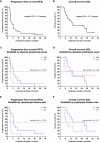
Figure 1
Figure 1
Kaplan-Meier survival curves. Dashes indicate censoring of subjects. (A) Overall survival and (B) progression-free survival of the overall population. (C) PFS and (D) OS in patients stratified by the median ALC. (E) PFS and (F) OS in patients stratified by the median size of lymphocyte fractions.
Figure 2
Figure 2
CD3+ T cell subpopulations and CD69 expression. (A) Heat map depicting mean of relative changes of CD3+ T cell subpopulations 3 months after therapy initiation compared to baseline. Mean of the relative changes were log10 transformed. (B) Percentage of CD3+ EM T cells in patients with therapy response. (C) Heat map depicting the relative change of CD69 expression on CD3+ T cells. Each box represents an individual patient and relative changes were log10 transformed. (D) Percentage of CD69 expression on CD3+ T cells in patients with therapy response. Percentage of CD69 expression on (E) CD3+/EMRA and (F) CD3+/EM T cells in patients with therapy response. Bars indicate standard error of the mean. *p<0.05, **p<0.01.
Figure 3
Figure 3
Expression of alternate immune checkpoints on T cells. (A) Heat map depicting the relative change of CTLA-4, LAG-3 and TIM-3 expression on CD3+ T cells after 3 months of PD-1 inhibition compared with treatment initiation. Each box represents an individual non-responding patient and relative changes were log10 transformed. (B) Percentage of CTLA-4 expression on CD3+ T cells in patients with resistance to therapy. Percentage of CTLA-4, LAG-3 and TIM-3 expression on (C) CD4+ and (D) CD8+ T cells in patients with therapy resistance. (E) Heat map depicting the relative change of CTLA-4, LAG-3 and TIM-3 expression on CD3+ T cells after 3 months of PD-1 inhibition compared with treatment initiation. Each box represents an individual responding patient and relative changes were log10 transformed. (F) Percentage of CTLA-4 expression on CD3+ T cells in patients with response to therapy. Percentage of CTLA-4, LAG-3 and TIM-3 expression on (G) CD4+ and (H) CD8+ T cells in patients with therapy response. Bars indicate standard error of the mean. ns non-significant, *p<0.05, **p<0.01.
Figure 4
Figure 4
CTLA-4 expression on CD4+ and CD8+ subpopulations. Percentage of CTLA-4 expression on different (A) CD4+ and (B) CD8+ T cell subsets in patients with therapy resistance. Percentage of CTLA-4 expression on different (C) CD4+ and (D) CD8+ T cell subsets in patients with therapy response. Bars indicate standard error of the mean. ns non-significant, * p<0.05, ** p<0.01.

References

    1. Johnson DE, Burtness B, Leemans CR, Lui VWY, Bauman JE, Grandis JR. Head and neck squamous cell carcinoma. Nat Rev Dis Primers (2020) 6(1):92. doi: 10.1038/s41572-020-00224-3 - DOI - PMC - PubMed
    1. Argiris A, Karamouzis MV, Raben D, Ferris RL. Head and neck cancer. Lancet (2008) 371(9625):1695–709. doi: 10.1016/S0140-6736(08)60728-X - DOI - PMC - PubMed
    1. Sung H, Ferlay J, Siegel RL, Laversanne M, Soerjomataram I, Jemal A, et al. . Global cancer statistics 2020: GLOBOCAN estimates of incidence and mortality worldwide for 36 cancers in 185 countries. CA A Cancer J Clin (2021) 71(3):209–49. doi: 10.3322/caac.21660 - DOI - PubMed
    1. Vermorken JB, Mesia R, Rivera F, Remenar E, Kawecki A, Rottey S, et al. . Platinum-based chemotherapy plus cetuximab in head and neck cancer. N Engl J Med (2008) 359(11):1116–27. doi: 10.1056/NEJMoa0802656 - DOI - PubMed
    1. Ferris RL, Blumenschein G, Fayette J, Guigay J, Colevas AD, Licitra L, et al. . Nivolumab for recurrent squamous-cell carcinoma of the head and neck. N Engl J Med (2016) 375(19):1856–67. doi: 10.1056/NEJMoa1602252 - DOI - PMC - PubMed