

Case Reports
doi: 10.1016/j.radcr.2024.02.049.
eCollection 2024 Jun.
Cholecystitis-related cystic artery pseudoaneurysm: Case report
Affiliations
- PMID: 38515769
- PMCID: PMC10950602
- DOI: 10.1016/j.radcr.2024.02.049
Item in Clipboard
Case Reports
Cholecystitis-related cystic artery pseudoaneurysm: Case report
Radiol Case Rep.
.
Abstract
The pseudoaneurysms of the cystic artery (CAP) are very uncommon. They usually develop as a result of an acute cholecystitis or after a cholecystectomy. Among the complications, we can find hemorrhaging, biliary blockage, and haemobilia. Limited understanding of the illness makes managing specific cases difficult. We describe a case of a cystic artery pseudoaneurysm complicating an acute cholecystitis that was successfully treated by transcatheter arterial embolization.
Keywords: Cholecystitis; Cystic artery; Interventional radiology; Pseudoaneurysm.
© 2024 The Authors. Published by Elsevier Inc. on behalf of University of Washington.
Figures
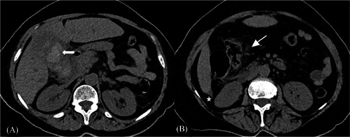
Unenhanced computed tomography shows a large intravesicular hematoma. (A) (white arrow). It also demonstrates a peri-vesicular fatty infiltration (arrowhead) and minimal sub-hepatic fluid collection (white star) (B).
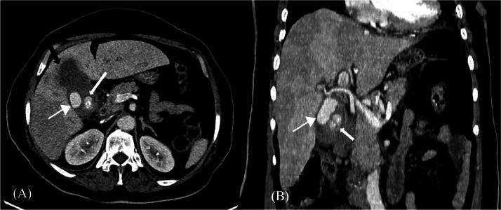
Contrast enhanced computed tomography scan showing a large cystic artery pseudoaneurysm (arrowhead) which is connected to the cystic artery associated with an infundibular gallstone (arrow) (A and B). Focal wall irregularity at the fundus suggesting a pre-perforating gallbladder (black arrows) (A).
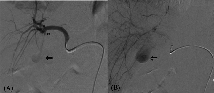
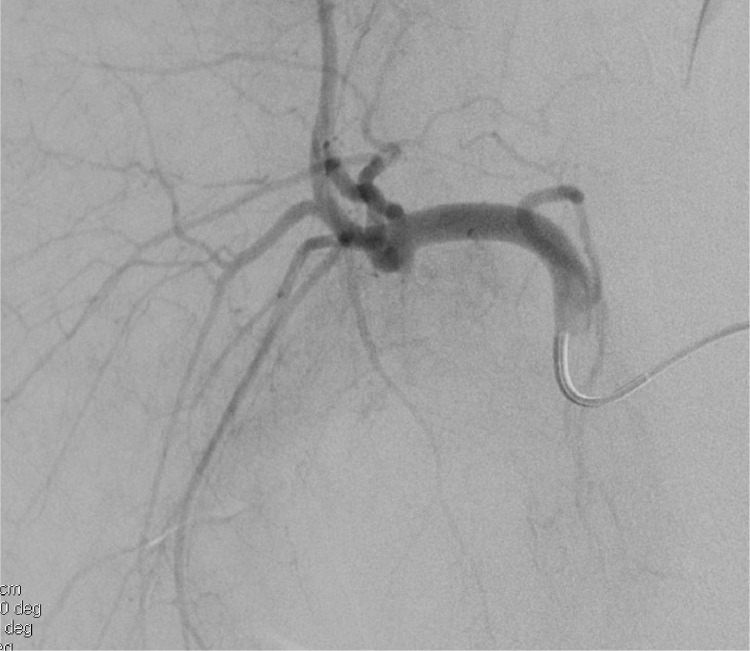
Hepatic artery Angiogram shows a cystic artery pseudoaneurysm with a diameter of 3cm (arrow) with a narrow neck (arrowhead).
Microcatheterism of the cystic artery shows a cystic artery pseudoaneurysm with a diameter of 3 cm (arrow) with a narrow neck (arrowhead).
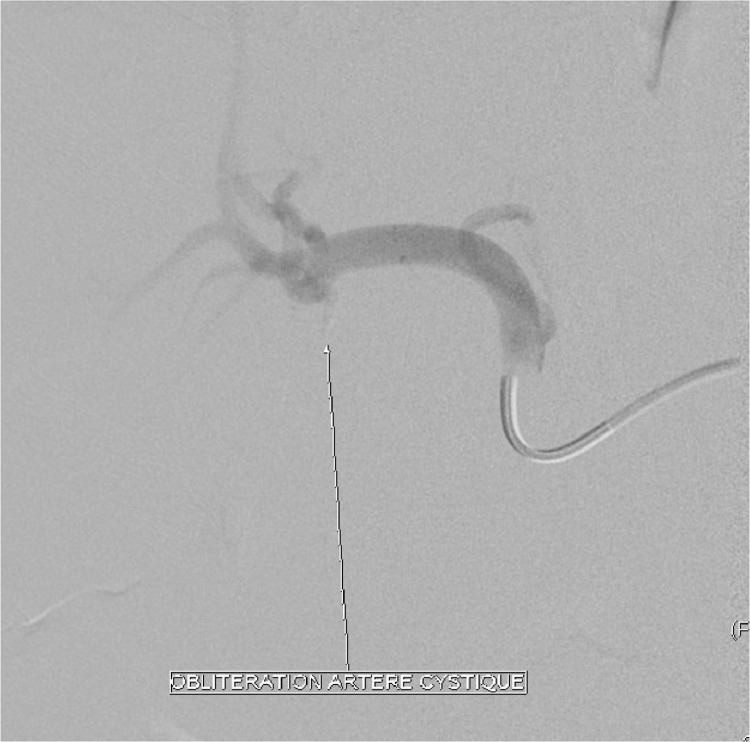
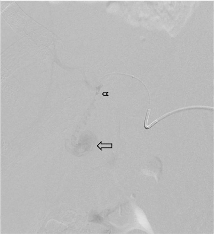
After embolization with glue, arterial inflow to the cystic artery is completely blocked.
The postembolization angiogram revealed CAP exclusion.
References
-
- Velázquez RM, Pérez JAC, Mérida MAA, Gómez JM. Non traumatic pseudoaneurysm of the cystic artery as a cause of haemobilia. Gastroenterol Hepatol. 2018;41(4):257–259. - PubMed
-
- Barba CA, Bret PM, Hinchey J. Pseudoaneurysm of the cystic artery: a rare cause of hemobilia. Can J Surg. 1994;37(1):64. - PubMed
Publication types
LinkOut - more resources
Full Text Sources
Miscellaneous
