. 2023 Aug 31;9(4):195-205.
doi: 10.52601/bpr.2023.230016.

Xenopus cell-free extracts and their applications in cell biology study

Affiliations

Xenopus cell-free extracts and their applications in cell biology study

Junjun Liu et al. Biophys Rep. .

Abstract

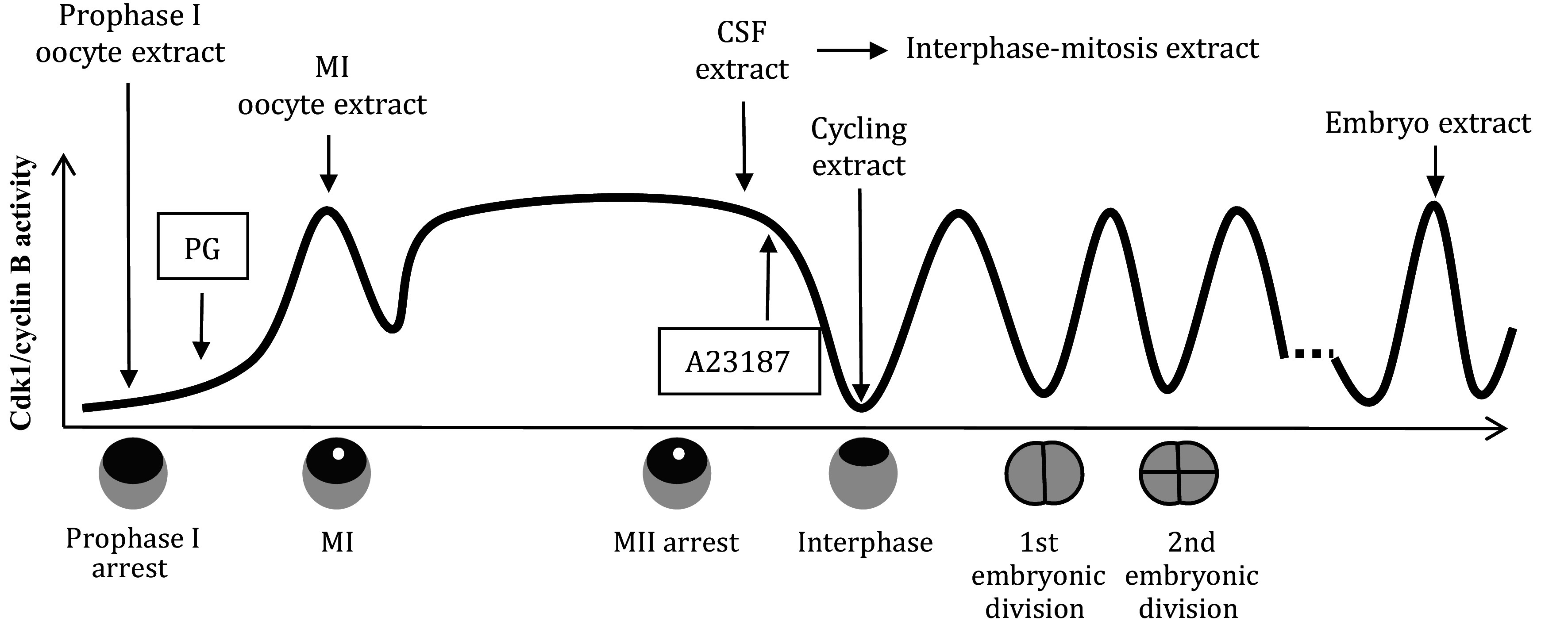
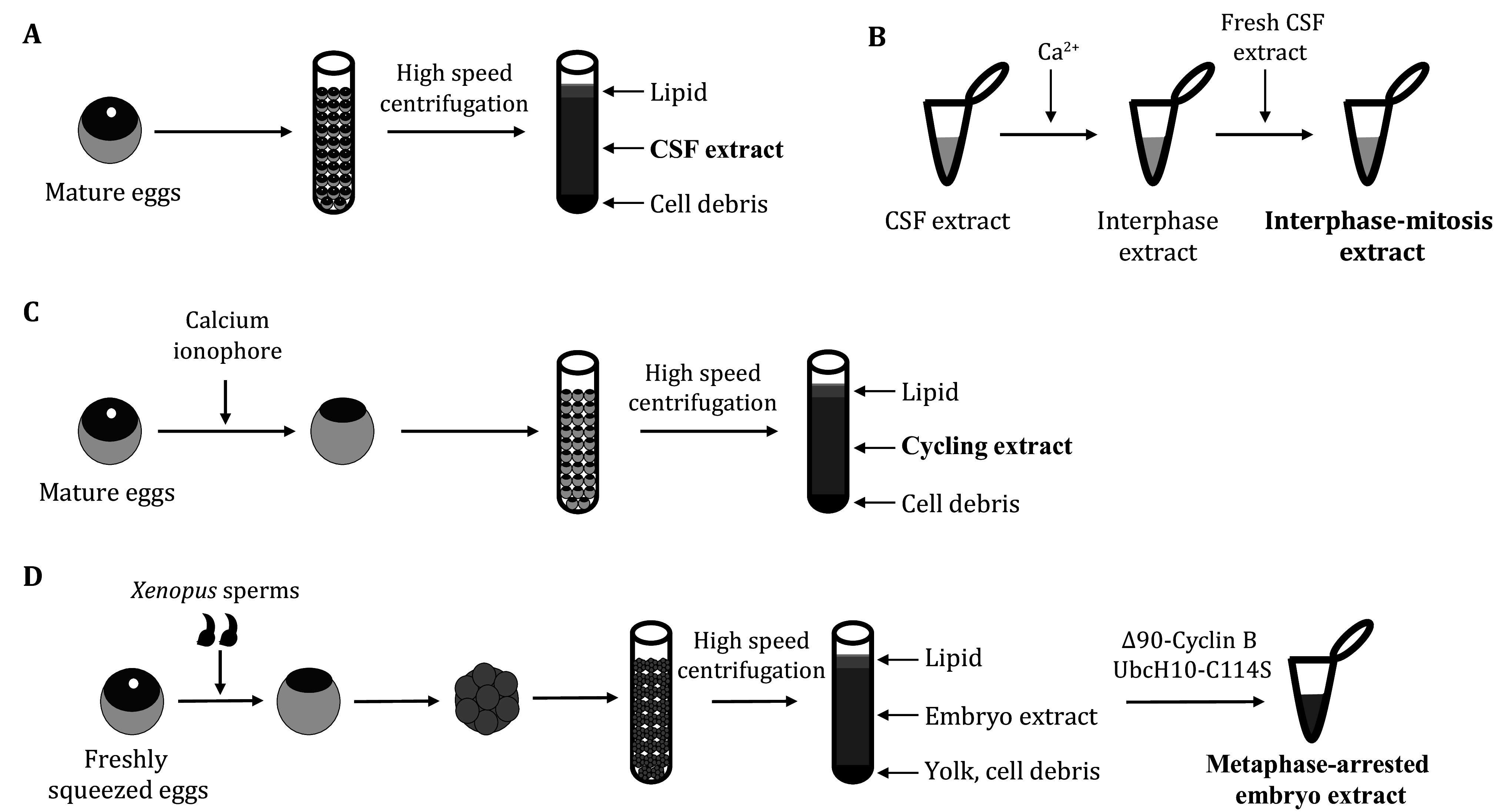
Xenopus has proven to be a remarkably versatile model organism in the realm of biological research for numerous years, owing to its straightforward maintenance in laboratory settings and its abundant provision of ample-sized oocytes, eggs, and embryos. The cell cycle of these oocytes, eggs, and early embryos exhibits synchrony, and extracts derived from these cells serve various research purposes. Many fundamental concepts in biochemistry, cell biology, and development have been elucidated through the use of cell-free extracts derived from Xenopus cells. Over the past few decades, a wide array of cell-free extracts has been prepared from oocytes, eggs, and early embryos of different Xenopus species at varying cell cycle stages. Each of these extracts possesses distinct characteristics. This review provides a concise overview of the Xenopus species employed in laboratory research, the diverse types of cell-free extracts available, and their respective properties. Furthermore, this review delves into the extensive investigation of spindle assembly in Xenopus egg extracts, underscoring the versatility and potency of these cell-free systems in the realm of cell biology.

Keywords: Cell-free extract; Egg; Embryo; Meiosis; Mitosis; Oocyte; Spindle; Xenopus.


Conflict of interest statement

Junjun Liu and Chuanmao Zhang declare that they have no conflict of interest.

Figures

Figure 1
Figure 1
Xenopus cell-free extracts. Progesterone (PG) induces prophase I-arrested Xenopus oocyte to mature. At GVBD, white spots appear at the animal hemisphere of the maturing oocytes, and the mature oocytes arrest at metaphase II due to CSF, which is fertilizable. After fertilization, the CSF is released, and the mature eggs enter interphase and start synchronous embryonic cell division until blastula stage VIII when the synchrony is lost. In the laboratory, the fertilization is simulated by the addition of calcium ionophore A23187. Prophase I-arrested oocytes are used to prepare prophase I oocyte extracts. MI-arrested (by cold treatment) oocytes are used to prepare MI oocyte extracts. MII-arrested eggs are used to prepare CSF extracts, and interphase-mitosis extracts are prepared from the freshly prepared CSF extracts with the addition of calcium. Activated eggs are used to prepare cycling extracts. Early-stage embryos are used to prepare embryo extracts, which can be arrested in mitosis with the addition of cell cycle regulators
Figure 2
Figure 2
Xenopus egg/embryo extracts. A MII-arrested Xenopus eggs are crushed by high-speed centrifugation to make CSF extract. B The CSF extract is added with calcium to release CSF arrest to allow the extract to enter interphase. With the addition of freshly prepared CSF extract, the system is driven back to and arrest at mitosis, and the extract is sometimes called interphase-mitosis extract. C MII-arrested Xenopus eggs are released to interphase with the addition of calcium ionophore A23187. Forty-five minutes after the calcium ionophore addition, the CSF-released eggs are used to prepare the cycling extract. D Mature Xenopus eggs are fertilized by sperm slurry prepared from Xenopus males and early embryonic development starts. These early Xenopus embryos at different development stages, e.g. Stage IV or Stage VIII, are used to prepare embryo extract. These extracts are added with cell cycle regulators such as Δ90-cyclin B and UbcH10-C114S to arrest the extracts at mitosis
Figure 3
Figure 3
Two pathways to spindle assembly in vitro. A In Xenopus CSF extract, each sperm nucleus contains one centrosome and directs the assembly of the one-half spindle. Two half spindles then fuse and form a bipolar spindle. B In extracts that go through interphase, the centrosome and DNA of the sperm nuclei will replicate, and each sperm nucleus is capable of the assembly of a bipolar spindle in this case

Similar articles

References

    1. Amaya E, Offield MF, Grainger RM Frog genetics: Xenopus tropicalis jumps into the future. Trends Genet. 1998;14(7):253–255. doi: 10.1016/S0168-9525(98)01506-6. - DOI - PubMed
    1. Benbow RM, Ford CC Cytoplasmic control of nuclear DNA synthesis during early development of Xenopus laevis: a cell-free assay. Proc Natl Acad Sci USA. 1975;72(6):2437–2441. doi: 10.1073/pnas.72.6.2437. - DOI - PMC - PubMed
    1. Bier E, De Robertis EM Embryo development. BMP gradients:a paradigm for morphogen-mediated developmental patterning. Science. 2015;348(6242):aaa5838. doi: 10.1126/science.aaa5838. - DOI - PubMed
    1. Blachon S, Khire A, Avidor-Reiss T The origin of the second centriole in the zygote of Drosophila melanogaster. Genetics. 2014;197(1):199–205. doi: 10.1534/genetics.113.160523. - DOI - PMC - PubMed
    1. Blow JJ, Laskey RA Initiation of DNA replication in nuclei and purified DNA by a cell-free extract of Xenopus eggs. Cell. 1986;47(4):577–587. doi: 10.1016/0092-8674(86)90622-7. - DOI - PubMed

LinkOut - more resources