

Deep learning approaches for breast cancer detection in histopathology images: A review
- PMID: 38517775
- PMCID: PMC11191493
- DOI: 10.3233/CBM-230251
Deep learning approaches for breast cancer detection in histopathology images: A review
Abstract
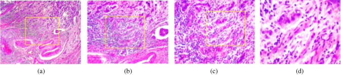
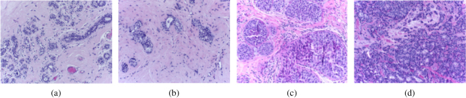
Background: Breast cancer is one of the leading causes of death in women worldwide. Histopathology analysis of breast tissue is an essential tool for diagnosing and staging breast cancer. In recent years, there has been a significant increase in research exploring the use of deep-learning approaches for breast cancer detection from histopathology images.
Objective: To provide an overview of the current state-of-the-art technologies in automated breast cancer detection in histopathology images using deep learning techniques.
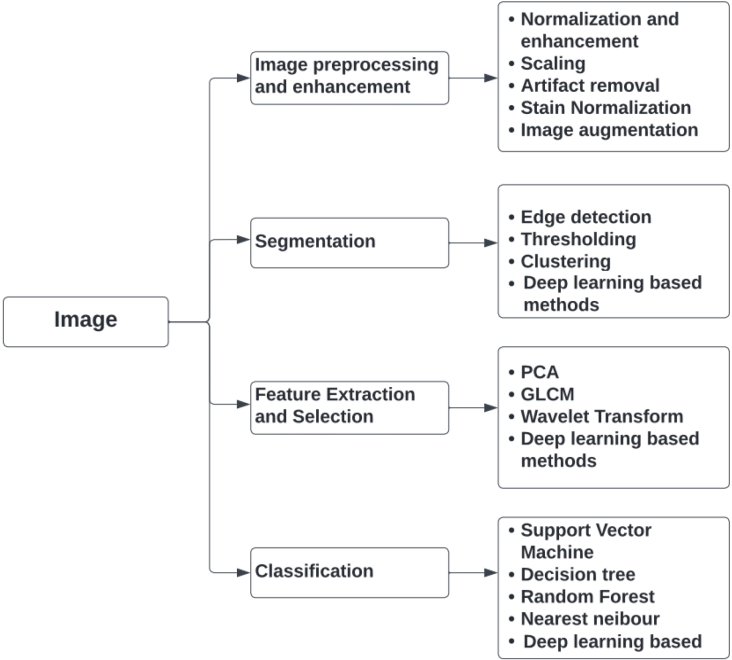
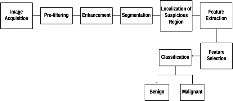
Methods: This review focuses on the use of deep learning algorithms for the detection and classification of breast cancer from histopathology images. We provide an overview of publicly available histopathology image datasets for breast cancer detection. We also highlight the strengths and weaknesses of these architectures and their performance on different histopathology image datasets. Finally, we discuss the challenges associated with using deep learning techniques for breast cancer detection, including the need for large and diverse datasets and the interpretability of deep learning models.
Results: Deep learning techniques have shown great promise in accurately detecting and classifying breast cancer from histopathology images. Although the accuracy levels vary depending on the specific data set, image pre-processing techniques, and deep learning architecture used, these results highlight the potential of deep learning algorithms in improving the accuracy and efficiency of breast cancer detection from histopathology images.
Conclusion: This review has presented a thorough account of the current state-of-the-art techniques for detecting breast cancer using histopathology images. The integration of machine learning and deep learning algorithms has demonstrated promising results in accurately identifying breast cancer from histopathology images. The insights gathered from this review can act as a valuable reference for researchers in this field who are developing diagnostic strategies using histopathology images. Overall, the objective of this review is to spark interest among scholars in this complex field and acquaint them with cutting-edge technologies in breast cancer detection using histopathology images.
Keywords: Computer-aided detection; Convolutional Neural Network (CNN); breast cancer; deep learning; histopathology images.
Figures
Similar articles
-
An explainable AI-driven deep neural network for accurate breast cancer detection from histopathological and ultrasound images.Sci Rep. 2025 May 20;15(1):17531. doi: 10.1038/s41598-025-97718-5. Sci Rep. 2025. PMID: 40394112 Free PMC article.
-
PViT-AIR: Puzzling vision transformer-based affine image registration for multi histopathology and faxitron images of breast tissue.Med Image Anal. 2025 Jan;99:103356. doi: 10.1016/j.media.2024.103356. Epub 2024 Sep 30. Med Image Anal. 2025. PMID: 39378568 Free PMC article.
-
Deep computational pathology in breast cancer.Semin Cancer Biol. 2021 Jul;72:226-237. doi: 10.1016/j.semcancer.2020.08.006. Epub 2020 Aug 17. Semin Cancer Biol. 2021. PMID: 32818626 Review.
-
An experimental study on breast lesion detection and classification from ultrasound images using deep learning architectures.BMC Med Imaging. 2019 Jul 1;19(1):51. doi: 10.1186/s12880-019-0349-x. BMC Med Imaging. 2019. PMID: 31262255 Free PMC article.
-
Deep Learning Approaches Towards Skin Lesion Segmentation and Classification from Dermoscopic Images - A Review.Curr Med Imaging. 2020;16(5):513-533. doi: 10.2174/1573405615666190129120449. Curr Med Imaging. 2020. PMID: 32484086 Review.
Cited by
-
Equilibrium Optimization-Based Ensemble CNN Framework for Breast Cancer Multiclass Classification Using Histopathological Image.Diagnostics (Basel). 2024 Oct 9;14(19):2253. doi: 10.3390/diagnostics14192253. Diagnostics (Basel). 2024. PMID: 39410657 Free PMC article.
-
Challenges and opportunities for Aotearoa/New Zealand's breast screening programme.J R Soc N Z. 2025 Feb 18;55(5):1295-1303. doi: 10.1080/03036758.2025.2463448. eCollection 2025. J R Soc N Z. 2025. PMID: 40718040 Free PMC article. Review.
-
A Pilot Study of Breast Cancer Histopathological Image Classification Using Google Teachable Machine: A No-Code Artificial Intelligence Approach.Cureus. 2025 Jul 4;17(7):e87301. doi: 10.7759/cureus.87301. eCollection 2025 Jul. Cureus. 2025. PMID: 40761997 Free PMC article.
References
-
- Abdollahi J., Davari N., Panahi Y., Gardaneh M. et al., Detection of metastatic breast cancer from whole-slide pathology images using an ensemble deep-learning method, Archives of Breast Cancer, 2022.
-
- Ahmad H.M., Ghuffar S. and Khurshid K., Classification of breast cancer histology images using transfer learning, in: 2019 16th International Bhurban Conference on Applied Sciences and Technology (IBCAST), IEEE, 2019, pp. 328–332.
-
- Ahmad N., Asghar S. and Gillani S.A., Transfer learning-assisted multi-resolution breast cancer histopathological images classification, The Visual Computer, 2021, 1–20.
-
- Anwar F., Attallah O., Ghanem N. and Ismail M.A., Automatic breast cancer classification from histopathological images, in: 2019 International Conference on Advances in the Emerging Computing Technologies (AECT), IEEE, 2020, pp. 1–6.
-
- Anwar S.M., Majid M., Qayyum A., Awais M., Alnowami M. and Khan M.K., Medical image analysis using convolutional neural networks: A review, Journal of Medical Systems 42 (2018), 1–13. - PubMed
Publication types
MeSH terms
LinkOut - more resources
Full Text Sources
Medical
