. 2024 Apr 23;43(4):113988.
doi: 10.1016/j.celrep.2024.113988. Epub 2024 Mar 22.

Evolution of chromosome-arm aberrations in breast cancer through genetic network rewiring

Affiliations

Evolution of chromosome-arm aberrations in breast cancer through genetic network rewiring

Elena Kuzmin et al. Cell Rep. .

Abstract

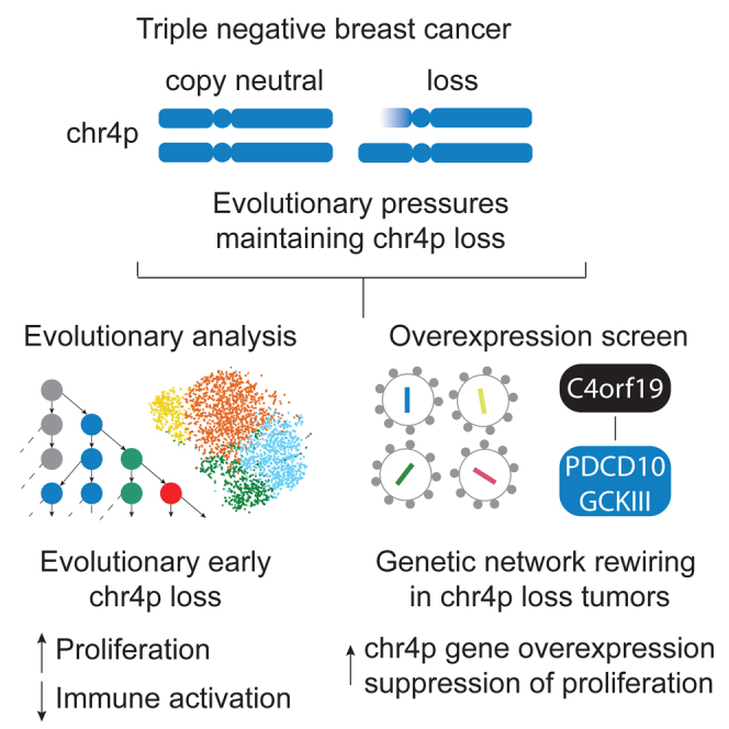
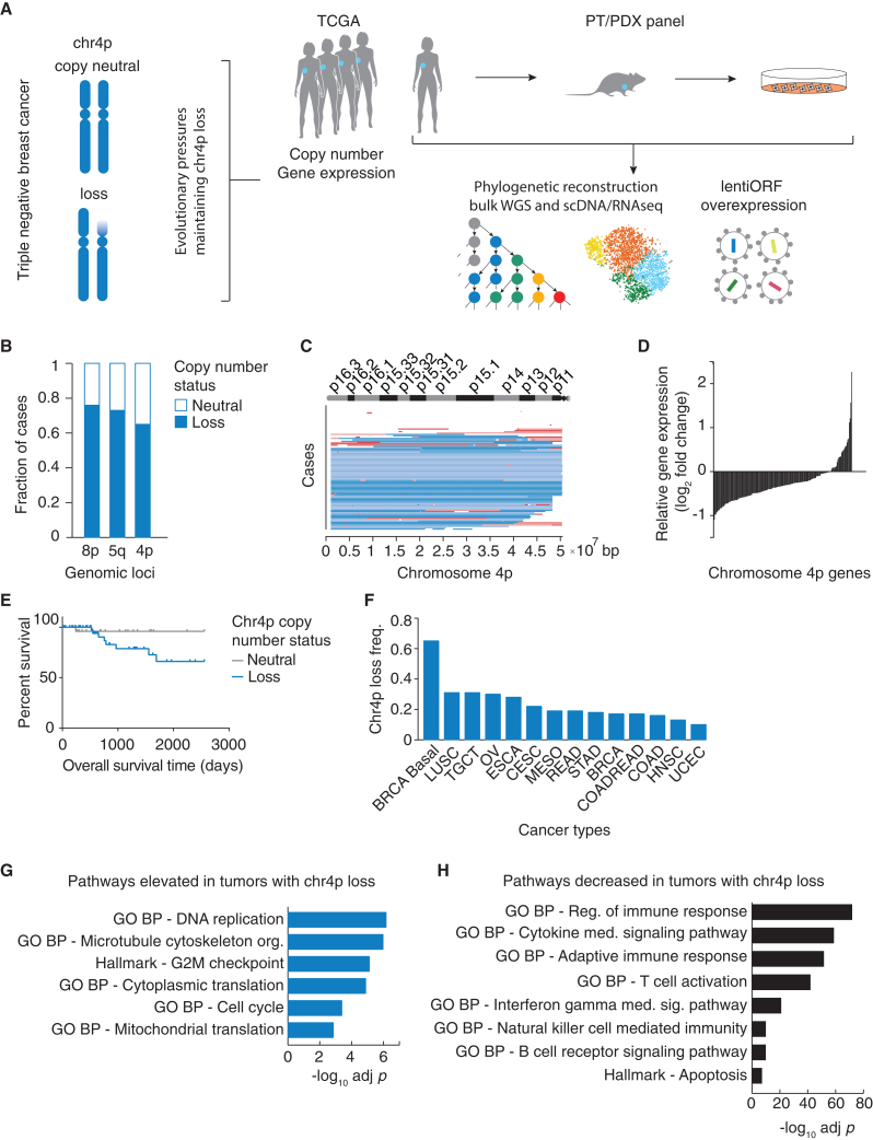
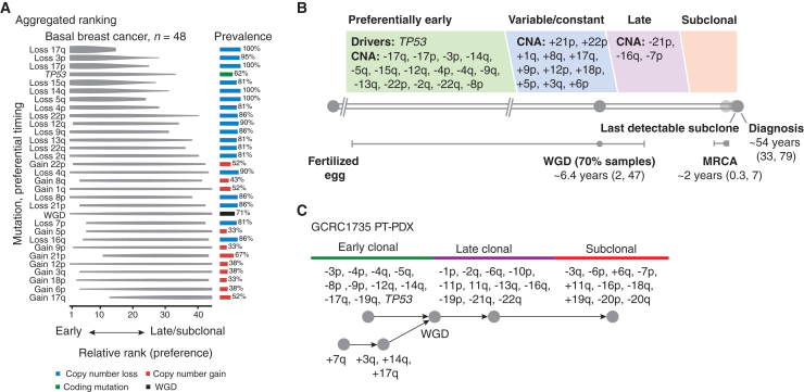
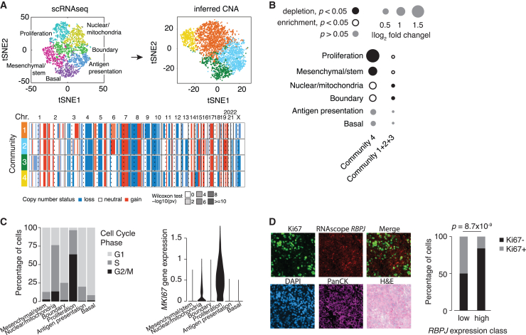
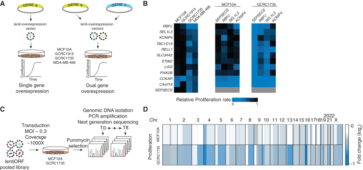
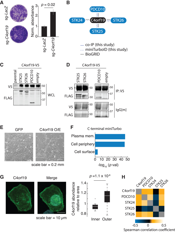
The basal breast cancer subtype is enriched for triple-negative breast cancer (TNBC) and displays consistent large chromosomal deletions. Here, we characterize evolution and maintenance of chromosome 4p (chr4p) loss in basal breast cancer. Analysis of The Cancer Genome Atlas data shows recurrent deletion of chr4p in basal breast cancer. Phylogenetic analysis of a panel of 23 primary tumor/patient-derived xenograft basal breast cancers reveals early evolution of chr4p deletion. Mechanistically we show that chr4p loss is associated with enhanced proliferation. Gene function studies identify an unknown gene, C4orf19, within chr4p, which suppresses proliferation when overexpressed-a member of the PDCD10-GCKIII kinase module we name PGCKA1. Genome-wide pooled overexpression screens using a barcoded library of human open reading frames identify chromosomal regions, including chr4p, that suppress proliferation when overexpressed in a context-dependent manner, implicating network interactions. Together, these results shed light on the early emergence of complex aneuploid karyotypes involving chr4p and adaptive landscapes shaping breast cancer genomes.

Keywords: CP: Cancer; CP: Genomics; GCK-III; PDCD10; aneuploidy; basal breast cancer; cancer evolution; chromosomal arm copy number aberrations; chromosome 4p; triple-negative breast cancer.


Conflict of interest statement

Declaration of interests The authors declare no competing interests.

Figures

None
Graphical abstract
Figure 1
Figure 1
Loss of chromosome 4p in basal breast cancer is recurrent and functionally significant (A) Experimental and analytic pipeline. (B) The Cancer Genome Atlas (TCGA) invasive breast carcinoma single nucleotide polymorphism array dataset analysis shows the three most frequent chromosomal-arm losses in basal breast cancer. (C) Regions of chr4p loss span a large fraction of the chromosome 4p. Segmented mean thresholds: dark blue, stringent threshold deletion < −0.3; light blue, lenient threshold −0.3 < deletion < −0.1; light red, lenient threshold 0.1 < gain <0.3; red, stringent threshold gain >0.3; white, copy-neutral state. (D) In TCGA basal breast cancer, ∼80% of genes along chr4p decrease in expression due to chr4p loss. (E) Overall survival of basal breast cancer patients with varying copy-number status of chr4p, p < 0.0997. (F) Chr4p copy-number status across pan-cancer TCGA datasets. (G and H) Gene set enrichment analysis showing representative terms that are enriched for genes displaying (G) elevated or (H) decreased expression due to chr4p loss in TCGA basal breast cancer.
Figure 2
Figure 2
Chromosome 4p loss is an early event in basal breast cancer evolution Basal breast cancer primary tumor/patient-derived xenograft (PT/PDX) panel was used for phylogenetic reconstruction. (A) Aggregated single-sample ordering reveals typical timing of chromosome-arm aberrations. Preferential ordering diagrams show probability distributions revealing uncertainty of timing for specific events in the cohort. The prevalence of the event type in the cohort is displayed as a bar plot on the right. Losses occurring in >80% and gains occurring in >30% of all cases and evolutionary stages are depicted. (B) Timeline representing the length of time, in years, between the fertilized egg and the median age of diagnosis for basal breast cancer. Real-time estimates for major events, such as whole-genome doubling (WGD) and the emergence of the most recent common ancestor (MRCA), are used to define early, variable, late, and subclonal stages of tumor evolution approximately in chronological time. Driver mutations and copy-number alterations (CNAs) are shown in each stage according to their preferential timing, as defined by relative ordering. (C) An example of individual patient (PT/PDX1735) trajectory (partial ordering relationships).
Figure 3
Figure 3
Chromosome 4p loss is associated with a proliferative state (A) Single-cell RNA-sequencing data of PDX1735 (top left) from a previous study were used to infer copy-number status (top right) and displayed using t-distributed stochastic neighbor-embedding plots, colored by shared gene expression or inferred copy-number profiles. Heatmap (bottom) shows the inferred copy-number profile of three communities harboring chr4p deletion (1–3) and community 4 enriched for cells with chr4p copy-neutral state. chr denotes chromosome, dashed line denotes centromere, and solid line denotes start/end of chromosome. Loss is blue, copy neutral is white, and gain is red. Likelihood of inferred copy-number change is represented by Wilcoxon test −log10 p value. (B) Frequency of cells inferred to harbor chromosome 4p deletion or copy-neutral state across “transcriptional program” clusters. The size of the circle reflects the fold increase over the background fraction of all cells in a specific gene expression cluster. Significance was assessed by a hypergeometric test; p < 0.05. Solid black circles, depletion; open black circles, enrichment; gray, no change. (C) Distribution of cells across (left) cell-cycle phases (light gray, G1 phase; medium gray, S phase; black, G2/M phase) and (right) MKI67 gene expression using “transcriptional program” clusters from (A). (D) Staining of paraffin-embedded fixed tissue section of PDX1735 using a combination of immunofluorescence (IF) for Ki67 (marker of proliferation), RNA in situ hybridization (ISH) for RBPJ (marker of chr4p), DAPI staining for nuclei, pan-cytokeratin (PanCK) IF for epithelial cancer cells, and H&E staining for cancer histology. Significance was assessed by Fisher’s exact test.
Figure 4
Figure 4
Overexpression of chromosome 4p genes leads to context-dependent suppression of proliferation (A) Schematic of gene overexpression strategy in chr4p copy-neutral cells MCF10A, GCRC1915, or chr4p deletion cells GCRC1735, MDA-MB-468. (B) Single or dual chr4p gene overexpression confers a proliferation defect in chr4p loss but not in chr4p copy-neutral cell lines. Blue, proliferation defect; black, no change relative to control. n = 3 with three technical replicates. (C) Schematic of the genome-wide pooled LentiORF overexpression screen. (D) Genome-wide pooled LentiORF overexpression screen revealed genomic regions with context-dependent suppression of proliferation, e.g., chr4p and 13q. Blue, proliferation defect; black, no change relative to T0 control.
Figure 5
Figure 5
C4orf19 (PGCKA1) is associated with the PDCD10-GCKIII module (A) Colony-formation assay for MCF10A cells treated with a control sgRNA-LacZ and sgRNA-C4orf19. A representative well per condition is shown and quantified. Significance was assessed using a Wilcoxon rank-sum test. Data are presented as mean ± SD, n = 3, two technical replicates. (B) Summary of coIP assay results from (D), miniTurbo ID conducted in MCF10A cells expressing C4rf19-miniTurbo from (F), and literature curation using BioGRID. (C) Western blot using whole-cell lysate shows heterologous expression of C4orf19-v5 and PDCD10-GCKIII module 3×FLAG-PDCD10/STK25/STK26 in MCF10A cells. n = 3. (D) CoIP assay using Anti-V5 for pull-down shows the presence of 3×FLAG-PDCD10/STK25/STK26 in MCF10A cells. n = 3. (E) Representative bright-field microscopy images of MCF10A cells overexpressing GFP control or C4orf19 48 h post-induction with doxycycline. n = 3. (F) Analysis of gene ontology molecular function of proteins in proximity to C4orf19 from miniTurbo ID from (C) shows enrichment of proteins at the plasma membrane. (G) Immunofluorescence of C4orf19 indicates its localization at the cell periphery and quantification using n = 3 biological replicates comprising 27 cells. Significance was assessed using a Wilcoxon rank-sum test. (H) Heatmap shows co-expression for the members of the C4orf19-PDCD10-GCKIII module using the Cancer Cell Line Encyclopedia mRNA expression dataset; Spearman correlation coefficient, p < 0.05 (see STAR Methods). Gray indicates missing data.

Comment in

References

    1. Hammond M.E.H., Hayes D.F., Dowsett M., Allred D.C., Hagerty K.L., Badve S., Fitzgibbons P.L., Francis G., Goldstein N.S., Hayes M., et al. American Society of Clinical Oncology/College Of American Pathologists guideline recommendations for immunohistochemical testing of estrogen and progesterone receptors in breast cancer. J. Clin. Oncol. 2010;28:2784–2795. - PMC - PubMed
    1. Haffty B.G., Yang Q., Reiss M., Kearney T., Higgins S.A., Weidhaas J., Harris L., Hait W., Toppmeyer D. Locoregional relapse and distant metastasis in conservatively managed triple negative early-stage breast cancer. J. Clin. Oncol. 2006;24:5652–5657. - PubMed
    1. Lehmann B.D., Bauer J.A., Chen X., Sanders M.E., Chakravarthy A.B., Shyr Y., Pietenpol J.A. Identification of human triple-negative breast cancer subtypes and preclinical models for selection of targeted therapies. J. Clin. Invest. 2011;121:2750–2767. - PMC - PubMed
    1. Lehmann B.D., Jovanović B., Chen X., Estrada M.V., Johnson K.N., Shyr Y., Moses H.L., Sanders M.E., Pietenpol J.A. Refinement of Triple-Negative Breast Cancer Molecular Subtypes: Implications for Neoadjuvant Chemotherapy Selection. PLoS One. 2016;11 - PMC - PubMed
    1. Sorlie T., Perou C.M., Tibshirani R., Aas T., Geisler S., Johnsen H., Hastie T., Eisen M.B., van de Rijn M. Jeffrey S.S.et al. Gene expression patterns of breast carcinomas distinguish tumor subclasses with clinical implications. Proc Natl Acad Sci USA. 2001;98:10869–10874. - PMC - PubMed

Publication types