. 2024 Mar 22;10(12):eadn4649.
doi: 10.1126/sciadv.adn4649. Epub 2024 Mar 22.

Childhood cancer mutagenesis caused by transposase-derived PGBD5

Affiliations

Childhood cancer mutagenesis caused by transposase-derived PGBD5

Makiko Yamada et al. Sci Adv. .

Abstract

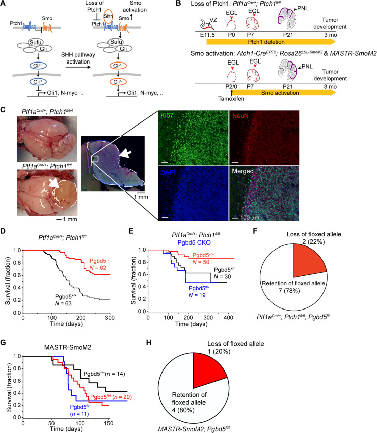
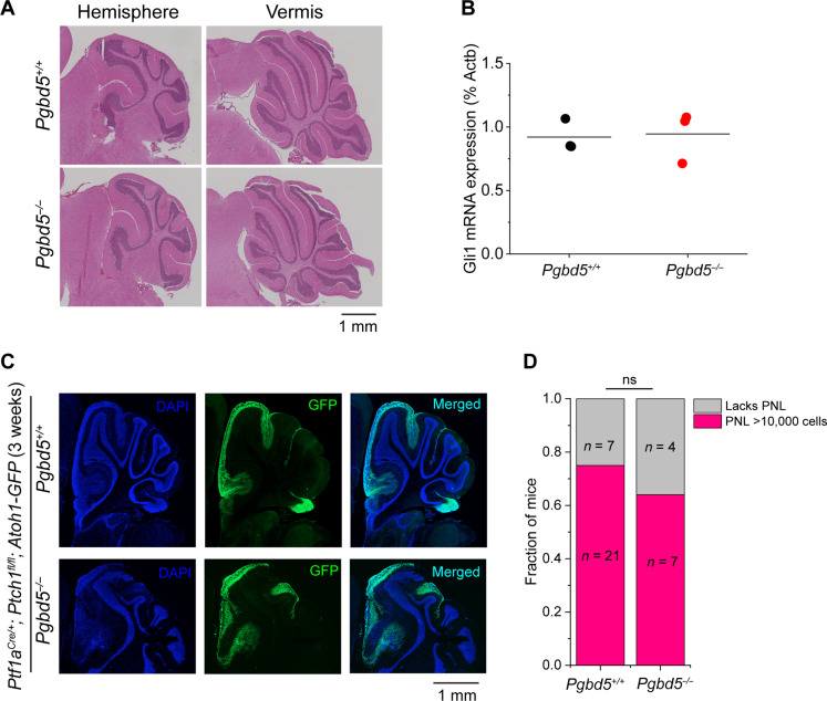
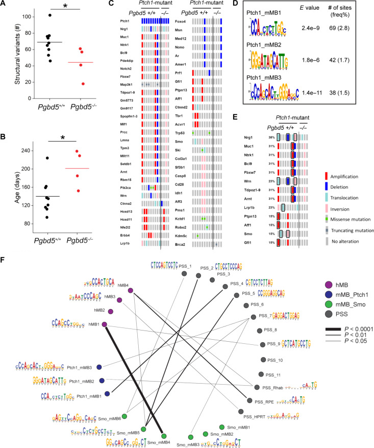
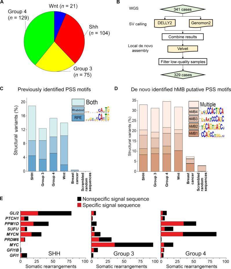
Genomic rearrangements are a hallmark of most childhood tumors, including medulloblastoma, one of the most common brain tumors in children, but their causes remain largely unknown. Here, we show that PiggyBac transposable element derived 5 (Pgbd5) promotes tumor development in multiple developmentally accurate mouse models of Sonic Hedgehog (SHH) medulloblastoma. Most Pgbd5-deficient mice do not develop tumors, while maintaining normal cerebellar development. Ectopic activation of SHH signaling is sufficient to enforce cerebellar granule cell progenitor-like cell states, which exhibit Pgbd5-dependent expression of distinct DNA repair and neurodevelopmental factors. Mouse medulloblastomas expressing Pgbd5 have increased numbers of somatic structural DNA rearrangements, some of which carry PGBD5-specific sequences at their breakpoints. Similar sequence breakpoints recurrently affect somatic DNA rearrangements of known tumor suppressors and oncogenes in medulloblastomas in 329 children. This identifies PGBD5 as a medulloblastoma mutator and provides a genetic mechanism for the generation of oncogenic DNA rearrangements in childhood cancer.


Figures

Fig. 1.
Fig. 1.. Pgbd5 promotes tumorigenesis in diverse developmentally accurate mouse models of SHH MB.
(A) Schematic of aberrant mechanisms of SHH signaling in cerebellar GCPs in MB development (left). In Ptch1-mutant Ptf1aCre/+;Ptch1fl/fl MB, by deletion of Ptch1 encoding a receptor for SHH, SMO signaling is disinhibited and highly activated, leading to the generation of activated GLI (GLIA). In Smo-mutant MASTR-SmoM2 or Atoh1-CreERT2;Rosa26LSL-SmoM2 MB, oncogenic constitutively activated form of SMO results in GLI activation and aberrant SHH signaling. (B) Schematic of cerebellar tumor development in Ptch1- (top) and SmoM2-mutant (bottom) MB. Red arrowheads mark conditionally gene-targeted cell populations. PNLs (purple) lead to MB development. E, embryonic day; P, postnatal day. (C) Representative photographs of dissected brains of Ptf1aCre/+;Ptch1fl/fl (bottom left) mice that develop MBs marked by white arrows and dashed circles, as compared to Ptf1aCre/+;Ptch1fl/wt mice (top left) that do not develop tumors. Immunofluorescence microscopy (right) shows high Ki67 (green) and low NeuN (red) expression in MB tumors, with nuclei marked with DAPI (blue). The edge of the tumor (white inset) is magnified with NeuN-positive cells on tumor edge corresponding to normal cerebellum. (D) Survival of Ptf1aCre/+;Ptch1fl/fl;Pgbd5+/+ (black) and Ptf1aCre/+;Ptch1fl/fl;Pgbd5−/− (red) mice. (E) Survival of Ptf1aCre/+;Ptch1fl/fl mice with conditional knockout (CKO) of Pgbd5fl/− (blue) or control Pgbd5+/− (black) or Pgbd5−/− (red) littermates. (F) Genomic PCR analysis of conditional Pgbd5 excision in Pgbd5fl/− CKO Ptch1-mutant tumors demonstrates that seven of nine (79%) analyzed tumors retain intact Pgbd5, as detailed in fig. S1D. (G) Survival of MASTR-SmoM2 mice with CKO of Pgbd5fl/fl (red), as compared to control Pgbd5+/+ (black) or Pgbd5fl/+ (blue) littermates. (H) Genomic PCR analysis of conditional Pgbd5 excision in MASTR-SmoM2; Pgbd5fl/fl tumors demonstrates that four of five (80%) analyzed tumors retain Pgbd5 floxed alleles, as detailed in fig. S2B.
Fig. 2.
Fig. 2.. Pgbd5 is dispensable for normal SHH signaling and cerebellar development.
(A) Representative immunohistochemistry micrographs of sagittal sections of cerebellum of Pgbd5+/+ (top) and Pgbd5−/− (bottom) mice at 6 weeks of age show normal cytoarchitecture and morphology of cerebellar hemispheres (left) and vermis (right). (B) Expression of Gli1 mRNA in purified cerebellar GCPs from 5-day-old Pgbd5+/+ (black) and Pgbd5−/− (red) mice. Bars represent means of three biologic replicates (P = 0.87). (C) Representative fluorescence images of cerebellar hemispheres of 3-week-old Pgbd5+/+ (top) versus Pgbd5−/− (bottom) Ptf1aCre/+;Ptch1fl/fl;Atoh1-GFP mice showing PNLs (green) marked by Atoh1-GFP expression, with nuclei marked with DAPI (blue). (D) Fraction of mice harboring PNLs (red) in Pgbd5wt/wt;Ptf1aCre/+;Ptch1fl/fl;Atoh1-GFP and Pgbd5−/−;Ptf1aCre/+;Ptch1fl/fl;Atoh1-GFP mice between 3 and 8 weeks of age. Both groups harbor similar fractions of PNLs that are defined as at least 10,000 Atoh1-GFP–positive cells (Fisher’s exact test P = 0.69); ns, not significant.
Fig. 3.
Fig. 3.. Pgbd5 promotes somatic mutagenesis of recurrently mutated tumor suppressor and oncogenes in mouse SHH MBs.
(A) Numbers of SVs in Ptf1aCre/+;Ptch1fl/fl tumors. Pgbd5+/+ tumors (black, n = 9) harbor more SVs than Pgbd5−/− (red, n = 4). Lines indicate mean (69 and 44, respectively), and significance is measured using t test (*P = 0.036). (B) Age of tumors (days) in Ptch1-mutant tumors. Pgbd5+/+ tumors (black, n = 9) are younger than Pgbd5−/− tumors (red, n = 4; mean 140 and 201 days, *P = 0.024). (C) Oncoprint showing genes recurrently affected by SVs and SNVs in independent Ptch1-mutant tumors. Genes are curated based on likelihood that SVs or SNVs affect gene function (see Materials and Methods for details). The left nine and right four columns indicate tumors from Pgbd5+/+ and Pgbd5−/− mice, respectively. Red, blue, light blue, pink, and gray symbols indicate amplifications, deletions, translocations, inversions, and no alteration, respectively. Green and dark gray squares in gray symbols indicate missense and truncating mutations, respectively. (D) Three Pgbd5+/+-specific motifs are identified at SV breakpoints in Ptch1-mutant tumors, using discriminative MEME with Pgbd5−/− tumors as controls. E values indicate MEME discriminative algorithm significance (see Materials and Methods for details). The frequency shown was calculated by dividing the number of sites by total numbers of 50-bp breakpoint sequences extracted from SVs. (E) These motifs in (D) and the previously identified PSS_RPE and PSS_Rhab motifs were identified at SV breakpoints affecting known tumor suppressor and oncogenes in six of nine (66%) of Pgbd5+/+ tumors. The affected tumor suppressors and oncogenes are boxed black. (F) Circos plot showing similarities among all motifs (see Materials and Methods for details).
Fig. 4.
Fig. 4.. PGBD5-associated sequence breakpoints recurrently affect somatic DNA rearrangements of known tumor suppressor and oncogenes in human MBs.
(A) Representation of human patient cohort showing the four major subgroups of MB that were included in the analysis (n = 329). (B) Pipeline to identify somatic SVs in human MB. (C) Previously identified PSS sequences (4, 24) are enriched at SV breakpoints in human MB as compared to somatic SVs in human breast carcinomas (χ2 test P = 7.7 × 10−117). Percentages represent the frequency of SVs (each SV has two breakpoints and 4 × 50-mers) where the motif was identified using a FIMO q value threshold of 0.3 based on a ROC curve analysis (fig. S14, B and C). Multiple indicates that more than one motif was identified at one SV, in either distinct or the same 50-mers. Scrambled sequences showed no enrichment and represent the background of the FIMO algorithm. (D) A set of four de novo motifs identified at SV breakpoints in human MB is enriched relative to breast carcinoma and scrambled sequences. hMB1 to hMB4 were identified as being specific using MEME and eliminating repetitive motifs. In addition, discriminative MEME, where control sequences were a set of 50,000 randomly selected 50-mers from the hg19 reference genome, was used to determine whether the motif was enriched at breakpoints relative to the genome (fig. S14). Percentages represent motif frequency among SVs as in (C) and are compared to SV breakpoints in human breast carcinoma (χ2 test P = 1.5 × 10−155) and scrambled sequences, which represent the background of the FIMO algorithm. (E) Recurrently mutated MB tumor suppressors and oncogenes in diverse tumor subtypes involve somatic DNA rearrangements with specific (red) sequence breakpoints, including PSS motifs. Numbers refer to the SVs detected in human patient cohort described in (A).
Fig. 5.
Fig. 5.. Pgbd5-dependent mechanisms of cerebellar GCP transformation.
(A) Schematic of experimental procedure for snRNA-seq of three Pgbd5+/+ and three Pgbd5−/− fresh-frozen Ptf1aCre/+;Ptch1fl/fl SHH MBs; same tumors as were analyzed by whole-genome DNA sequencing were used. (B) Uniform Manifold Approximation and Projection (UMAP) plots of the cerebellar reference (N = 62,040) used to annotate the cells from Pgbd5+/+ and Pgbd5−/− Ptf1aCre/+;Ptch1fl/fl SHH MBs. Cells are colored by age (left) and by refined cell class ontology (right). (C) Cell class and cluster consensus predictions for Pgbd5+/+ (orange) and Pgbd5−/− (blue) MB tumor cells. Left: UMAP plots of the cerebellar reference highlighting the mapped clusters of malignant cells of each genotype. Middle: Bar plot depicting the cell class consensus predictions for the tumor cells for each genotype. Right: Bar plot depicting the cluster-specific consensus predictions for tumor cells for each genotype. Only clusters with more than 10 cells mapped to by either genotype are shown. (D) Volcano plot showing differential gene expression between Pgbd5+/+ and Pgbd5−/− malignant GCPs and granule cells. Genes significantly up-regulated in Pgbd5+/+ MBs are highlighted in orange, while significantly up-regulated genes in Pgbd5−/− tumor cells are highlighted in blue (log2FC > 0.25, adjusted P < 0.05). (E) Model of PGBD5-dependent tumorigenesis, illustrating how pathogenic SHH signaling is associated with hyperplasia of cerebellar granule cell progenitor cells, leading to PNLs that undergo PGBD5-dependent somatic mutagenesis and malignant transformation.

References

    1. Hanahan D., Weinberg R. A., Hallmarks of cancer: The next generation. Cell 144, 646–674 (2011). - PubMed
    1. Anderson N. D., de Borja R., Young M. D., Fuligni F., Rosic A., Roberts N. D., Hajjar S., Layeghifard M., Novokmet A., Kowalski P. E., Anaka M., Davidson S., Zarrei M., Id Said B., Schreiner L. C., Marchand R., Sitter J., Gokgoz N., Brunga L., Graham G. T., Fullam A., Pillay N., Toretsky J. A., Yoshida A., Shibata T., Metzler M., Somers G. R., Scherer S. W., Flanagan A. M., Campbell P. J., Schiffman J. D., Shago M., Alexandrov L. B., Wunder J. S., Andrulis I. L., Malkin D., Behjati S., Shlien A., Rearrangement bursts generate canonical gene fusions in bone and soft tissue tumors. Science 361, eaam8419 (2018). - PMC - PubMed
    1. Henssen A. G., Henaff E., Jiang E., Eisenberg A. R., Carson J. R., Villasante C. M., Ray M., Still E., Burns M., Gandara J., Feschotte C., Mason C. E., Kentsis A., Genomic DNA transposition induced by human PGBD5. eLife 4, e10565 (2015). - PMC - PubMed
    1. Henssen A. G., Koche R., Zhuang J., Jiang E., Reed C., Eisenberg A., Still E., MacArthur I. C., Rodríguez-Fos E., Gonzalez S., Puiggròs M., Blackford A. N., Mason C. E., de Stanchina E., Gönen M., Emde A. K., Shah M., Arora K., Reeves C., Socci N. D., Perlman E., Antonescu C. R., Roberts C. W. M., Steen H., Mullen E., Jackson S. P., Torrents D., Weng Z., Armstrong S. A., Kentsis A., PGBD5 promotes site-specific oncogenic mutations in human tumors. Nat. Genet. 49, 1005–1014 (2017). - PMC - PubMed
    1. Henssen A. G., Reed C., Jiang E., Garcia H. D., von Stebut J., MacArthur I. C., Hundsdoerfer P., Kim J. H., de Stanchina E., Kuwahara Y., Hosoi H., Ganem N. J., dela Cruz F., Kung A. L., Schulte J. H., Petrini J. H., Kentsis A., Therapeutic targeting of PGBD5-induced DNA repair dependency in pediatric solid tumors. Sci. Transl. Med. 9, eaam9078 (2017). - PMC - PubMed

Publication types