Comment
. 2024 Apr 9;57(4):890-903.e6.
doi: 10.1016/j.immuni.2024.03.001. Epub 2024 Mar 21.

Convergent evolution and targeting of diverse E2 epitopes by human broadly neutralizing antibodies are associated with HCV clearance

Affiliations
Comment

Convergent evolution and targeting of diverse E2 epitopes by human broadly neutralizing antibodies are associated with HCV clearance

Clinton O Ogega et al. Immunity. .

Abstract

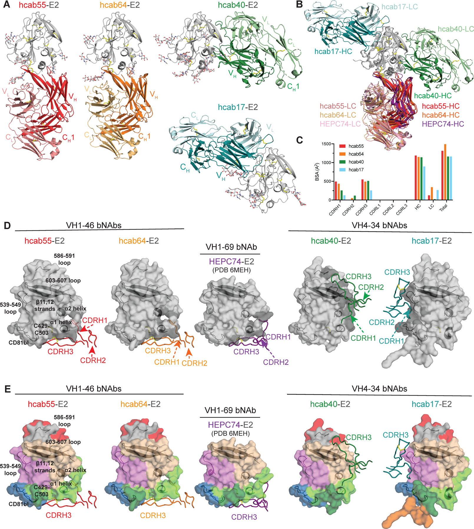
The early appearance of broadly neutralizing antibodies (bNAbs) in serum is associated with spontaneous hepatitis C virus (HCV) clearance, but to date, the majority of bNAbs have been isolated from chronically infected donors. Most of these bNAbs use the VH1-69 gene segment and target the envelope glycoprotein E2 front layer. Here, we performed longitudinal B cell receptor (BCR) repertoire analysis on an elite neutralizer who spontaneously cleared multiple HCV infections. We isolated 10,680 E2-reactive B cells, performed BCR sequencing, characterized monoclonal B cell cultures, and isolated bNAbs. In contrast to what has been seen in chronically infected donors, the bNAbs used a variety of VH genes and targeted at least three distinct E2 antigenic sites, including sites previously thought to be non-neutralizing. Diverse front-layer-reactive bNAb lineages evolved convergently, acquiring breadth-enhancing somatic mutations. These findings demonstrate that HCV clearance-associated bNAbs are genetically diverse and bind distinct antigenic sites that should be the target of vaccine-induced bNAbs.

Keywords: B cells; BCR sequencing; HCV; antibody evolution; broadly neutralizing antibodies; hepatitis C virus; monoclonal antibodies; neutralizing epitopes; vaccine.


Conflict of interest statement

Declaration of interests J.R.B. and C.O.O. have filed a provisional patent 63/470,326 related to monoclonal antibodies described in this manuscript.

Figures

Figure 1.
Figure 1.. Elite neutralizer plasma is broadly neutralizing against genetically and antigenically diverse HCVpp.
(A) Neutralizing breadth of early infection plasma from 63 BBAASH participants with either spontaneous clearance of infection (n=21, prefix “C”) or persistence of infection (n=42, prefix “P”). The 10% of participants with the greatest neutralizing breadth (n=6) were designated elite neutralizers (EN). (B) Neutralizing activity of 6 EN against each of 17 Tier 1 (sensitive) to Tier 4 (resistant) HCVpp. Values for subject C110 are indicated in red. Error bars indicate standard deviations. (C) Viral RNA levels of subject C110 demonstrating spontaneous clearance of 3 infections (delineated by distinct colors). The timepoint used for mAb isolation (D2178) is indicated (arrow) along with the four timepoints used for BCR-seq analysis (red dots). (D) C110 plasma neutralizing breadth at the 5 timepoints used for BCR-seq and mAb analysis. Neutralization was measured in duplicate in one experiment.
Figure 2.
Figure 2.. BCRs show E2 cross-reactivity, increased somatic mutation, and longer CDRH3 sequences.
(A) E2-reactive B cell staining with a mixture of three antigenically distinct soluble E2 proteins. (B) B cells cultured at varying cell density, with frequency (%) of wells positive for human IgG or reactive with each E2 variant. (C) Frequency (%) of IgG positive 2 or 1 B cell supernatants reactive with each E2 variant or cross-reactive with multiple E2 variants. (D) CDRH3 lengths of E2-nonreactive or longitudinal E2-reactive BCRs or mAbs. (E) CDRL3 lengths of E2-nonreactive or longitudinal E2-reactive BCRs or mAbs. (F) VH-gene nucleotide identity to inferred germline sequences. (G) VK or VL-gene nucleotide identity to inferred germline sequences. For D-G, each dot represents an individual BCR sequence. Distribution is shown via violin plot, with horizontal lines indicating medians. For D-G, statistical comparisons were made using the Kruskal-Wallis test followed by the Dunn post-hoc test with the Benjamini-Hochberg correction applied for multiple comparisons. **** p<0.0001; *** p<0.001, ** p<0.01, * p<0.05. See also Figure S1.
Figure 3.
Figure 3.. VH1–69 and D2–2/D2–15 usage favor broad neutralizing activity but are not required.
Frequency of usage of VH (A) or VK/VL (B) gene segments among E2-nonreactive or E2-reactive BCRs. Only V-genes used by at least one mAb are shown here, with the remainder in Supplemental Figure 2. Statistical significance is denoted only for E2-reactive BCRs relative to E2-nonreactive BCRs. * p<0.05 using Fisher’s exact test with the Benjamini-Hochberg correction for multiple comparisons. (C) Neutralizing breadth of 55 E2-reactive mAbs measured using the panel of 17 HCVpp. (D) Neutralizing breadth of mAbs (# of 17 HCVpp neutralized) segregated by VH-gene usage. Each point represents one mAb, with the number of mAbs encoded by each VH-gene (n) indicated. (E) Neutralizing breadth of mAbs (# of 17 HCVpp neutralized) segregated by D-gene usage, with the number of mAbs encoded by each D-gene (n) indicated. NR=not resolved. (F) Neutralizing breadth of mAbs using both VH1–69 and D2–2/D2–15, either, or neither. ns=not significant, * p<0.05 by Kruskal Wallis test for non-normally distributed data, adjusted for multiple comparisons using the Benjamini, Krieger and Yekutieli method. In D-F, dashed line indicates neutralization of 9 HCVpp, the threshold for bNAb designation. Solid horizontal lines indicate medians. Neutralization values are the average of two independent experiments performed in duplicate. See also Figures S2-3.
Figure 4.
Figure 4.. HCV bNAbs target at least three major antigenic sites on the E2 protein.
(A) bNAbs were segregated based on >50% loss of binding to E2 with multiple mutations in the front layer (FRLY KO) or in the AS412 epitope (AS412 KO), relative to binding to wild-type E2 protein. Six reference mAbs (bold) with known binding epitopes as well as previously characterized C110 bNAb HEPC74 were included as controls. Curves were fit and area under the curve (AUC) was calculated from binding of 5-fold serial dilutions of each mAb, performed in duplicate (B) Data from Bio-Layer Interferometry epitope binning experiments using 17 hcabs and 5 reference antibodies (italicized). Numbers indicate the percentage binding of second mAb in the presence of the first mAb compared to binding of un-competed second mAb. mAbs were judged to compete for the same antigenic site if the maximum binding of the second mAb was reduced to <30% of its un-competed binding (black boxes with white numbers). The mAbs were considered non-competing if the maximum binding of the second mAb was >60% of its un-competed binding (white boxes with red numbers). Gray boxes with black numbers indicate an intermediate phenotype (competition resulted in between 30% and 60% of un-competed binding). Dashed lines in red (contains FRLY reference mAbs), dark blue (contains a β-sheet reference mAb), and green (contains no reference mAbs) indicate three major competition groups. The light blue dashed line indicates a minor antigenic group that partially overlaps with the FRLY and β-sheet antigenic groups. A reference bNAb HCV1 is a single member of the AS412 competition group, indicated by yellow dashed lines.
Figure 5.
Figure 5.. Crystal structures of hcab55-E2, hcab64-E2, hcab40-E2, and hcab17-E2 complexes reveal neutralizing epitopes of non-VH1–69 HCV bNAbs.
(A) Crystal structures of E2 (gray, N-glycans shown as sticks and disulfide bonds shown as yellow sticks) in complex with hcab55 (red), hcab64 (orange), hcab40 (green), and hcab17 (cyan) Fabs. (B) Superposition of E2 (gray) in complex with hcab55 (red), hcab64 (orange), hcab40 (green), and hcab17 (cyan), and HEPC74 (PDB 6MEH) (purple) Fabs structures. Structures were superimposed on the E2 head domains. Disulfide bonds are shown as yellow sticks. (C) Comparison of buried surface areas (BSAs). (D) Heavy-chain CDR (CDRH) loops of hcab55, hcab64, hcab40, hcab17, and HEPC74 (PDB 6MEH) Fabs mapped onto the E2 surface. CDRH loops are colored as in (A), and HC interacting residues are colored in dark gray on the E2 surface. Epitopes (surface representation) were defined as residues in E2 containing an atom within 4 Å of the bound Fab. The location of α1-helix, α2-helix, ß11,12-strands, CD81bl, loop 539–549, loop 586–591, and loop 603–607 in E2 are indicated by black cartoon representations, and the C429–C503 disulfide bond is indicated by yellow sticks. (E) Comparison of the CDRH loop positions in E2 complex structures with hcab55, hcab64, hcab40, and HEPC74. E2 structures are shown as surface representation and CDRHs as tubes. CDRH loops are colored as in (A), and important features of E2 and the position of CDRH3 are indicated. E2 surfaces are colored by structural components: HVR1, orange; AS412, dark green; front layer, light green; VR2, red; β sandwich, violet; CD81bl, blue; VR3, gray; back layer, tan. See also Figures S4-5, Tables S1-3.
Figure 6.
Figure 6.. HCV FRLY bNAbs display convergent evolution.
(A) CDRH1–2 substitutions shared by multiple FRLY bNAbs and significantly enriched (bold) relative to E2-nonreactive BCRs from the same participant. E2-nonreactive BCRs were matched to FRLY bNAbs based on V-gene usage and % homology to germline sequences. CDRH3 amino acids shared by multiple FRLY bNAbs (A), β-sheet mAbs (B), or Back layer bNAbs (C) and significantly enriched (bold) relative to E2-nonreactive BCRs. E2-nonreactive BCRs were matched to FRLY, β-sheet, or back layer hcabs based on V-gene usage, % homology to germline, and CDRH3 length. Germline-encoded amino acids are in green, somatic mutation-encoded substitutions are in pink, and junctional nucleotide-encoded amino acids are in blue. P values were determined using Fischer’s exact test, adjusted for multiple comparisons using the Benjamini-Hochberg correction. Numbering according to the Kabat method. (D) Neutralizing breadth across a panel of 17 HCVpp by wild-type (WT) front-layer-reactive hcabs or hcabs with a single amino acid altered by site-directed mutagenesis. Each pair of linked points represents a single HCVpp. Values are the average of two independent experiments, tested in duplicate. For each mAb pair, HCVpp that were not neutralized by either WT or mutant mAbs were excluded from the analysis. Significance determined by Wilcoxon paired signed rank test. NS=not significant, * p<0.05, ** p<0.01, *** p<0.001. (E) Neutralizing potency against heterologous HCVpp by wild type (WT) hcabs or hcabs with a single amino acid altered by site-directed mutagenesis. The HCVpp used for each test is indicated. Graphs are representative of two independent experiments performed in duplicate. Points are means and error bars represent standard deviations. See also Figure S6.

Comment on

References

    1. van der Meer AJ, Veldt BJ, Feld JJ, Wedemeyer H, Dufour JF, Lammert F, Duarte-Rojo A, Heathcote EJ, Manns MP, Kuske L, et al. (2012). Association between sustained virological response and all-cause mortality among patients with chronic hepatitis C and advanced hepatic fibrosis. JAMA 308, 2584–2593. 1487498 [pii];10.1001/jama.2012.144878 [doi]. - DOI - PubMed
    1. Davis GL, Alter MJ, El-Serag H, Poynard T, and Jennings LW (2010). Aging of hepatitis C virus (HCV)-infected persons in the United States: a multiple cohort model of HCV prevalence and disease progression. Gastroenterology 138, 513–521. S0016-5085(09)01885-X [pii];10.1053/j.gastro.2009.09.067 [doi]. - DOI - PubMed
    1. Prevention, C.f.D.C.a. (2018). Viral Hepatitis Surveillance Report 2018 — Hepatitis C.
    1. Suryaprasad AG, White JZ, Xu F, Eichler BA, Hamilton J, Patel A, Hamdounia SB, Church DR, Barton K, Fisher C, et al. (2014). Emerging Epidemic of Hepatitis C Virus Infections Among Young Non-Urban Persons who Inject Drugs in the United States, 2006–2012. Clin. Infect. Dis 10.1093/cid/ciu643. - DOI - PubMed
    1. Osburn WO, Fisher BE, Dowd KA, Urban G, Liu L, Ray SC, Thomas DL, and Cox AL (2010). Spontaneous Control of Primary Hepatitis C Virus Infection and Immunity Against Persistent Reinfection. Gastroenterology 138, 315–324. S0016-5085(09)01658-8 [pii];10.1053/j.gastro.2009.09.017 [doi]. - DOI - PMC - PubMed

Publication types