. 2024 Mar 21;12(3):e007538.
doi: 10.1136/jitc-2023-007538.

Targeting pediatric cancers via T-cell recognition of the monomorphic MHC class I-related protein MR1

Affiliations

Targeting pediatric cancers via T-cell recognition of the monomorphic MHC class I-related protein MR1

Annelisa M Cornel et al. J Immunother Cancer. .

Abstract

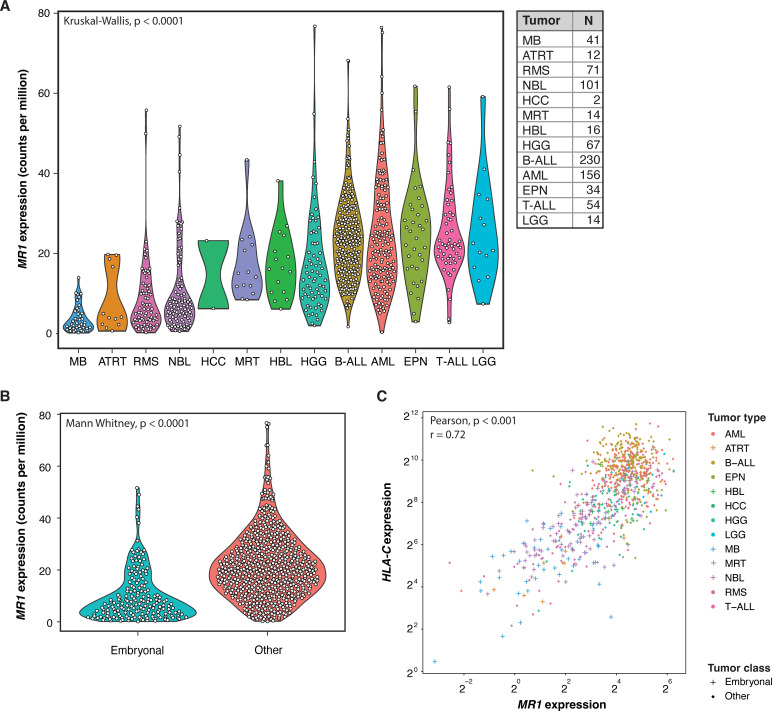
Human leukocyte antigen (HLA) restriction of conventional T-cell targeting introduces complexity in generating T-cell therapy strategies for patients with cancer with diverse HLA-backgrounds. A subpopulation of atypical, major histocompatibility complex-I related protein 1 (MR1)-restricted T-cells, distinctive from mucosal-associated invariant T-cells (MAITs), was recently identified recognizing currently unidentified MR1-presented cancer-specific metabolites. It is hypothesized that the MC.7.G5 MR1T-clone has potential as a pan-cancer, pan-population T-cell immunotherapy approach. These cells are irresponsive to healthy tissue while conferring T-cell receptor(TCR) dependent, HLA-independent cytotoxicity to a wide range of adult cancers. Studies so far are limited to adult malignancies. Here, we investigated the potential of MR1-targeting cellular therapy strategies in pediatric cancer. Bulk RNA sequencing data of primary pediatric tumors were analyzed to assess MR1 expression. In vitro pediatric tumor models were subsequently screened to evaluate their susceptibility to engineered MC.7.G5 TCR-expressing T-cells. Targeting capacity was correlated with qPCR-based MR1 mRNA and protein overexpression. RNA expression of MR1 in primary pediatric tumors varied widely within and between tumor entities. Notably, embryonal tumors exhibited significantly lower MR1 expression than other pediatric tumors. In line with this, most screened embryonal tumors displayed resistance to MR1T-targeting in vitro MR1T susceptibility was observed particularly in pediatric leukemia and diffuse midline glioma models. This study demonstrates potential of MC.7.G5 MR1T-cell immunotherapy in pediatric leukemias and diffuse midline glioma, while activity against embryonal tumors was limited. The dismal prognosis associated with relapsed/refractory leukemias and high-grade brain tumors highlights the promise to improve survival rates of children with these cancers.

Keywords: cell engineering; immunotherapy; pediatrics.


Conflict of interest statement

Competing interests: ZS and JK are inventors on different patents for γδ TCR sequences, recognition mechanisms and isolation strategies. JK is scientific cofounder and shareholder of Gadeta (www.gadeta.nl). The remaining authors declare no competing interests.

Figures

Figure 1
Figure 1
MR1 mRNA expression across pediatric primary tumor entities. (A) MR1 mRNA expression levels (counts per million) across pediatric tumor entities around diagnosis. Primary tumor biopsies are routinely bulk RNA sequenced at time of diagnosis. Statistical differences between tumor entities were calculated using a Kruskal-Wallis test. (B) MR1 RNA expression differences (counts per million) between embryonal (MB, ATRT, MRT, NBL, and HBL) and other tumor entities. Statistical differences were calculated using a Mann-Whitney U test. (C) Correlation between MR1 and HLA-C expression in pediatric primary tumors. + : embryonal tumors, · : other tumors. Log2 transformed data are shown. Correlation was determined using Pearson correlation. AML, acute myeloid leukemia; ATRT, atypical teratoid rhabdoid tumors; B-ALL, B-cell acute lymphoblastic leukemia; EPN, ependymoma; HBL, hepatoblastoma; HCC, hepatocellular carcinoma; HGG, high-grade glioma; LGG, low-grade glioma; MB, medulloblastoma; MHC-I, major histocompatibility complex I; MR1, MHC-I related protein 1; MRT, malignant rhabdoid tumors; NBL, neuroblastoma; RMS, rhabdomyosarcoma; T-ALL, T-cell acute lymphoblastic leukemia.
Figure 2
Figure 2
Heterogeneity in MC.7.G5 MR1T-cell targeting of pediatric tumor models. (A) Standardized % killing after overnight coculture of MC.7.G5 MR1T-cells at an effector-to-target ratio of 10:1 (and 1:1 for Jurkats & Nalm-6, as aspecific killing was observed in controls at higher ratios). Killing was standardized to cocultures with untransduced, donor-matched T-cells. Striped bars represent patient-derived organoid or PDX samples, filled bars represent cell lines. Tumor types marked with * are considered embryonal. K562 cells were taken along as a positive control in every assay. 039, JD081T, JD041T, 78T2: n=2, rest: n≥3. (B) Standardized % killing in embryonal tumors (MRT, NBL, and HB) compared to the other screened tumor entities. Statistical differences were calculated using a Mann-Whitney U test. ***p<0.001 (C) Correlation between standardized killing % and relative MR1 expression levels (2−ΔCt) quantified by qPCR. Colors are matched to (A). Correlation was determined using simple linear regression. (D) Relative MR1 expression (2−ΔCt) in MC.7.G5 targeted and non-targeted pediatric cancer models. Susceptibility to targeting was defined as >15% standardized killing. Statistical differences were calculated using a Mann-Whitney U test. **p<0.01 (E) Standardized % killing after overnight coculture of MC.7.G5 MR1T-cells with wildtype or MR1 overexpressing target cells at an effector-to-target ratio of 10:1. Killing was standardized to cocultures with untransduced, donor-matched T-cells. K562 cells were taken along as a positive control. Colors are matched to (A). n=2, five replicates. Statistical differences were calculated using a Mann-Whitney U test. *p<0.05, **p<0.01 (F) Immune activation score calculated from single cell RNAseq data from MC.7.G5-resistant HB13E and MC.7.G5-sensitive HB13F HBL cells. Statistical differences were calculated using a Mann-Whitney U test.****p<0.0001 (G) Gene set enrichment analysis of gene sets involved in metabolism comparing single cell sequencing data from MC.7.G5 resistant HB13E and MC.7.G5 sensitive HB13F HBL organoids. Significantly enriched processes are shown (p<0.05). AML, acute myeloid leukemia; B-ALL, B-cell acute lymphoblastic leukemia; DMG, diffuse-midline glioma (a type of high-grade glioma); HB, hepatoblastoma; MRTs, malignant rhabdoid tumors; NBL, neuroblastoma; PDX, patient-derived xenograft; T-ALL, T-cell acute lymphoblastic leukemia.

References

    1. Rozemuller E, Eckle SBG, McLaughlin I, et al. . MR1 encompasses at least six allele groups with coding region alterations. HLA 2021;98:509–16. 10.1111/tan.14390 - DOI - PubMed
    1. Riegert P, Wanner V, Bahram S. Genomics, isoforms, expression, and phylogeny of the MHC class I-related MR1 gene. J Immunol 1998;161:4066–77. - PubMed
    1. Lepore M, Kalinichenko A, Calogero S, et al. . Functionally diverse human T cells recognize non-microbial antigens presented by MR1. Elife 2017;6:e24476. 10.7554/eLife.24476 - DOI - PMC - PubMed
    1. Lamichhane R, Ussher JE. Expression and trafficking of MR1. Immunology 2017;151:270–9. 10.1111/imm.12744 - DOI - PMC - PubMed
    1. Vacchini A, Chancellor A, Spagnuolo J, et al. . MR1-restricted T cells are unprecedented cancer fighters. Front Immunol 2020;11:751. 10.3389/fimmu.2020.00751 - DOI - PMC - PubMed

Publication types

Substances