. 2024 Mar 21;12(3):e008338.
doi: 10.1136/jitc-2023-008338.

Molecular assessment of intratumoral immune cell subsets and potential mechanisms of resistance to odronextamab, a CD20×CD3 bispecific antibody, in patients with relapsed/refractory B-cell non-Hodgkin lymphoma

Affiliations

Molecular assessment of intratumoral immune cell subsets and potential mechanisms of resistance to odronextamab, a CD20×CD3 bispecific antibody, in patients with relapsed/refractory B-cell non-Hodgkin lymphoma

Jurriaan Brouwer-Visser et al. J Immunother Cancer. .

Abstract

Background: Patients with relapsed/refractory B-cell non-Hodgkin lymphoma (R/R B-NHL) have a significant need for effective treatment options. Odronextamab is an Fc-silenced, human, CD20×CD3 bispecific antibody that targets CD20-expressing cells via T-cell-mediated cytotoxicity independent of T-cell/major histocompatibility complex interaction. Phase I results in patients with R/R B-NHL demonstrated that odronextamab monotherapy could achieve deep and durable responses with a generally manageable safety profile (ELM-1; NCT02290951). As part of a biomarker analysis of the same study, we investigated potential biomarkers and mechanisms of resistance to odronextamab.

Methods: Patients with R/R B-NHL enrolled in ELM-1 received one time per week doses of intravenous odronextamab for 4×21 day cycles, then doses every 2 weeks thereafter. Patient tumor biopsies were obtained at baseline, on-treatment, and at progression. Immune cell markers were analyzed by immunohistochemistry, flow cytometry, single-cell RNA sequencing, and whole genome sequencing.

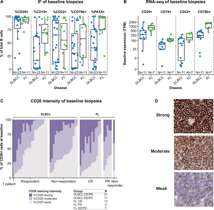
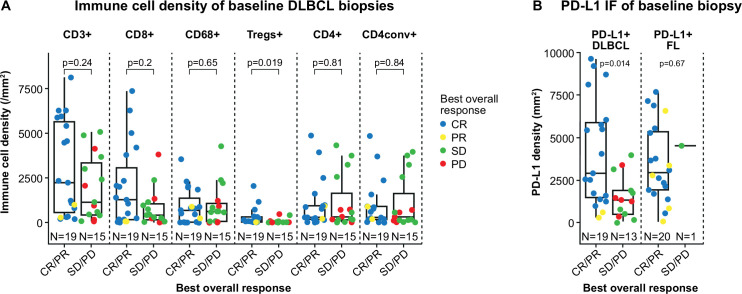
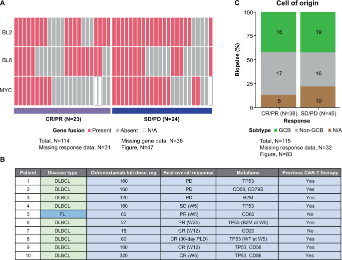
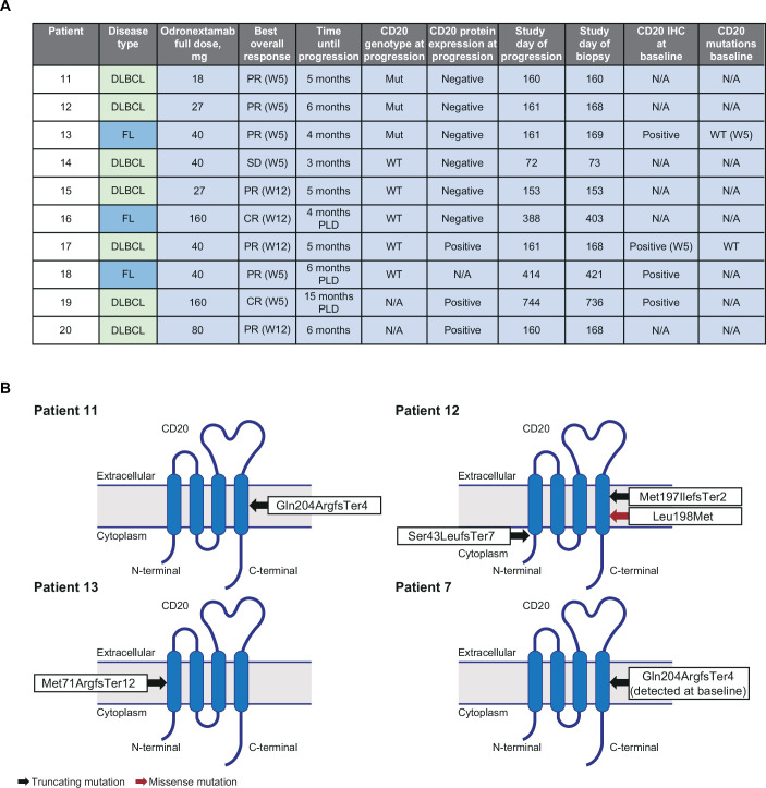
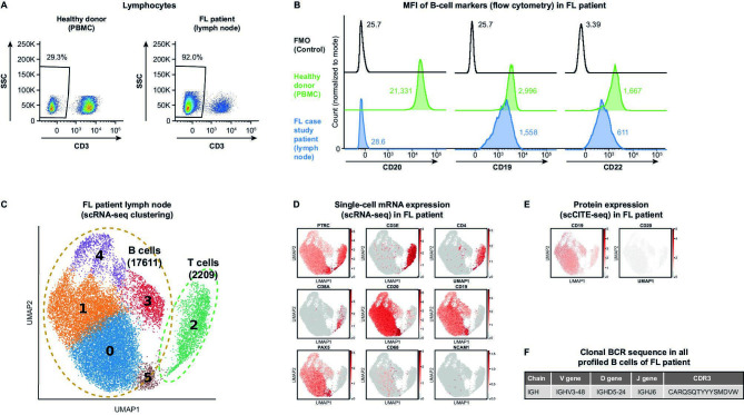
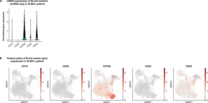
Results: Baseline tumor biopsies showed that almost all patients had high proportions of B cells that expressed the CD20 target antigen, whereas expression of other B-cell surface antigens (CD19, CD22, CD79b) was more variable. Responses to odronextamab in patients with diffuse large B-cell lymphoma were not related to the relative level of baseline CD20 expression, cell of origin, or high-risk molecular subtype. A potential link was observed between greater tumor programmed cell death-ligand 1 expression and increased likelihood of response to odronextamab. Similarly, a trend was observed between clinical response and increased levels of CD8 T cells and regulatory T cells at baseline. We also identified an on-treatment pharmacodynamic shift in intratumoral immune cell subsets. Finally, loss of CD20 expression through inactivating gene mutations was identified as a potential mechanism of resistance in patients who were treated with odronextamab until progression, as highlighted in two detailed patient cases reported here.

Conclusions: This biomarker analysis expands on clinical findings of odronextamab in patients with R/R B-NHL, providing verification of the suitability of CD20 as a therapeutic target, as well as evidence for potential mechanisms of action and resistance.

Keywords: Antibodies, Bispecific; Biomarkers, Tumor.


Conflict of interest statement

Competing interests: JB-V, NF, RPD, KJC, DS, SJ, LB, NTG, SG, CA, DMF, AC, GT, SRA, and VJ are employees of, and hold stock in, Regeneron Pharmaceuticals, Inc. MST: research funding from Regeneron Pharmaceuticals, Inc.; consulting fees from Gilead, Janssen, Kite, Novartis, Regeneron Pharmaceuticals Inc., and Roche; and support for attending meetings or travel from Amgen and Janssen. RB: employee of Ipsen; and his spouse holds stock options in and is an employee of Sanofi-Pasteur. JD: patient fees to the institution from MorphoSys. RHA: advisory board membership from ADC Therapeutics, Bristol Myers Squibb, Epizyme, Genentech, Gilead, Incyte, Karyopharm, Portola, and Seattle Genetics; and institutional research funding from Cyteir, Forty Seven, Genentech, Gilead, Janssen, Regeneron Pharmaceuticals Inc., and Roche.

Figures

Figure 1
Figure 1
Baseline expression of cell surface markers within B cells using multiplex immunofluorescence (A) and RNA-seq (B) in patients with R/R B-NHL. Relative quantification of baseline CD20 levels by IHC in patients with R/R B-NHL (C; only including patients with at least one response assessment) with a representative staining example (D). B-NHL, B-cell non-Hodgkin lymphoma; CR, complete response; DLBCL, diffuse large B-cell lymphoma; FL, follicular lymphoma; IF, immunofluorescence; IHC, immunohistochemistry; PD, progressive disease; PR, partial response; RNA-seq, RNA sequencing; R/R, relapsed/refractory; SD, stable disease; TPM, transcripts per million.
Figure 2
Figure 2
T-cell infiltration density within tumors (A) and PD-L1 expression within tumors at baseline and its association with clinical responses to odronextamab (B). Only patients with at least one response assessment were included in these analyses. P values were calculated using the unpaired Wilcoxon test and paired Wilcoxon test for Parts A and B, respectively. CR, complete response; DLBCL, diffuse large B-cell lymphoma; FL, follicular lymphoma; IF, immunofluorescence; PD, progressive disease; PD-L1, programmed cell death-ligand 1; PR, partial response; SD, stable disease; Treg, regulatory T cell.
Figure 3
Figure 3
Odronextamab responses among different molecular subtypes (A). Common oncogenic variants observed in biopsy samples and outcomes in those patients (B). Odronextamab responses among different cells of origin in R/R DLBCL (C). Only patients with at least one response assessment were included in these analyses. CAR, chimeric antigen receptor; CR, complete response; DLBCL, diffuse large B-cell lymphoma; FL, follicular lymphoma; GCB, germinal center B-cell-like; N/A, not available; PD, progressive disease; PLD, post-last dose; PR, partial response; R/R, relapsed/refractory; SD, stable disease; W, week; WT, wild-type.
Figure 4
Figure 4
Proportion of total immune cells at baseline and Week 5 within tumor biopsies. Percentage of total immune cells from lymph node biopsies (*total [100%] immune cell number comprises CD20+ plus CD3+ plus CD68+ cells) (A). Target:effector (CD20:CD3) ratio (B). P values were calculated using the unpaired Wilcoxon test and paired Wilcoxon test for Parts A and B, respectively. CR, complete response; DLBCL, diffuse large B-cell lymphoma; FL, follicular lymphoma; IF, immunofluorescence; PD, progressive disease; PR, partial response; SCR, screening; SD, stable disease; W, week.
Figure 5
Figure 5
Summary of CD20 expression and presence of gene variants in progression biopsies (A). Summary of inactivating CD20 mutations detected (B). CR, complete response; DLBCL, diffuse large B-cell lymphoma; FL, follicular lymphoma; IHC, immunohistochemistry; Mut, mutant; N/A, not available; PD, progressive disease; PLD, post-last dose; PR, partial response; SD, stable disease; W, week; WT, wild-type.
Figure 6
Figure 6
Flow cytometry analysis of dissociated immune cells from a fresh lymph node biopsy collected from a patient with FL with disease progression, relative to a healthy donor PBMC control. Flow plot gates indicate CD45+ cells that are CD3− (A). Flow plots demonstrating the MFI of B-cell surface markers CD20, CD19, and CD22 gated on cells that are CD3−CD19+ (B). UMAP of total cells captured with scRNA-seq for the patient with FL dissociated lymph node biopsy sample. Of the six clusters, groups of T cells and B cells are indicated by dashed lines (C). Feature plots showing normalized gene expression of the immune cell markers PTPRC/CD45, CD3E, CD4, CD8A, CD20, CD19, PAX5, CD68, and NCAM1 (D). Feature plots showing CD20 and CD19 normalized cell surface protein expression by scCITE-seq analysis (E). A clonal BCR sequence was found in all B cells profiled by scBCR-seq (F). BCR, B-cell receptor; CDR3, complementary determining region 3; FL, follicular lymphoma; FMO, fluorescence minus one; MFI, mean fluorescence intensity; PBMC, peripheral blood mononuclear cell; scBCR-seq, single-cell B-cell receptor sequencing; scRNA-seq, single-cell RNA sequencing; SSC, side scatter; UMAP, Uniform Manifold Approximation and Projection.
Figure 7
Figure 7
Analysis of immune cells from the biopsy of a patient with DLBCL with disease progression. Distribution of normalized gene expression by scRNA-seq of B-cell markers CD19, CD20, CD79b, CD22, and PAX5 (A). Feature plots showing normalized gene expression of B-cell markers CD19, CD20, CD79b, CD22, and PAX5 (B). DLBCL, diffuse large B-cell lymphoma; mRNA, messenger RNA; scRNA-seq, single-cell RNA sequencing; scCITE-seq, single-cell Cellular Indexing of Transcriptomes and Epitopes sequencing; UMAP, Uniform Manifold Approximation and Projection.

References

    1. Sehn LH, Salles G. Diffuse large B-cell lymphoma. N Engl J Med 2021;384:842–58. 10.1056/NEJMra2027612 - DOI - PMC - PubMed
    1. Kanters S, Ball G, Kahl B, et al. . Clinical outcomes in patients relapsed/refractory after ≥2 prior lines of therapy for follicular lymphoma: a systematic literature review and meta-analysis. BMC Cancer 2023;23:74. 10.1186/s12885-023-10546-6 - DOI - PMC - PubMed
    1. Shirley M. Glofitamab: first approval. Drugs 2023;83:935–41. 10.1007/s40265-023-01894-5 - DOI - PMC - PubMed
    1. Nierengarten MB. FDA grants accelerated approval of mosunetuzumab for relapsed, refractory follicular lymphoma. Cancer 2023;129:1465–6. 10.1002/cncr.34810 - DOI - PubMed
    1. Smith EJ, Olson K, Haber LJ, et al. . A novel, native-format bispecific antibody triggering T-cell killing of B-cells is robustly active in mouse tumor models and cynomolgus monkeys. Sci Rep 2015;5:17943. 10.1038/srep17943 - DOI - PMC - PubMed

Publication types

MeSH terms