. 2024 Jan 8;18(1):wrae051.
doi: 10.1093/ismejo/wrae051.

Chronic bee paralysis virus exploits host antimicrobial peptides and alters gut microbiota composition to facilitate viral infection

Affiliations

Chronic bee paralysis virus exploits host antimicrobial peptides and alters gut microbiota composition to facilitate viral infection

Yanchun Deng et al. ISME J. .

Erratum in

Abstract

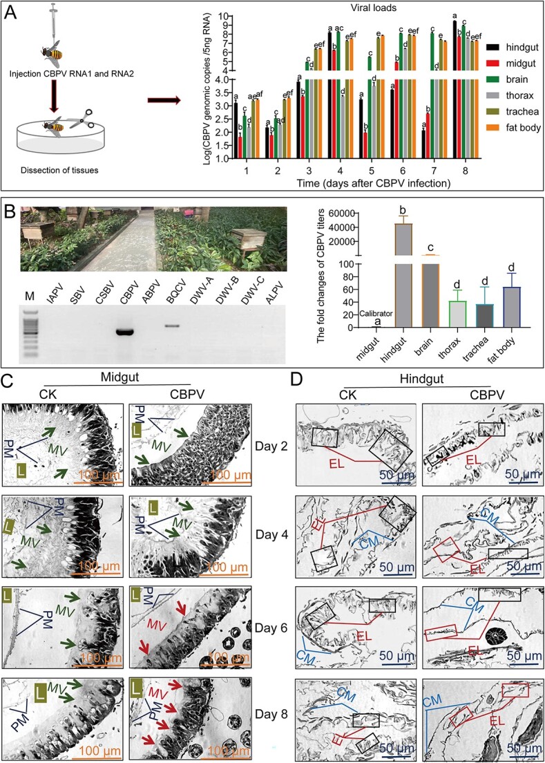
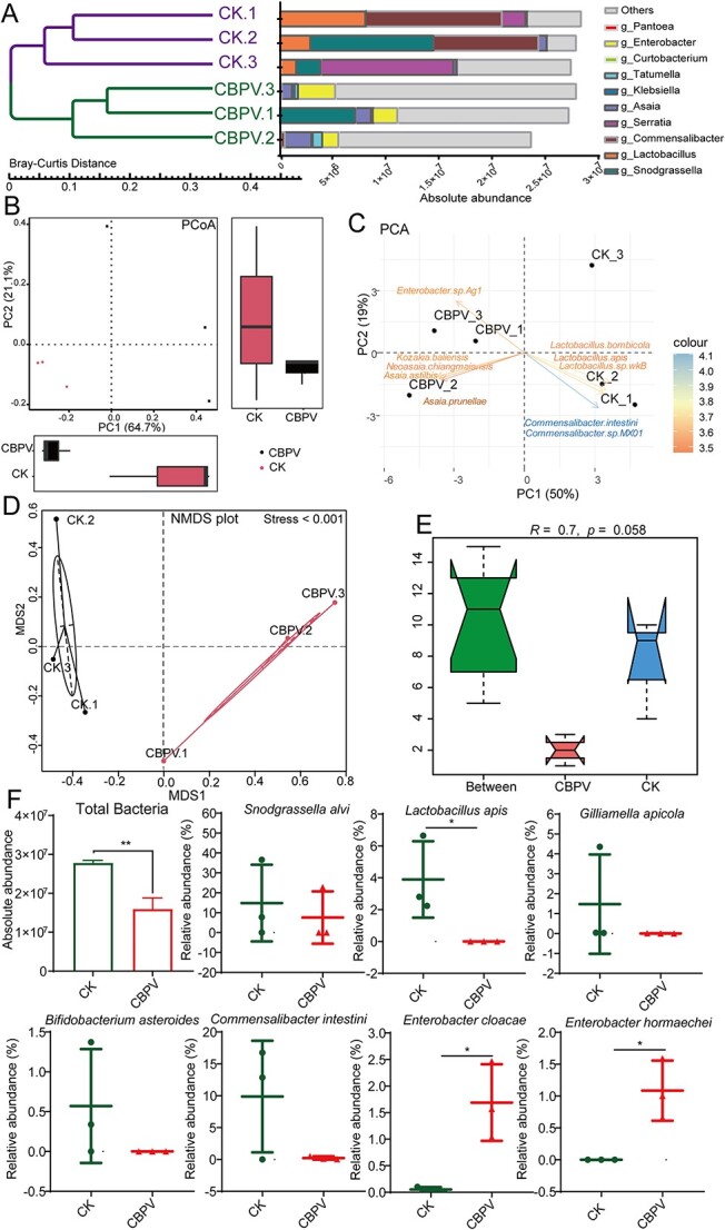
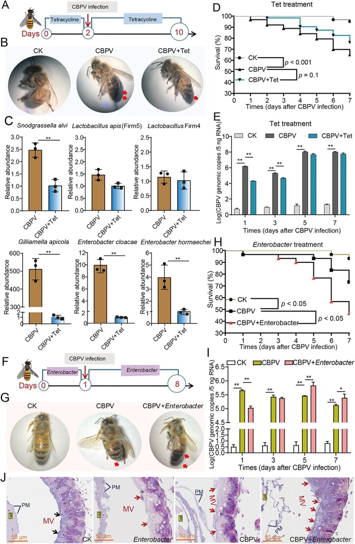
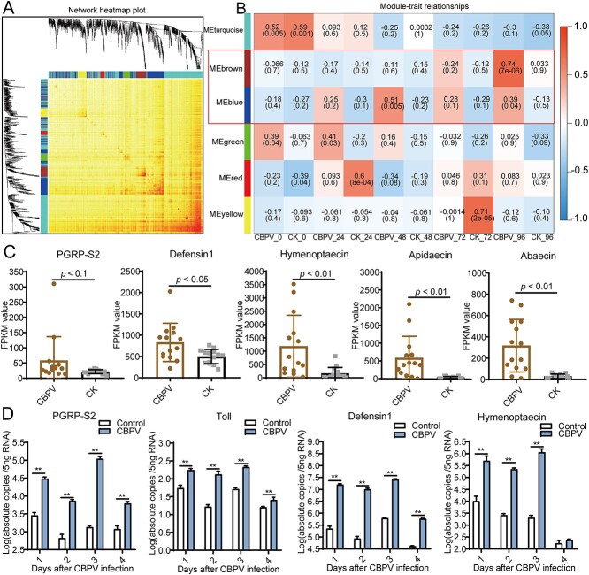
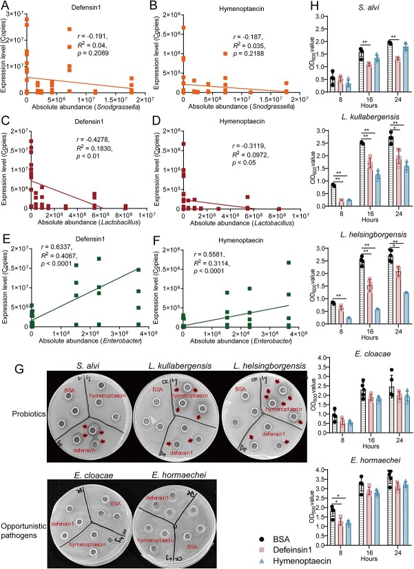
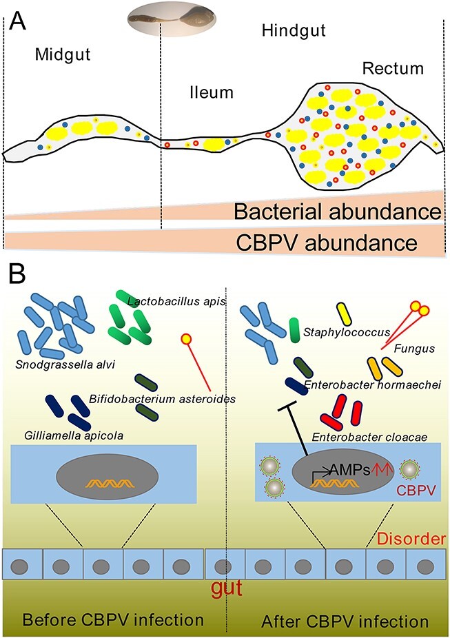
The significance of gut microbiota in regulating animal immune response to viral infection is increasingly recognized. However, how chronic bee paralysis virus (CBPV) exploits host immune to disturb microbiota for its proliferation remains elusive. Through histopathological examination, we discovered that the hindgut harbored the highest level of CBPV, and displayed visible signs of damages. The metagenomic analysis showed that a notable reduction in the levels of Snodgrassella alvi and Lactobacillus apis, and a significant increase in the abundance of the opportunistic pathogens such as Enterobacter hormaechei and Enterobacter cloacae following CBPV infection. Subsequent co-inoculation experiments showed that these opportunistic pathogens facilitated the CBPV proliferation, leading to accelerated mortality in bees and exacerbation of bloated abdomen symptoms after CBPV infection. The expression level of antimicrobial peptide (AMP) was found to be significantly up-regulated by over 1000 times in response to CBPV infection, as demonstrated by subsequent transcriptome and quantitative real-time PCR investigations. In particular, through correlation analysis and a bacteriostatic test revealed that the AMPs did not exhibit any inhibitory effect against the two opportunistic pathogens. However, they did demonstrate inhibitory activity against S. alvi and L. apis. Our findings provide different evidence that the virus infection may stimulate and utilize the host's AMPs to eradicate probiotic species and facilitate the proliferation of opportunistic bacteria. This process weakens the intestinal barrier and ultimately resulting in the typical bloated abdomen.

Keywords: CBPV; antimicrobial peptide; bloated abdomen; gut microbiota; honey bees.


Conflict of interest statement

The authors declare that they have no conflict ofinterest.

Figures

Figure 1
Figure 1
Viral titers in different tissues of honey bees injected with the infectious clone of CBPV at various time points and the damage caused by CBPV infection in different tissues of A. mellifera. (A) Quantification of CBPV titers in six tissues: trachea, fat body, hindgut, thorax, brain, and midgut of honey bees at Days 1, 2, 3, 4, 5, 6, 7, and 8. (B) Quantification of CBPV titers on trachea, fat body, hindgut, thorax, brain, and midgut of bees from naturally infected predominantly with CBPV in field. (C) The histology of midgut of honey bee after CBPV infection at Days 2, 4, 6, and 8. (D) The histology of hindgut of honey bee after CBPV infection at Days 2, 4, 6, and 8. L, lumen; PM, peritrophic membranes; MV, microvilli; CM, circular muscle.
Figure 2
Figure 2
Metagenome sequencing analysis showed that viral infection affects the gut microbiota composition in A. mellifera at Day 5. (A) The UPGMA analysis showed the absolute abundance of gut bacteria species in healthy and CBPV-infected honey bees at the genus level. (B) PCoA analysis exhibited a variation in microbial diversity between healthy and CBPV-infected honey bees. (C) PCA analysis exhibited the difference in microbial diversity between healthy and CBPV-infected honey bees. (D) NMDS analysis exhibited the difference in microbial diversity between healthy and CBPV-infected honey bees. (E) ANOSIM analysis exhibited variation in microbial diversity between healthy and CBPV-infected honey bees. (F) The column graph depicted the absolute abundance of gut bacteria species, and the dot plot displayed the relative abundances of gut microbiota core species in healthy and CBPV-infected honey bees. Each group has three replicates. Significant variations in each gene from the different groups are indicated by asterisks. *P < 0.05.
Figure 3
Figure 3
The effect of tetracycline or opportunistic pathogens treatment on gut microbiota, CBPV loads and physiology of A. mellifera. (A) Schematic illustration of the tetracycline experimental design. (B) The photo of the newly emerging bees after CBPV infection and tetracycline treatment. (C) The relative abundances of gut microbiota were quantified by qPCR with specific bacterial species, and normalized by 16S rRNA gene. (D) Survival rates of the CBPV-infected newly emerging bees with and without tetracycline treatment. (E) The effect of tetracycline on CBPV genomic copies in the newly emerging bees after viral infection. (F) Schematic illustration of the opportunistic pathogens experimental design. (G) The photo of the newly emerging bees after CBPV infection and opportunistic pathogens treatment. (H) Survival rates of the CBPV-infected newly emerging bees after opportunistic pathogens treatment. (I) The effect of opportunistic pathogens on CBPV genomic copies in the newly emerging bees after viral infection. (J) The histology of gut of the newly emerging bees after CBPV infection and opportunistic pathogens treatment. Asterisks indicate significant differences between groups. *P < 0.05, **P < 0.01. L, lumen; PM, peritrophic membranes; MV, microvilli.
Figure 4
Figure 4
The AMPs were significantly induced after CBPV infection. (A) The co-expression modules of the expressed genes in healthy and CBPV-infected honey bees, and the clustering dendrogram of 30 samples. (B) The relationship of 10 traits and 6 modules. (C) The FPKM values of five immune genes, peptidoglycan recognition protein S2 (PGRP-S2), hymenoptaecin, defensin1, apidaecin, and abaecin. (D) The expression levels of the immune genes in Toll/Imd pathway in the newly emerged CBPV-infected A. mellifera at Days 1, 2, 3, and 4. *P < 0.05, **P < 0.01.
Figure 5
Figure 5
The inhibition activity of AMPs induced by CBPV infection against core probiotic species and opportunistic pathogens. (A) The correlation analysis between the expression level of defensin1 and the absolute abundance of the genus Snodgrassella. (B) The correlation analysis between the expression level of hymenoptaecin and the absolute abundance of the genus Snodgrassella. (C) The correlation analysis between the expression level of defensin1 and the absolute abundance of the genus Lactobacillus. (D) The correlation analysis between the expression level of hymenoptaecin and the absolute abundance of the genus Lactobacillus. (E) The correlation analysis between the expression level of defensin1 and the absolute abundance of the genus Enterobacter. (F) The correlation analysis between the expression level of hymenoptaecin and the absolute abundance of the genus Enterobacter. (G) The representative LB/MRS plates showed the inhibition activity of Defensin1 or Hymenoptaecin against the gut microbiota. The core probiotic species (S. alvi, L. kullabergensis, L. helsingborgensis) and opportunistic pathogens (E. cloacae, E. hormaechei) were isolated from the gut of CBPV-infected A. mellifera, was spotted onto the LB/MRS plates and then incubated with BSA (0.5 mg/ml), Defensin1 (0.5 mg/ml), or Hymenoptaecin (0.5 mg/ml), culturing for 24 hours at 37°C. (H) The growth curves showed the inhibition activity of Defensin1 or Hymenoptaecin (0.02 mg/ml) against the gut microbiota.
Figure 6
Figure 6
A model for gut immune modulation and CBPV infection amplification by the hindgut microbiota has been proposed. (A) A diagram of the bee gut, showing the midgut and hindgut. The regions of the hindgut known as the ileum and rectum exhibit greatest bacteria densities and abundance of CBPV. (B) CBPV infection induces AMPs expression to change gut microbiota composition, including decreasing the abundance of probiotic species (such as S. alvi, L. apis, G. apicola, and B. asteroides) and increasing the abundance of opportunistic bacteria (E. cloacae, E. hormaechei, Staphylococcus spp.).

References

    1. Zheng H, Steele MI, Leonard SP et al. Honey bees as models for gut microbiota research. Lab Anim 2018;47:317–25. 10.1038/s41684-018-0173-x - DOI - PMC - PubMed
    1. Bonilla-Rosso G, Steiner T, Wichmann F et al. Honey bees harbor a diverse gut virome engaging in nested strain-level interactions with the microbiota. Proc Natl Acad Sci USA 2020;117:7355–62. 10.1073/pnas.2000228117 - DOI - PMC - PubMed
    1. Motta EVS, Raymann K, Moran NA. Glyphosate perturbs the gut microbiota of honey bees. Proc Natl Acad Sci USA 2018;115:10305–10. 10.1073/pnas.1803880115 - DOI - PMC - PubMed
    1. Raymann K, Shaffer Z, Moran NA. Antibiotic exposure perturbs the gut microbiota and elevates mortality in honeybees. PLoS Biol 2017;15:e2001861. 10.1371/journal.pbio.2001861 - DOI - PMC - PubMed
    1. Zheng J, Wittouck S, Salvetti E et al. A taxonomic note on the genus Lactobacillus: description of 23 novel genera, emended description of the genus Lactobacillus Beijerinck 1901, and union of Lactobacillaceae and Leuconostocaceae. Int J Syst Evol Microbiol 2020;70:2782–858. 10.1099/ijsem.0.004107 - DOI - PubMed

Substances