

Partial facial duplication (diprosopus): a case report and review of the literature
- PMID: 38519951
- PMCID: PMC10960485
- DOI: 10.1186/s13256-024-04423-4
Partial facial duplication (diprosopus): a case report and review of the literature
Abstract
Background: Diprosopus, or craniofacial duplication, is a rare entity that occurs in approximately 1 in 180,000 to 15 million live births. The degree of duplication varies from complete facial duplication to small facial structure duplication like the nose and eye. The cause of diprosopus is unknown though there are proposed factors.
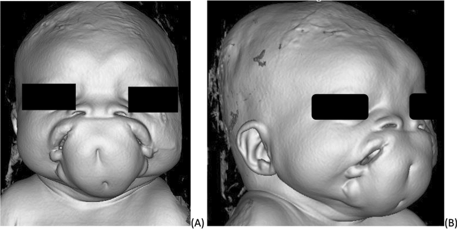
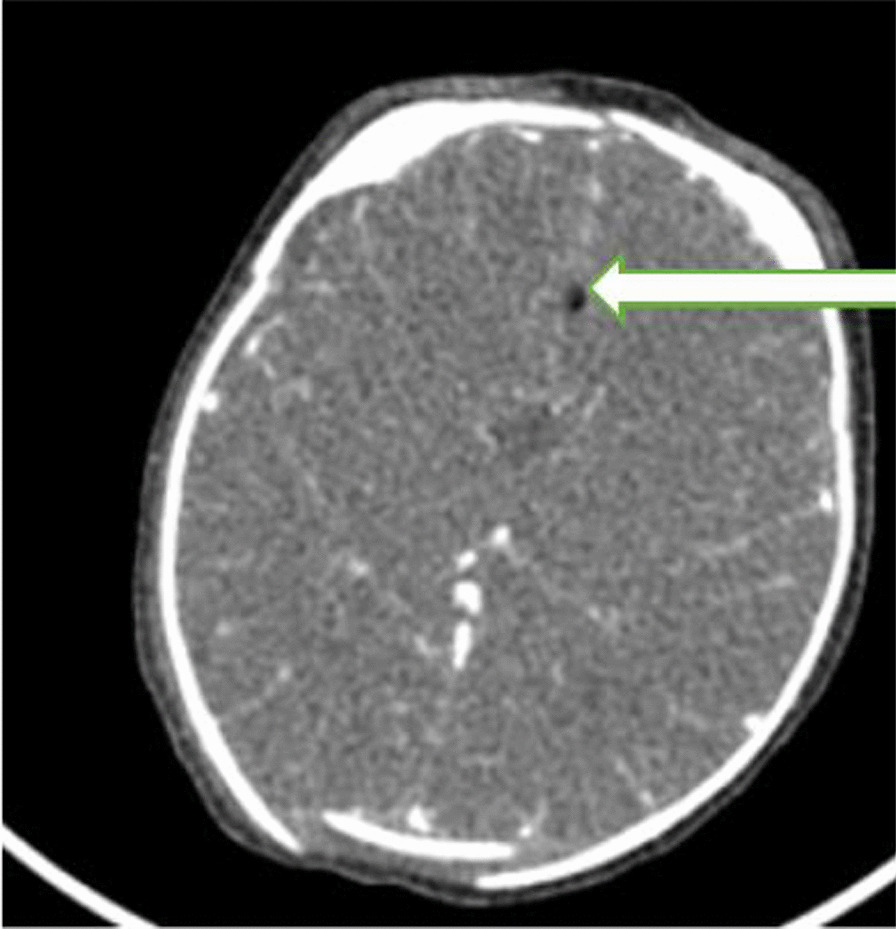
Case presentation: Our African patient was a term 72 hours old female neonate who was referred to our center with impression of lower facial duplication with two oral cavity that are located side to side separated by large soft tissue, she also had flat nasal bridge with widely separated nostrils and widely spaced eyes. Besides the facial malformation she had multiple episodes of vomiting with aspiration. Her blood tests were normal. Precontract brain computed tomography (CT) scan confirmed partially duplicated mandible and maxilla, two oral cavity separated by large fatty tissue, brain tissue were well formed and the only abnormality was corpus callosum agenesis and interhemispheric lipoma. In her stay at hospital nasogastric tube (NG) tube feed was initiated and started with antibiotics for aspiration pneumonia. After 25th day the neonatal passed away with possible cause of death being respiratory failure.
Conclusion: Craniofacial duplication is a very rare anomaly with only a few cases reported. Most of these patients are stillborn, even if they survive the prognosis is often poor. Early prenatal diagnosis is very important as termination of pregnancy can sometimes be considered an option.
Keywords: Corpus callosum agenesis; Craniofacial duplication; Diprosopus; Interhemispheric lipoma.
© 2024. The Author(s).
Conflict of interest statement
The authors declare that they have no competing interests.
Figures


Similar articles
-
[Partial facial duplication (a rare diprosopus): Case report and review of the literature].Rev Stomatol Chir Maxillofac Chir Orale. 2015 Dec;116(6):376-9. doi: 10.1016/j.revsto.2015.09.004. Epub 2015 Nov 12. Rev Stomatol Chir Maxillofac Chir Orale. 2015. PMID: 26586597 Review. French.
-
Partial craniofacial duplication: a review of the literature and case report.J Craniomaxillofac Surg. 2014 Jun;42(4):290-6. doi: 10.1016/j.jcms.2013.05.016. Epub 2013 Aug 19. J Craniomaxillofac Surg. 2014. PMID: 23969147 Review.
-
Facial duplication: case, review, and embryogenesis.Teratology. 1982 Apr;25(2):153-9. doi: 10.1002/tera.1420250205. Teratology. 1982. PMID: 6808690
-
Diprosopus: A Rare Case of Craniofacial Duplication and a Systematic Review of the Literature.Genes (Basel). 2023 Aug 31;14(9):1745. doi: 10.3390/genes14091745. Genes (Basel). 2023. PMID: 37761885 Free PMC article.
-
Accessory oral cavity associated with duplication of the tongue and the mandible in a newborn: a rare case of Diprosopus. Multi-row detector computed tomography diagnostic role.J Craniomaxillofac Surg. 2014 Dec;42(8):1924-8. doi: 10.1016/j.jcms.2014.07.013. Epub 2014 Aug 15. J Craniomaxillofac Surg. 2014. PMID: 25218149
References
Publication types
MeSH terms
LinkOut - more resources
Full Text Sources
