. 2024 Mar 23;22(1):126.
doi: 10.1186/s12951-024-02390-y.

Construction of programmed time-released multifunctional hydrogel with antibacterial and anti-inflammatory properties for impaired wound healing

Affiliations

Construction of programmed time-released multifunctional hydrogel with antibacterial and anti-inflammatory properties for impaired wound healing

Yuan Peng et al. J Nanobiotechnology. .

Abstract

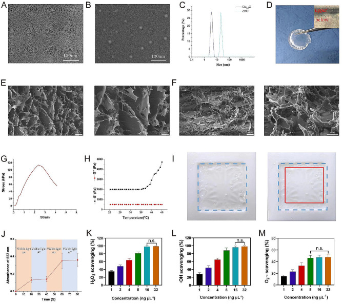
The successful reprogramming of impaired wound healing presents ongoing challenges due to the impaired tissue microenvironment caused by severe bacterial infection, excessive oxidative stress, as well as the inappropriate dosage timing during different stages of the healing process. Herein, a dual-layer hydrogel with sodium alginate (SA)-loaded zinc oxide (ZnO) nanoparticles and poly(N-isopropylacrylamide) (PNIPAM)-loaded Cu5.4O ultrasmall nanozymes (named programmed time-released multifunctional hydrogel, PTMH) was designed to dynamically regulate the wound inflammatory microenvironment based on different phases of wound repairing. PTMH combated bacteria at the early phase of infection by generating reactive oxygen species through ZnO under visible-light irradiation with gradual degradation of the lower layer. Subsequently, when the upper layer was in direct contact with the wound tissue, Cu5.4O ultrasmall nanozymes were released to scavenge excessive reactive oxygen species. This neutralized a range of inflammatory factors and facilitated the transition from the inflammatory phase to the proliferative phase. Furthermore, the utilization of Cu5.4O ultrasmall nanozymes enhanced angiogenesis, thereby facilitating the delivery of oxygen and nutrients to the impaired tissue. Our experimental findings indicate that PTMHs promote the healing process of diabetic wounds with bacterial infection in mice, exhibiting notable antibacterial and anti-inflammatory properties over a specific period of time.

Keywords: Anti-inflammation; Antibacterial; Nanozymes; Time-released hydrogel; Wound healing.


Conflict of interest statement

The authors declare that they have no known competing financial interests or personal relationships that could have appeared to influence the work reported in this paper.

Figures

Fig. 1
Fig. 1
Schematic of the design, structure, and therapeutic process of PTMH for impaired wound healing. (A) A dual-layer hydrogel with sodium alginate (SA)-loaded zinc oxide (ZnO) nanoparticles and poly(N-isopropylacrylamide) (PNIPAM)-loaded Cu5.4O ultrasmall nanozymes was designed. (B) The programmed regulation process in the mouse model of diabetic wounds with P. aeruginosa infection, including anti-infection with ZnO via generation of reactive oxygen species (ROS) and anti-inflammation, as well as angiogenesis promotion by Cu5.4O ultrasmall nanozymes. PTMH, programmed time-released multifunctional hydrogel
Fig. 2
Fig. 2
Preparation and characterization of PTMHs. (A) TEM images of Cu5.4O and (B) ZnO. (Scale bar: 100 nm). (C) The particle size distribution of Cu5.4O and ZnO nanoparticles. (D) Optical image of PTMH. (E) SEM images of PTMH. (Scale bar, left: 100 µm; right: 100 nm) (F) SEM images of ZnO@SA layer view after 3 days of incubation at 35°C. (Scale bar, left: 100 nm; right: 50 nm). (G) The stress‒strain curve of PNIPAM. (H) The elastic modulus (G’) and loss modulus (G’’) of PNIPAM. (I) Images of PNIPAM following incubation at 35 °C. (J) The test of ROS production of ZnO. (K) H2O2, (L) ·OH, and (M) O2· scavenging capacity of different concentrations of Cu5.4O@PNIPAM. Data in K, L, and M represent the mean ± standard deviation (n = 5). n.s., no significance; one-way ANOVA. TEM, transmission electron microscopy; SA, sodium alginate; PNIPAM, poly(N-isopropylacrylamide); PTMH, programmed time-released multifunctional hydrogel; ANOVA, analysis of variance
Fig. 3
Fig. 3
Evaluation of the antibacterial activity of PTMH in vitro. (A) Images of bacterial colonies of P. aeruginosa and (B) MRSA following treatment with Cu5.4O@PNIPAM, ZnO@SA, and PTMH with or without VL irradiation. (C) The number of bacterial colonies formed by P. aeruginosa and MRSA under different treatments. (D) OD600 values of P. aeruginosa and (E) MRSA under different treatments. (F) Representative confocal digital images and (G) fluorescence intensity for DCFH-DA (green) staining of 3T3 cells under different treatments. (Scale bar: 50 μm) (H) ROS levels and (I) statistical analysis in 3T3 cells under different treatments. Data in C–E, G, and I represent the mean ± standard deviation (n = 5). *p < 0.05; **p < 0.01; one-way ANOVA. P. aeruginosa, Pseudomonas aeruginosa; MRSA, methicillin-resistant Staphylococcus aureus; VL, visible light; SA, sodium alginate; PNIPAM, poly(N-isopropylacrylamide); PTMH, programmed time-released multifunctional hydrogel; OD, optical density; DCFH-DA, 2′,7′-dichlorofluorescin diacetate; FSC, forward scatter; SSC, side scatter; ROS, reactive oxygen species; ANOVA, analysis of variance
Fig. 4
Fig. 4
Efficiency of PTMHs for the healing of mouse diabetic wounds with P. aeruginosa infection. (A) Schematic of the experimental design for the therapeutic effect evaluation of PTMH in mouse STZ-induced diabetic wounds with P. aeruginosa infection. (B) Digital images of diabetic wounds at various time intervals, with a blue disc measuring 6 millimeters in diameter for scale reference. (C) Schematic images of diabetic wounds under different treatments. (D) The proportions of wound healing achieved through various treatment on days 0, 3, 7, and 14 (n = 5). (E) Area percentages of closed wounds. (F) Ultimate healing time of each group. (G) Representative histological images of H&E staining in the wound site on day 14. Black arrows represent the thickness of the granulation tissue. (Scale bar: 100 μm) (H) Statistical analysis of the thickness of the granulation tissue. (I) The images of Masson’s trichrome staining in diabetic wounds through various treatment. (Scale bar: 100 μm) (J) Statistical analysis of the percentage of collagen volume fraction. (K) Images and (L) quantitative counts of bacterial colonies formed by P. aeruginosa harvested from wound tissues at various time intervals. Data in E, F, H, J, and L represent the mean ± standard deviation (n = 5). *p < 0.05; **p < 0.01; n.s., no significance; one-way ANOVA. SA, sodium alginate; PNIPAM, poly(N-isopropylacrylamide); PTMH, programmed time-released multifunctional hydrogel; P. aeruginosa, Pseudomonas aeruginosa; STZ, streptozotocin; ANOVA, analysis of variance; CFU, colony-forming unit; H&E, hematoxylin and eosin
Fig. 5
Fig. 5
Regulatory effects of PTMH on the microenvironment in vivo. (A) Fluorescent images and (B) statistical analysis of PCNA (red) and DAPI (blue) in mouse diabetic wounds tissue. (Scale bar: 100 μm) (C) Fluorescent images and (D) statistical analysis of CD31 (red) and DAPI (blue) in mouse diabetic wounds tissue. (Scale bar: 100 μm) (E) Fluorescent images and (F) statistical analysis of DHE (red) and DAPI (blue) on days 3 and 14 post-treatment wound tissue. (G) The distribution of cells in wound tissue obtained from different groups on days 3 and 14 post-treatment, stained with rat anti-mouse CD11b-Brilliant Violet 650™, LY6G-FITC, and CD86-Brilliant Violet 421™ (1:200). (H) Statistical analysis of CD86-Brilliant Violet 421TM-positive cells. (I) The concentrations of TNF-α, (J) IL-6, (K) IL-1β, (L) IL-10, (M) VEGF, (N) SOD and (O) MPO in wound tissue were determined using ELISA. Data in B, D, F, H and I–O represent the mean ± standard deviation (n = 5). *p < 0.05; **p < 0.01; one-way ANOVA. SA, sodium alginate; PNIPAM, poly(N-isopropylacrylamide); PTMH, programmed time-released multifunctional hydrogel; PCNA, proliferation cell nuclear antigen; DHE, dihydroethidium; CD, cluster of differentiation; ELISA, enzyme-linked immunosorbent assay; TNF-α, tumor necrosis factor-α; IL, interleukin; VEGF, vascular endothelial growth factor; SOD, superoxide dismutase; MPO, myeloperoxidase; ANOVA, analysis of variance; DAPI, 4’,6-diamidino-2-phenylindole; SSC-A, side scatter area
Fig. 6
Fig. 6
Mechanisms underlying the therapeutic effects of PTMH in impaired wound healing. (A) Principal Component Analysis (PCA) was performed on the differentially expressed genes in the wound tissues of the mice subjected to treatment with PTMH (PTMH group) or PBS (Control group). Each data point within the two groups represents an independent replicate. (B) Venn diagram was constructed based on the transcriptomic data to illustrate the overlap between the two groups. (C) Volcano plots were generated to visualize the genes that were upregulated and downregulated after PTMH treatment. (D) KEGG pathway enrichment analysis of the differentially expressed genes (downregulation). (E) Heatmaps illustrating the noteworthy downregulation of genes implicated in inflammation and oxidative stress subsequent to PTMH treatment. (F) KEGG pathway enrichment analysis of the differentially expressed genes (upregulated). (G) Chord diagram showing the genes involved in inflammation and oxidative stress that were significantly upregulated following PTMH treatment. (H) Relative expression of genes involved in inflammation and wound regeneration. Data in h represent the mean ± standard deviation from three independent replicates (n = 3). *p < 0.05; **p < 0.01; n.s., no significance; t-test. PTMH, programmed time-released multifunctional hydrogel; PCA, principal component analysis; IL, interleukin; KEGG, Kyoto Encyclopedia of Genes and Genomes; ECM, extracellular matrix; Nod1, nucleotide-binding oligomerization domain containing 1
Fig. 7
Fig. 7
Schematic of the underlying mechanism of the effects of PTMH in impaired wounds healing. PTMH could combat bacterial infection by ZnO and alleviate inflammation via Cu5.4O ultrasmall nanozymes to improve the tissue repairing microenvironment, and ultimately promote impaired wound healing. IL, interleukin; PTMH, programmed time-released multifunctional hydrogel; ROS, reactive oxygen species; TNF-α, tumor necrosis factor-α

Similar articles

Cited by

References

    1. Sun BK, Siprashvili Z, Khavari PA. Advances in skin grafting and treatment of cutaneous wounds. Science. 2014;346(6212):941–5. doi: 10.1126/science.1253836. - DOI - PubMed
    1. Martin P. Wound healing–aiming for perfect skin regeneration. Science. 1997;276(5309):75–81. doi: 10.1126/science.276.5309.75. - DOI - PubMed
    1. Eming SA, Wynn TA, Martin P. Inflammation and metabolism in tissue repair and regeneration. Science. 2017;356(6342):1026–30. doi: 10.1126/science.aam7928. - DOI - PubMed
    1. Maschalidi S, Mehrotra P, Keceli BN, De Cleene HKL, Lecomte K, Van der Cruyssen R, Janssen P, Pinney J, van Loo G, Elewaut D, Massie A, Hoste E, Ravichandran KS. Targeting SLC7A11 improves efferocytosis by dendritic cells and wound healing in diabetes. Nature. 2022;606(7915):776–84. doi: 10.1038/s41586-022-04754-6. - DOI - PubMed
    1. Cao P, Fleming D, Moustafa DA, Dolan SK, Szymanik KH, Redman WK, Ramos A, Diggle FL, Sullivan CS, Goldberg JB, Rumbaugh KP, Whiteley M. A Pseudomonas aeruginosa small RNA regulates chronic and acute infection. Nature. 2023;618(7964):358–64. doi: 10.1038/s41586-023-06111-7. - DOI - PMC - PubMed