. 2024 Jun;11(22):e2309917.
doi: 10.1002/advs.202309917. Epub 2024 Mar 23.

Design of PD-L1-Targeted Lipid Nanoparticles to Turn on PTEN for Efficient Cancer Therapy

Affiliations

Design of PD-L1-Targeted Lipid Nanoparticles to Turn on PTEN for Efficient Cancer Therapy

Yelee Kim et al. Adv Sci (Weinh). 2024 Jun.

Abstract

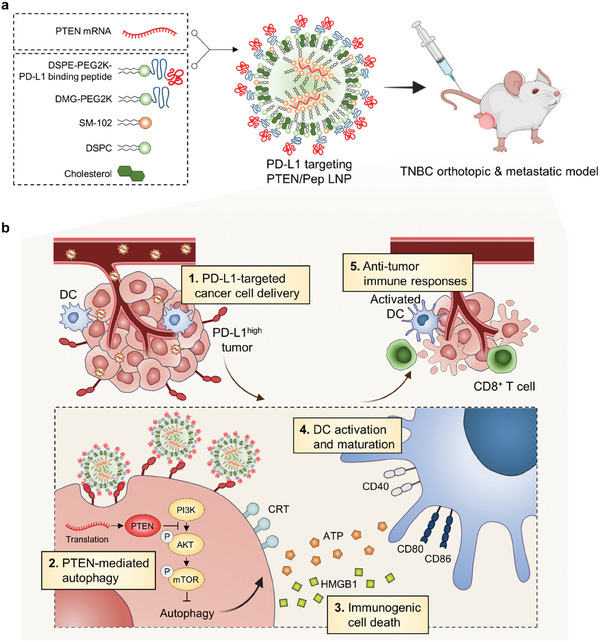
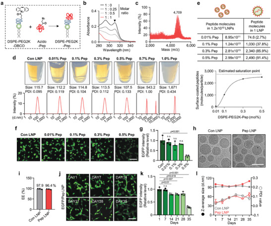
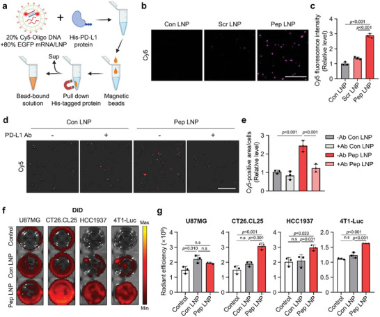
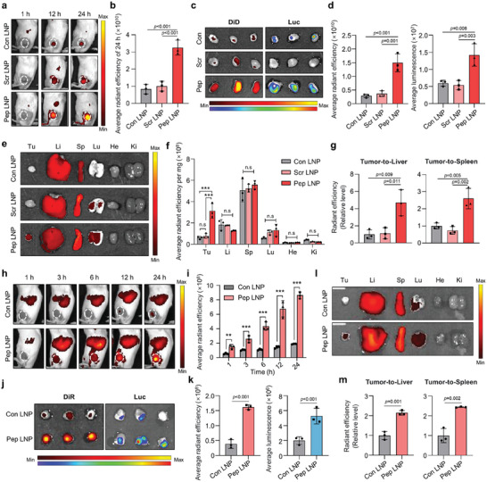
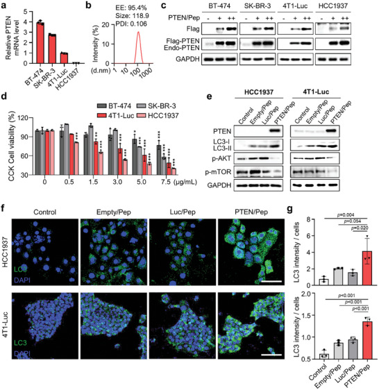
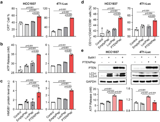
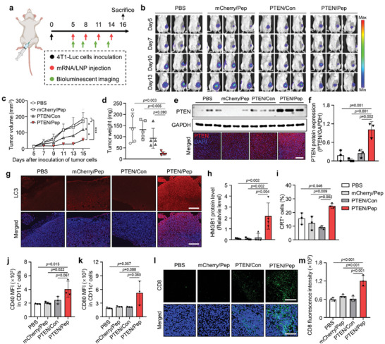
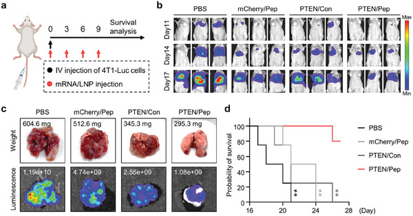
Lipid nanoparticles (LNPs) exhibit remarkable mRNA delivery efficiency, yet their majority accumulate in the liver or spleen after injection. Tissue-specific mRNA delivery can be achieved through modulating LNP properties, such as tuning PEGylation or varying lipid components systematically. In this paper, a streamlined method is used for incorporating tumor-targeting peptides into the LNPs; the programmed death ligand 1 (PD-L1) binding peptides are conjugated to PEGylated lipids via a copper-free click reaction, and directly incorporated into the LNP composition (Pep LNPs). Notably, Pep LNPs display robust interaction with PD-L1 proteins, which leads to the uptake of LNPs into PD-L1 overexpressing cancer cells both in vitro and in vivo. To evaluate anticancer immunotherapy mediated by restoring tumor suppressor, mRNA encoding phosphatase and tensin homolog (PTEN) is delivered via Pep LNPs to PTEN-deficient triple-negative breast cancers (TNBCs). Pep LNPs loaded with PTEN mRNA specifically promotes autophagy-mediated immunogenic cell death in 4T1 tumors, resulting in effective anticancer immune responses. This study highlights the potential of tumor-targeted LNPs for mRNA-based cancer therapy.

Keywords: cancer immunotherapy; lipid nanoparticle; mRNA delivery; tumor‐targeted delivery.


Conflict of interest statement

The authors declare no conflict of interest.

Figures

Figure 1
Figure 1
Schematic illustration of PD‐L1‐targeted PTEN/Pep LNPs for immunotherapy of TNBC. a) Scheme for the assembly of Pep LNPs with mRNA. b) Targeted delivery of PTEN/Pep LNPs to PD‐L1‐high tumors induces PTEN‐mediated autophagy and ICD, promotes DC maturation, and stimulates CD8+ T cell infiltration, enhancing antitumor immunity.
Figure 2
Figure 2
Synthesis condition and characterization of Pep LNPs. a) Schematic illustration showing the synthesis of DSPE‐PEG2K‐Pep. b) UV–vis absorption spectra obtained from click reaction of DSPE‐PEG2K‐DBCO: Azido‐Pep with different molar ratios (1:0, 1:0.25, 1:0.5, 1:1). c) MALDI‐TOFTOF mass spectrometric analysis of DSPE‐PEG2K‐Pep after a 1:1 molar ratio reaction. d) Pictures, size distribution diagram, and average PDI value of Pep LNPs with different DSPE‐PEG2K‐Pep mol% were measured by DLS. e) Quantification of peptides attached to the surface of a single LNP and assessment of coating efficiency through NTA analysis. f) Representative fluorescence images and g) a quantified graph showing the transfection efficiency of EGFP/Pep LNPs in CT26.CL25 cells. Scale bar: 100 µm. h) Representative cryo‐TEM image of Con LNPs and 0.3 mol% Pep LNPs. Scale bar: 100 nm. i) Encapsulation efficiency (EE) of mRNA assessed by RiboGreen assay. j) Representative fluorescence images and k) a quantified graph showing the transfection efficiency of EGFP/Pep LNPs in CT26.CL25 cells at various time points after storage at 4 °C. Scale bar: 100 µm. l) Z‐average size and PDI value of Con LNPs and Pep LNPs measured at different time points after storage at 4 °C. All data presented as mean ± SD, n = 3. n.s, not significant; g,k) One‐way ANOVA with Tukey's post‐hoc test.
Figure 3
Figure 3
PD‐L1 binding ability of Pep LNPs in vitro. a) Experimental scheme for in vitro PD‐L1 protein binding assay with Cy5‐labeled LNPs. b) Cy5 fluorescence image capturing the bead‐bound solution following incubation of Cy5‐labeled LNPs with His‐PD‐L1 protein. Scale bar: 50 µm. c) The Cy5 fluorescence intensity of bead‐bound solution. d) Cy5 fluorescence image showing the cellular binding of Cy5‐labeled Con or Pep LNPs in CT26.CL25 cells, with or without pre‐treatment of PD‐L1 antibody. Scale bar: 100 µm. e) Quantification of cellular binding assessed by measuring the Cy5‐positive area normalized to the number of cells. f) Well plate images and g) quantification of DiD radiant efficiency in cells treated with DiD‐labeled Luc/Con or Pep LNPs. All data presented as mean ± SD, n = 3. n.s, not significant; c,e,g) One‐way ANOVA with Tukey's post‐hoc test.
Figure 4
Figure 4
PD‐L1‐high tumor‐targeting of Pep LNPs. a) DiD fluorescence imaging of CT26.CL25 subcutaneous BALB/c mouse model after intravenous injection of DiD‐labeled Luc/Con, Scr, or Pep LNPs (Normalization was performed together for 1 h and 12, 24 h). b) The quantification of tumor‐localized average radiant efficiency of 24 h. c) Fluorescence and luminescence image of tumors harvested at 24 h post‐injection. d) The average radiant efficiency and luminescence of harvested tumors. e) DiD fluorescence imaging of harvested organs and f) quantified graphs of organ DiD radiant efficiency normalized to organ weight (mg). g) Quantified graphs of tumor DiD radiant efficiency normalized to that of the liver or spleen. h) DiR fluorescence imaging and i) quantification of in vivo tumor localized radiant efficiency after intravenous injection of DiR‐labeled Luc/Con or Pep LNPs in BALB/c mice bearing 4T1 tumor. j) DiR fluorescence or luminescence imaging and k) quantification of tumors harvested at 24 h postinjection. l) DiR fluorescence imaging of harvested organs and m) quantification of tumor DiR radiant efficiency with normalized to that of liver or spleen. Li: liver, Sp: spleen, Lu: lung, He: heart. Ki: kidney, Tu: tumor. All data presented as mean ± SD, n = 3. n.s, not significant; **p < 0.01, ***p < 0.001. b,d,g) One‐way ANOVA with Tukey's post‐hoc test, f, i) Two‐way ANOVA with Tukey's post‐hoc test. k,m) Unpaired t‐test.
Figure 5
Figure 5
PTEN‐deficient cancer cells experience autophagy via PTEN/Pep LNPs. a) Relative PTEN mRNA expression levels in BT‐474, SK‐BR‐3, 4T1‐Luc, and HCC1937 cells by qPCR. b) The size distribution diagram, average PDI value, and encapsulation efficiency (EE) of PTEN/Pep LNPs. c) Western blot images of cells treated with PTEN/Pep LNPs (0.5, 1 µg mL−1). d) CCK cell viability assay for PTEN mRNA‐mediated cell death effect. e) Western blot, f) immunofluorescence images, and g) quantification of LC3 fluorescence intensity per cell numbers of HCC1937 and 4T1‐Luc cells treated with different LNPs. Scale bar: 100 µm. All data presented as mean ± SD, n = 3. n.s, not significant; *p < 0.05, **p < 0.01, ***p < 0.001. d,g) One‐way ANOVA with Tukey's post‐hoc test.
Figure 6
Figure 6
PTEN/Pep LNP‐mediated ICD and DC maturation in TNBC. a–c) Analysis of ICD markers in HCC1937 and 4T1‐Luc cells 24 h (CRT) or 48 h (ATP and HMGB1) after treatment with different LNPs. a) CRT expression on the cell surface was evaluated by flow cytometry. b) Extracellular ATP release was measured by an ATP bioluminescence detection kit. c) Extracellular HMGB1 level was evaluated by western blot analysis. d) BMDCs were added with supernatants from HCC1937 and 4T1‐Luc cells, which had been treated with Empty/Pep LNPs, Luc/Pep LNPs, PTEN/Pep LNPs, or left untreated for 48 h. Then, the percentage of matured DCs (CD11c+CD40+CD86+) was analyzed by flow cytometry. e) HCC1937 and 4T1‐Luc cells were either untreated or treated with PTEN/Pep LNPs for 24 h. Cells were then exposed to BafA1 or left untreated for an additional 12 h before undergoing western blot analysis and extracellular ATP release assay. All data presented as mean ± SD, n = 3. a–e) One‐way ANOVA with Tukey's post‐hoc test.
Figure 7
Figure 7
In vivo antitumor immune response of PTEN/Pep LNPs in orthotopic TNBC model. a) Experimental scheme for in vivo study of PTEN/Pep LNPs. 4T1‐Luc tumor‐bearing mice were treated with different LNPs (0.6 mg kg−1). b) Bioluminescence imaging of orthotopic 4T1‐Luc tumor‐bearing mice. Imaging was obtained every 3 d from the initial injection day (day 5 after tumor inoculation) until day 13. c) The average tumor growth curve. The size of the tumor was measured on days 5, 7, 9, 11, 13, and 15 post‐inoculation of cancer cells. d) Excised tumor weight. e) Expression of PTEN in tumor tissues, assessed by western blotting and immunofluorescence imaging. Scale bar: 200 µm. f) Quantification of PTEN protein expression levels normalized by the expression of GAPDH. g) Tumor tissues stained with LC3 to evaluate autophagy. Scale bar: 200 µm. h) Relative amounts of HMGB1 in the tumor supernatants were analyzed by western blot and normalized by total protein. i) CRT‐positive cancer cells in tumor tissues were assessed by flow cytometry. j,k) Flow cytometry analysis of CD40 and CD80 expression in CD11c+ cells in the TDLN by quantification of median fluorescence intensity (MFI). l) Immunofluorescence imaging of tumor tissues stained with FITC‐conjugated CD8 antibody and m) quantification of CD8 fluorescence intensity. Scale bar: 200 µm. c,d) Data presented as mean ± SD, n = 5 mice, h) n = 4 mice, f,i–l) n = 3. *p < 0.05, ***p < 0.001. c) Two‐way ANOVA with Tukey's post‐hoc test. d,f,h–k,m) One‐way ANOVA with Tukey's post‐hoc test.
Figure 8
Figure 8
Anti‐metastatic effect of PTEN/Pep LNPs in TNBC lung metastatic model. a) Experimental scheme for in vivo study in a metastatic TNBC model. Mice were injected with 4T1‐Luc cells via the tail vein and treated with different LNPs (0.6 mg kg−1). b) Metastatic 4T1‐Luc tumor growth monitored by bioluminescence imaging. c) Ex vivo lung imaging with the bioluminescence flux and weight. d) Mice survival during treatment. Data presented as mean ± SD, n = 4 mice; **p < 0.01. d) Log‐rank (Mantel‐Cox) test.

References

    1. Han J., Lim J., Wang C.‐P. J., Han J.‐H., Shin H. E., Kim S.‐N., Jeong D., Lee S. H., Chun B.‐H., Park C. G., Park W., Nano Convergence 2023, 10, 36. - PMC - PubMed
    1. Goyal F., Chattopadhyay A., Navik U., Jain A., Reddy P. H., Bhatti G. K., Bhatti J. S., Adv. Ther. 2024, 7, 2300255.
    1. a) Wang Q. T., Nie Y., Sun S. N., Lin T., Han R. J., Jiang J., Li Z., Li J. Q., Xiao Y. P., Fan Y. Y., Yuan X. H., Zhang H., Zhao B. B., Zeng M., Li S. Y., Liao H. X., Zhang J., He Y. W., Cancer Immunol. Immunother. 2020, 69, 1375; - PMC - PubMed
    2. b) Sahin U., Oehm P., Derhovanessian E., Jabulowsky R. A., Vormehr M., Gold M., Maurus D., Schwarck‐Kokarakis D., Kuhn A. N., Omokoko T., Kranz L. M., Diken M., Kreiter S., Haas H., Attig S., Rae R., Cuk K., Kemmer‐Brück A., Breitkreuz A., Tolliver C., Caspar J., Quinkhardt J., Hebich L., Stein M., Hohberger A., Vogler I., Liebig I., Renken S., Sikorski J., Leierer M., et al., Nature 2020, 585, 107; - PubMed
    3. c) Kreiter S., Vormehr M., van de Roemer N., Diken M., Lower M., Diekmann J., Boegel S., Schrors B., Vascotto F., Castle J. C., Tadmor A. D., Schoenberger S. P., Huber C., Tureci O., Sahin U., Nature 2015, 520, 692; - PMC - PubMed
    4. d) Balachandran V. P., Rojas L. A., Sethna Z., Soares K., Derhovanessian E., Mueller F., Yadav M., Basturk O., Gonen M., Wei A. C.‐c., D'Angelica M. I., Kingham T. P., Greenbaum B., Merghoub T., Jarnagin W. R., Drebin J. A., Sahin U., Tuereci O., Wolchok J. D., O'Reilly E. M., J. Clin. Oncol. 2022, 40, 2516;
    5. e) Kopetz S., Morris V. K., Alonso‐Orduña V., Garcia‐Alfonso P., Reboredo M., Montes A. F., Maurel J., Paez D., Reinacher‐Schick A. C., Höhler T., Carrasco J., Galligan B. M., Manning L., Preußner L., Tureci Ö., Sahin U., J. Clin. Oncol. 2022, 40, TPS3641.
    1. Kong B., Kim Y., Kim E. H., Suk J. S., Yang Y., Adv. Drug Delivery Rev. 2023, 199, 114993. - PMC - PubMed
    1. a) Xiao Y., Chen J., Zhou H., Zeng X., Ruan Z., Pu Z., Jiang X., Matsui A., Zhu L., Amoozgar Z., Chen D. S., Han X., Duda D. G., Shi J., Nat. Commun. 2022, 13, 758; - PMC - PubMed
    2. b) Lin Y. X., Wang Y., Ding J., Jiang A., Wang J., Yu M., Blake S., Liu S., Bieberich C. J., Farokhzad O. C., Mei L., Wang H., Shi J., Sci. Transl. Med. 2021, 13, eaba9772. - PMC - PubMed