

Tattoos: risks and complications, clinical and histopathological approach
- PMID: 38521707
- PMCID: PMC11221160
- DOI: 10.1016/j.abd.2023.07.004
Tattoos: risks and complications, clinical and histopathological approach
Abstract
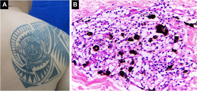
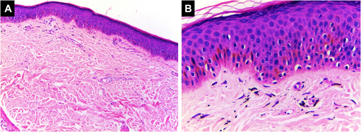
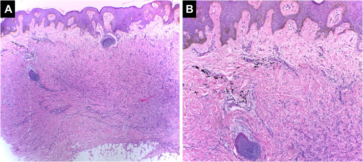
Background: Skin modification through tattoos is as old as humanity itself. However, this trend is on the rise, and with the use of different types of pigments and application practices, both cutaneous and systemic complications can arise. Adverse reactions can be grouped into five classes: inflammatory, infectious, neoplastic, aesthetic, and miscellaneous. On histopathology, inflammatory reactions can exhibit a lichenoid pattern or present as spongiotic dermatitis, granulomatous reactions, pseudolymphoma, pseudoepitheliomatous hyperplasia, or scleroderma/morphea-like changes. This article reviews tattoo complications, including their clinical and histopathological characteristics.
Methods: An open search was conducted on PubMed using the terms "tattoo", "complications", and "skin". No limits were set for period, language, or publication type of the articles.
Results: Reactions to tattoos are reported in up to 67% of people who get tattooed, with papulonodular and granulomatous reactions being the most common. Some neoplastic complications have been described, but their causality is still debated. Any pigment can cause adverse reactions, although red ink is more frequently associated with them. Patients with pre-existing dermatoses may experience exacerbation or complications of their diseases when getting tattoos; therefore, this procedure is not recommended for this patient group.
Conclusions: Dermatological consultation is recommended before getting a tattoo, as well as a histopathological examination in case of complications. In patients who develop cutaneous inflammatory reactions following tattooing, additional studies are recommended to investigate systemic diseases such as sarcoidosis, pyoderma gangrenosum, atopic dermatitis, and neoplasms. It is important for physicians to be trained in providing appropriate care in case of complications.
Keywords: Complications; Inks; Punctures; Skin; Tattooing.
Copyright © 2024 Sociedade Brasileira de Dermatologia. Published by Elsevier España, S.L.U. All rights reserved.
Figures
References
-
- McIlwee B.E., Alster T.S. Treatment of cosmetic tattoos: a review and case analysis. Dermatol Surg. 2018;44:1565–1570. - PubMed
-
- Vassileva S., Hristakieva E. Medical applications of tattooing. Clin Dermatol. 2007;25:367–374. - PubMed
-
- De Cuyper C. Permanent makeup: indications and complications. Clin Dermatol. 2008;26:30–34. - PubMed
-
- Islam P.S., Chang C., Selmi C., Generali E., Huntley A., Teuber S.S., et al. Medical complications of tattoos: a comprehensive review. Clin Rev Allergy Immunol. 2016;50:273–286. - PubMed
Publication types
MeSH terms
Substances
LinkOut - more resources
Full Text Sources
Medical
