. 2024 Jul;26(7):101125.
doi: 10.1016/j.gim.2024.101125. Epub 2024 Mar 21.

Homozygous missense variants in YKT6 result in loss of function and are associated with developmental delay, with or without severe infantile liver disease and risk for hepatocellular carcinoma

Affiliations

Homozygous missense variants in YKT6 result in loss of function and are associated with developmental delay, with or without severe infantile liver disease and risk for hepatocellular carcinoma

Mengqi Ma et al. Genet Med. 2024 Jul.

Abstract

Purpose: YKT6 plays important roles in multiple intracellular vesicle trafficking events but has not been associated with Mendelian diseases.

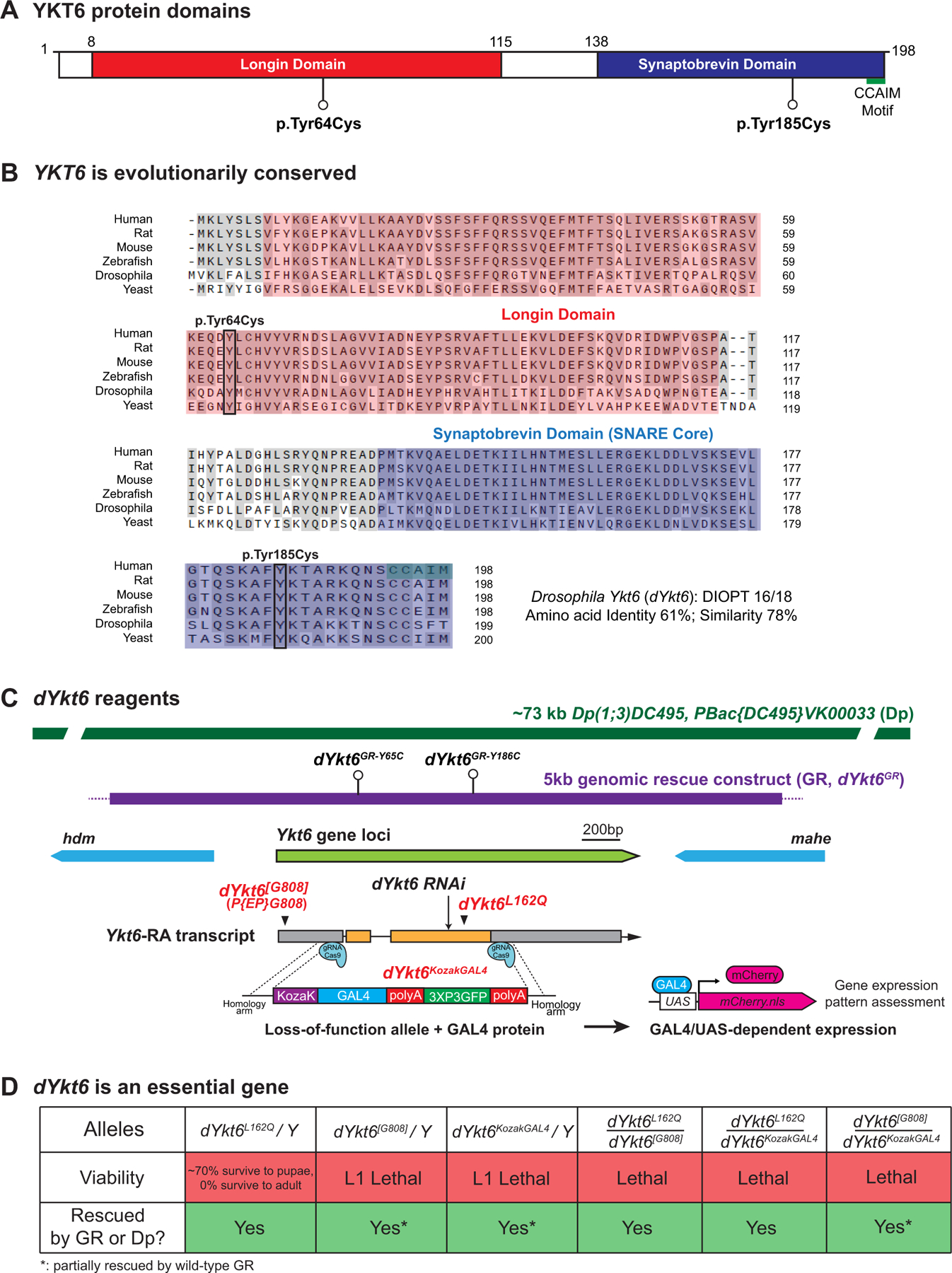
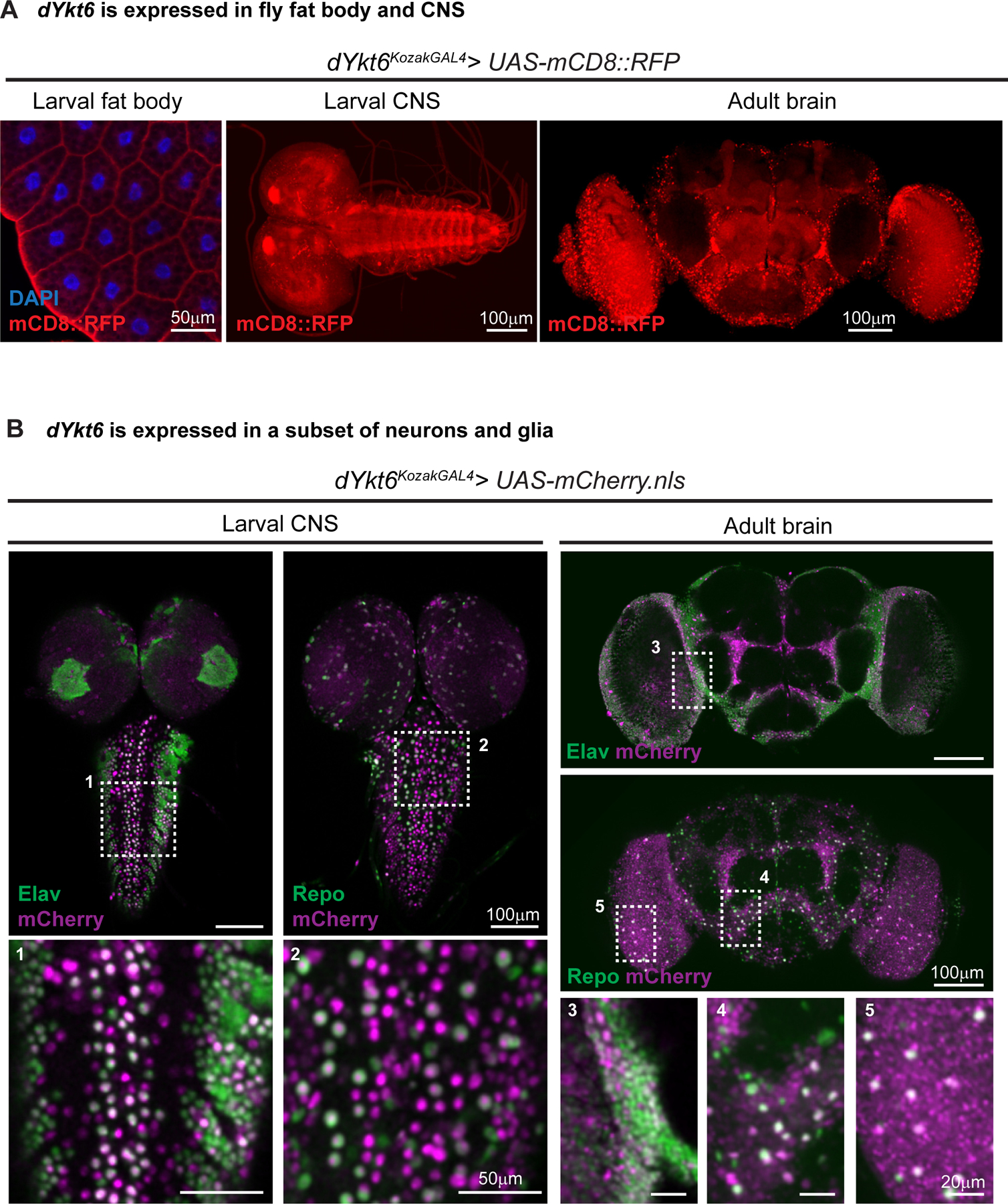
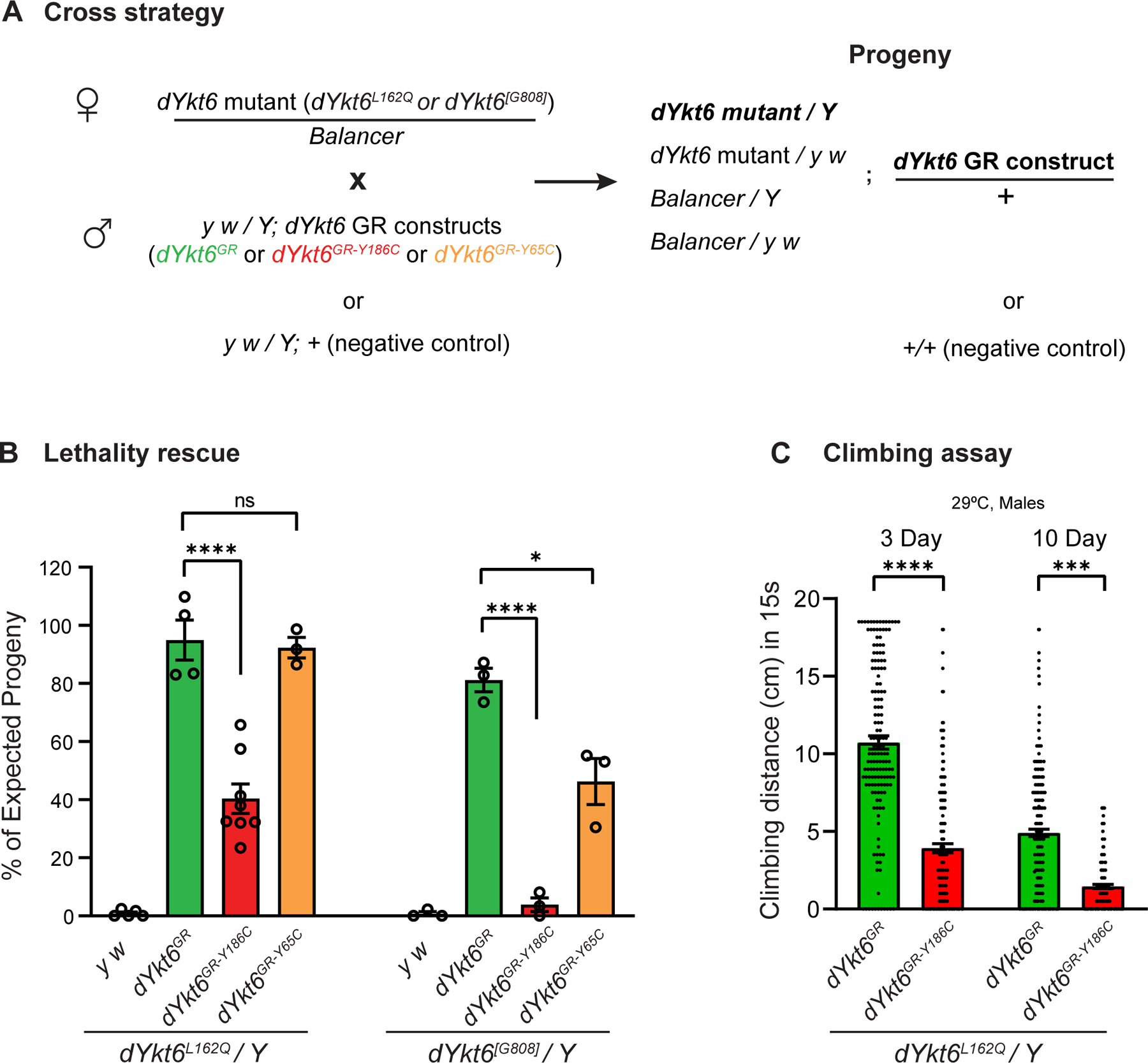
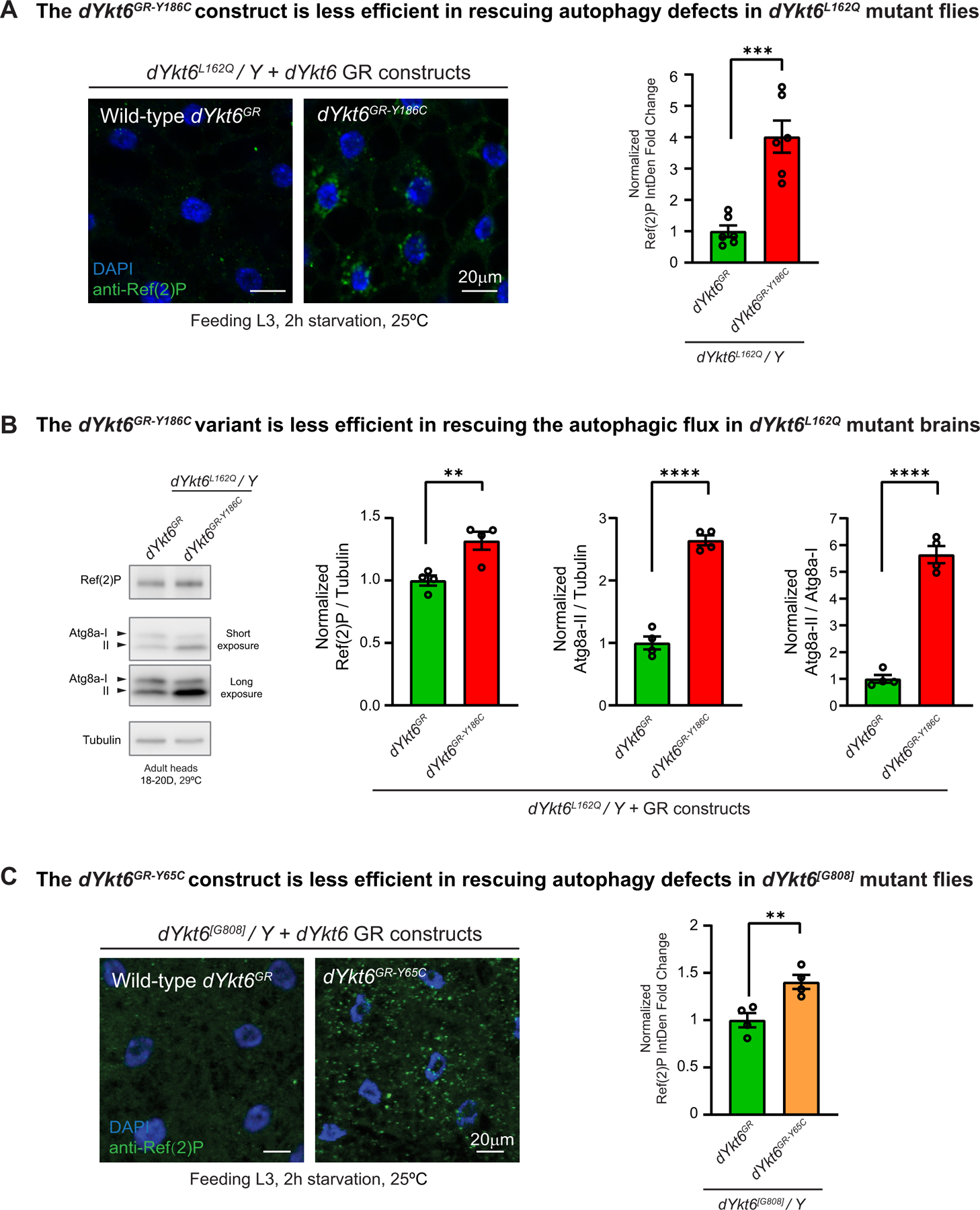
Methods: We report 3 unrelated individuals with rare homozygous missense variants in YKT6 who exhibited neurological disease with or without a progressive infantile liver disease. We modeled the variants in Drosophila. We generated wild-type and variant genomic rescue constructs of the fly ortholog dYkt6 and compared their ability in rescuing the loss-of-function phenotypes in mutant flies. We also generated a dYkt6KozakGAL4 allele to assess the expression pattern of dYkt6.

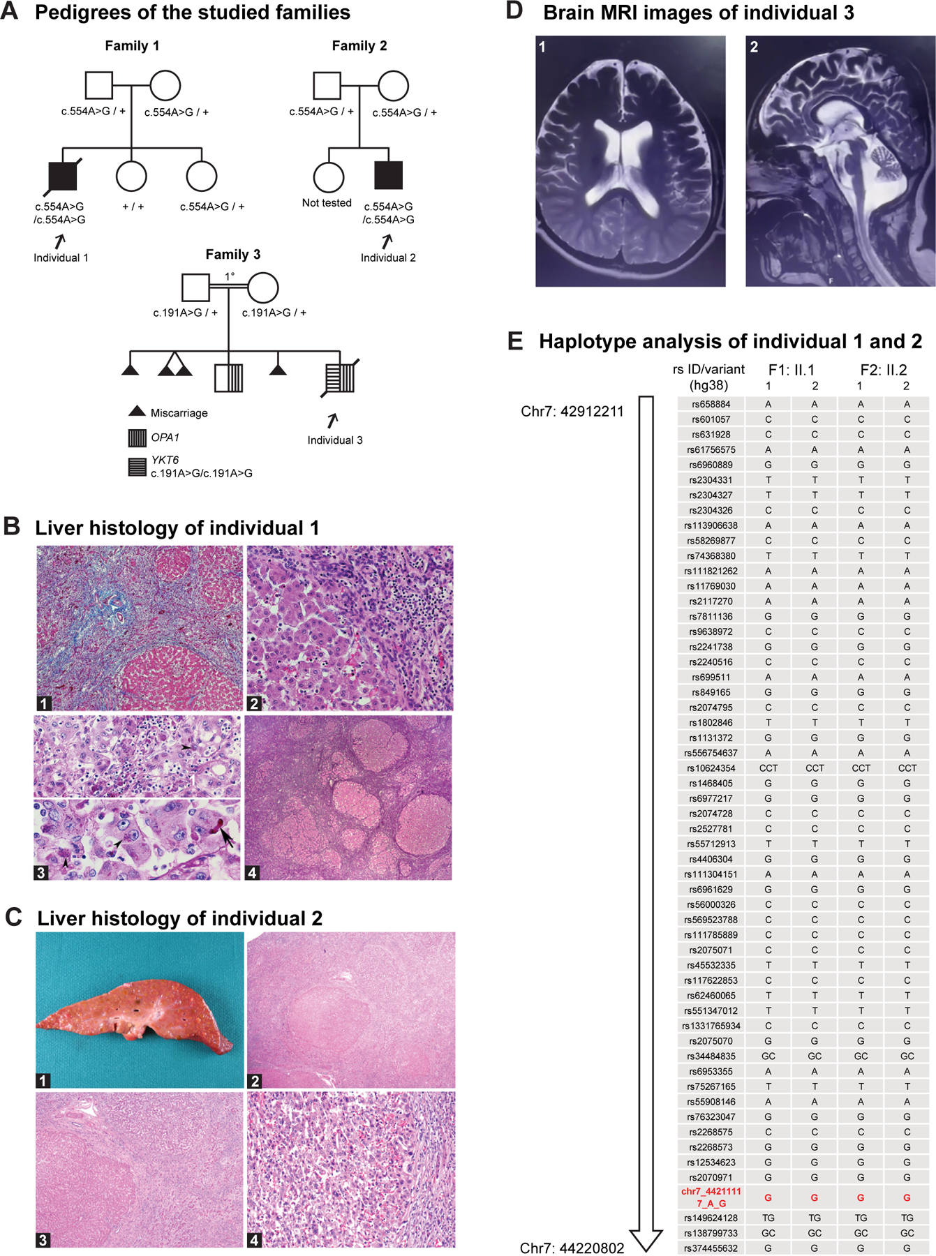
Results: Two individuals are homozygous for YKT6 [NM_006555.3:c.554A>G p.(Tyr185Cys)] and exhibited normal prenatal course followed by failure to thrive, developmental delay, and progressive liver disease. Haplotype analysis identified a shared homozygous region flanking the variant, suggesting a common ancestry. The third individual is homozygous for YKT6 [NM_006555.3:c.191A>G p.(Tyr64Cys)] and exhibited neurodevelopmental disorders and optic atrophy. Fly dYkt6 is essential and is expressed in the fat body (analogous to liver) and central nervous system. Wild-type genomic rescue constructs can rescue the lethality and autophagic flux defects, whereas the variants are less efficient in rescuing the phenotypes.

Conclusion: The YKT6 variants are partial loss-of-function alleles, and the p.(Tyr185Cys) is more severe than p.(Tyr64Cys).

Keywords: Autophagy; Drosophila; Failure to thrive; Fat body; Syrian Christians of India.


Conflict of interest statement

Conflict of Interest BCM and Miraca Holdings have formed a joint venture with shared ownership and governance of BG, which performs clinical microarray analysis, clinical ES (cES), and clinical biochemical studies. James R. Lupski serves on the Scientific Advisory Board of the BG. James R. Lupski has stock ownership in 23andMe, is a paid consultant for Genomics International, and is a coinventor on multiple United States and European patents related to molecular diagnostics for inherited neuropathies, eye diseases, genomic disorders, and bacterial genomic fingerprinting. Nhu Thao Nguyen Galván serves as a consultant for 3DSystems. Davut Pehlivan provides consulting service for Ionis Pharmaceuticals. Wendy K. Chung is on the Board of Directors of Prime Medicine and Rallybio. The Department of Molecular and Human Genetics at Baylor College of Medicine receives revenue from clinical genetic testing conducted at Baylor Genetics Laboratories. All other authors declare no conflicts of interest.

Figures

Figure 1
Figure 1. Three affected individuals have biallelic variants in YKT6.
A. Pedigrees of the studied families. The individuals with homozygous YKT6 variants are affected. Individual 1 and 2 have the c.554A>G p.(Tyr185Cys) variant; individual 3 has the c.191A>G p.(Tyr64Cys) variant. The parents in family 3 are first cousins. B. Liver histology of individual 1. B1. Liver parenchyma shows massive swaths of collapsed parenchyma in which regenerative tubules are intermixed with a mononuclear cell infiltrate and nodules of surviving hepatocytes. H&E stain ×50. B2. Margin of large residual nodule along the zone of collapse shows preserved hepatocytes in which reactive or regressive features are rare. Interface inflammatory activity is lacking. H&E stain ×200. B3. Upper panel: Progressive local loss of hepatocytes within parenchymal nodules is associated with mononuclear inflammatory infiltrate, prominent pigment granules in the cytoplasm of regressing hepatocytes (arrowhead), and aggregates of pigment-laden Kupffer cells. The pigment is interpreted as ceroid. PAS diastase stain ×400. Lower panel: reactive enlarged hepatocytes have prominent nucleoli, occasional canalicular bile plugs (arrow), and focal aggregates of ceroid pigment (arrowheads). PAS diastase stain ×400. B4. Generalized zones of parenchymal collapse and nodular regeneration of residual parenchyma, features of early cirrhosis, coexist with signs of ongoing hepatocyte degeneration and loss. Reticulin stain ×25. C. Liver histology of individual 2. C1. Liver explant 203 g (expected for age: 288 g) cut surface with diffuse, variable sized nodularity ranging in size from less than 0.1 to 1.5 cm. C2. Liver parenchyma with nodules of hepatocytes surrounded by thick fibrous bands without central veins. C3. Focus of hepatocellular carcinoma showing perinodular sclerotic rim H&E stain ×200. C4. Numerous diffuse regenerative and dysplastic nodules. H&E stain ×40. D. Brain MRI of individual 3. D1. Axial T2 MRI image showing enlarged lateral ventricles and extraaxial space increase in the frontotemporal regions. D2. Midsagittal T2 MRI image showing cerebellar atrophy and extraaxial space increase in the frontal regions. E. Analysis of the SNPs on chromosome 7 revealed a shared contiguous stretch of homozygosity (Chr7:42912211–44220802, hg38, 1.3Mb; SNPs n = 100) including YKT6, in the 2 affected individuals from the unrelated families 1 and 2. For each individual, F represents the family, and the 2 alleles are represented as 1 and 2. The YKT6 variant seen in the 2 families is shown in red (Chr7:44211117A>G, hg38).
Figure 2
Figure 2. YKT6 is evolutionally conserved and the fly ortholog is dYkt6.
A. Schematic of human YKT6 protein domains and the position of the variants identified from the affected individuals. Domain annotation is based on the PROSITE database. B. Alignment of YKT6 and the homologous proteins. The Longin domain is in red, the Synaptobrevin domain is in blue, and the CCAIM motif is marked in green. The 2 variants are marked with boxes. The variants affect the residues with conserved amino acid across species. The following isoforms were used for alignment: NP_006546.1 (Human), NP_113880.2 (Rat), NP_062635.2 (Mouse), NP_957386.1 (Zebrafish), NP_572423.1 (Drosophila), and NP_012725.1 (Baker’s yeast). C. Schematic of fly dYkt6 genomic span, transcript, and the reagents, including a P-element insertion allele dYkt6[G808], a point mutation allele dYkt6L162Q, a CRIMIC allele dYkt6KozakGAL4, a 73 kb duplication (Dp) construct, a 5 kb genomic rescue (GR) construct (dYkt6GR), and 2 GR constructs that carry the corresponding variants (dYkt6GR-Y65C and dYkt6GR-Y186C). D. The flies with hemizygous/trans-heterozygous dYkt6 alleles are lethal. The lethality can be rescued by either the Dp or the wild-type GR constructs.
Figure 3
Figure 3. dYkt6 is expressed in fly fat body and CNS, analogs of human liver and CNS.
A. The L3 fat body, larval CNS and adult brain of the dYkt6KozakGAL4 > UAS-CD8::RFP (membrane-targeted RFP labeling the dYkt6 expressing cells in red) flies showing that dYkt6 is expressed in both tissues. Nuclei are labeled by DAPI (blue). Scale bars, 50 μm in the fat body image, 100 μm in the CNS images. B. The larval CNS and adult brain of the dYkt6KozakGAL4 > UAS-mCherry.nls (nuclear-localized mCherry labeling the dYkt6 expressing cells in magenta) flies documenting the expression pattern of dYkt6 in the CNS. The tissues are costained with the pan-neuronal marker Elav or panglia marker Repo (green). Higher magnification images of the regions indicated by dashed rectangles (1–5) are shown. Several z-stack sections were processed. Scale bars, 50 μm in the magnified larval CNS images (1 and 2), 20 μm in the magnified adult brain images (3, 4, and 5), and 100 μm in other images.
Figure 4
Figure 4. dYKT6GR-Y186C is a more severe hypomorphic allele than dYKT6GR-Y65C.
A. The cross strategy of the lethality rescue experiments. Heterozygous female flies carrying dYkt6 mutant alleles were crossed with male transgenic flies with wild-type or variant GR constructs, or with the y w/Y flies (as a negative control). Based on Mendelian ratio, the number of the dYkt6 mutant hemizygotes with GR constructs should be one-quarter of the total number of progeny flies. B. Graph showing the observed/expected percentage of the progeny flies. The dYkt6GR-Y186C construct is significantly less efficient than the wilt-type dYkt6GR in rescuing the lethality caused by the dYkt6L162Q (left panel) or the dYkt6[G808] (right panel) allele. The dYkt6GR-Y65C construct can rescue the lethality caused by the dYkt6L162Q allele (left panel) but only partially rescue the lethality caused by the dYkt6[G808] allele (right panel). The flies were raised at 25 °C. Each dot in the graph represents 1 independent cross. One-way ANOVA with Tukey’s multiple comparisons test, *P < .05, ****P < .0001, mean ± SEM. C. Climbing assay of dYkt6L162Q mutant flies rescued by dYkt6GR or dYkt6GR-Y186C. The climbing ability of the mutant flies rescued by dYkt6GR-Y186C is significantly poorer than the ones rescued by wild-type dYkt6GR. The flies were kept at 29 °C and were tested 3 and 10 days after eclosion. Each dot represents 1 tested fly. Unpaired t test, ***P < .001, ****P < .0001, mean ± SEM.
Figure 5
Figure 5. The YKT6 variants identified from the affected individuals are associated with autophagic flux defects.
A. Fat body cells of the starved L3 larvae (feeding stage) were stained with the autophagic cargo adaptor Ref(2)P (green). The dYkt6L162Q mutant hemizygotes with wild-type dYkt6GR reduce the Ref(2)P level, whereas the ones with dYkt6GR-Y186C still show Ref(2)P accumulation. Nuclei are labeled by DAPI (blue). Scale bars, 20 μm. For the quantification, each dot represents 1 image as shown on the left. The average integrated density was calculated for 5–7 cells randomly selected in each image. At least 3 animals were dissected for imaging for each genotype. Unpaired t test, ***P < .001, mean ± SEM. B. Immunoblots of anti-Ref(2)P and anti-Atg8a of protein lysates from adult heads with quantification of Ref(2)P/Tubulin ratio, Atg8a-II/Tubulin ratio, and Atg8a-II/Atg8a-I ratio. Different parts of the blotting membranes were incubated with Ref(2)P or Atg8a antibodies. Subsequently, the membranes were stripped and incubated with Tubulin antibody. Low-exposure images were used for quantification. The eclosed flies were kept at 29 °C, and the heads were collected 18 to 20 days after eclosion. Each dot represents 1 biologically independent samples. Unpaired t test, **P < .01, ****P < .0001, mean ± SEM. C. Fat body cells of the starved L3 larvae (feeding stage) were stained with Ref(2)P (green). The dYkt6[G808] mutant hemizygotes with wild-type dYkt6GR reduce the Ref(2)P level, whereas the ones with dYkt6GR-Y65C cannot fully rescue the Ref(2)P accumulation phenotype. Nuclei are labeled by DAPI (blue). Scale bars, 20 μm. For the quantification, each dot represents 1 image. The average integrated density was calculated for 5 to 7 cells randomly selected in each image. At least 3 animals were dissected for imaging for each genotype. Unpaired t test, **P < .01, mean ± SEM.

Similar articles

References

    1. Jahn R, Scheller RH. SNAREs–engines for membrane fusion. Nat Rev Mol Cell Biol 2006;7(9):631–643. 10.1038/nrm2002 - DOI - PubMed
    1. Söllner T, Whiteheart SW, Brunner M, et al. SNAP receptors implicated in vesicle targeting and fusion. Nature. 1993;362(6418):318–324. 10.1038/362318a0 - DOI - PubMed
    1. Tochio H, Tsui MM, Banfield DK, Zhang M. An autoinhibitory mechanism for nonsyntaxin SNARE proteins revealed by the structure of Ykt6p. Science. 2001;293(5530):698–702. 10.1126/science.1062950 - DOI - PubMed
    1. Bas L, Papinski D, Licheva M, et al. Reconstitution reveals Ykt6 as the autophagosomal SNARE in autophagosome-vacuole fusion. J Cell Biol 2018;217(10):3656–3669. 10.1083/jcb.201804028 - DOI - PMC - PubMed
    1. Gao J, Reggiori F, Ungermann C. A novel in vitro assay reveals SNARE topology and the role of Ykt6 in autophagosome fusion with vacuoles. J Cell Biol 2018;217(10):3670–3682. 10.1083/jcb.201804039 - DOI - PMC - PubMed

MeSH terms

LinkOut - more resources