. 2024 Aug;24(8):1382-1394.
doi: 10.1016/j.ajt.2024.03.028. Epub 2024 Mar 24.

Nanoparticle targeting of neutrophil glycolysis prevents lung ischemia-reperfusion injury

Affiliations

Nanoparticle targeting of neutrophil glycolysis prevents lung ischemia-reperfusion injury

Fuyi Liao et al. Am J Transplant. 2024 Aug.

Abstract

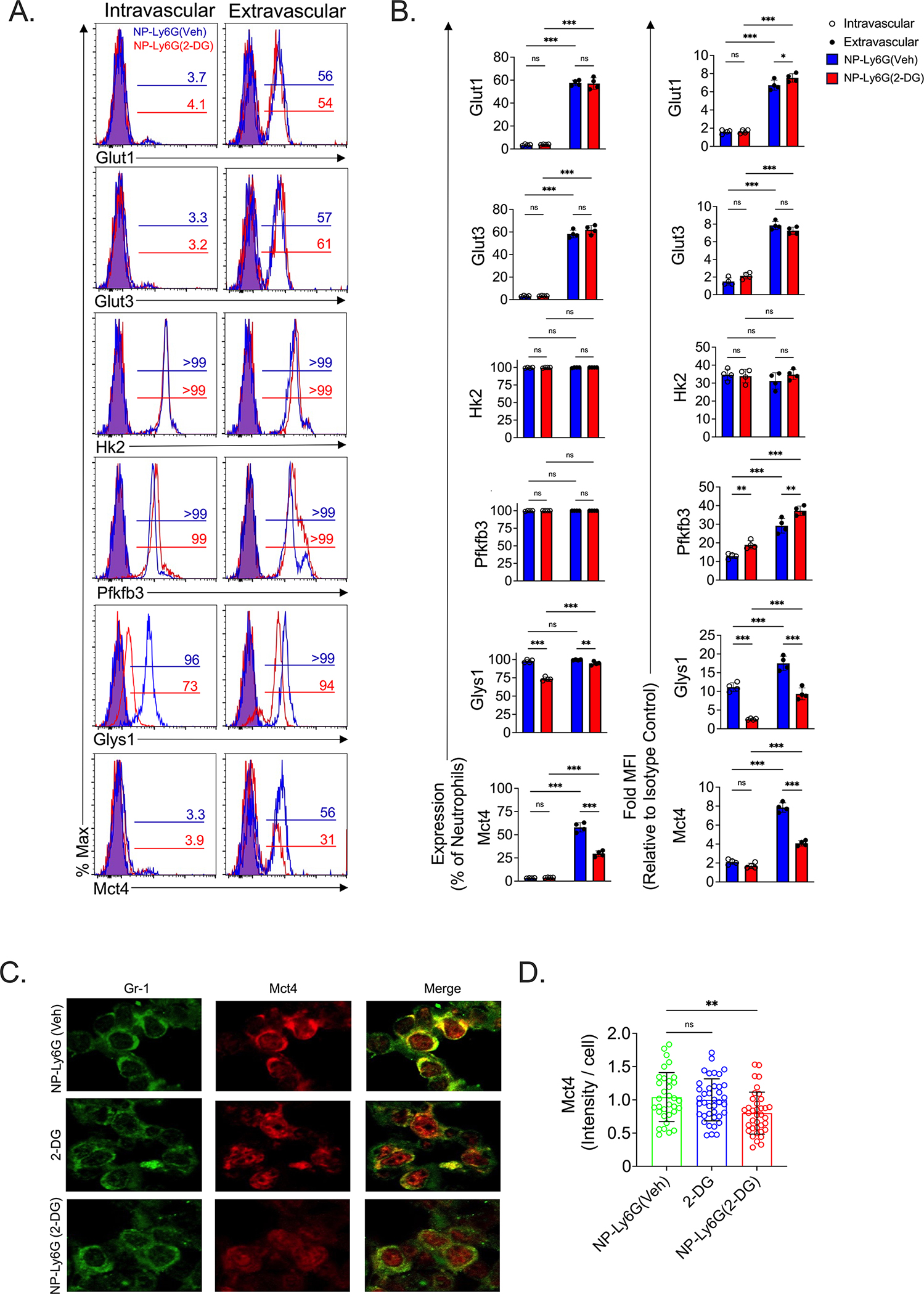
Neutrophils exacerbate pulmonary ischemia-reperfusion injury (IRI) resulting in poor short and long-term outcomes for lung transplant recipients. Glycolysis powers neutrophil activation, but it remains unclear if neutrophil-specific targeting of this pathway will inhibit IRI. Lipid nanoparticles containing the glycolysis flux inhibitor 2-deoxyglucose (2-DG) were conjugated to neutrophil-specific Ly6G antibodies (NP-Ly6G[2-DG]). Intravenously administered NP-Ly6G(2-DG) to mice exhibited high specificity for circulating neutrophils. NP-Ly6G(2-DG)-treated neutrophils were unable to adapt to hypoglycemic conditions of the lung airspace environment as evident by the loss of demand-induced glycolysis, reductions in glycogen and ATP content, and an increased vulnerability to apoptosis. NP-Ly6G(2-DG) treatment inhibited pulmonary IRI following hilar occlusion and orthotopic lung transplantation. IRI protection was associated with less airspace neutrophil extracellular trap generation, reduced intragraft neutrophilia, and enhanced alveolar macrophage efferocytotic clearance of neutrophils. Collectively, our data show that pharmacologically targeting glycolysis in neutrophils inhibits their activation and survival leading to reduced pulmonary IRI.

Keywords: acute lung injury and immunosuppression; efferocytosis; glycolysis; immunometabolism; ischemia-reperfusion injury; lung transplant; nanoparticles; neutrophils; primary graft dysfunction.


Conflict of interest statement

Declaration of competing interest The authors of this manuscript have no conflicts of interest to disclose as described by the American Journal of Transplantation.

Figures

Figure 1:
Figure 1:. Development and characterization of Ly6G antigen-targeting nanoparticles.
(A) Diagram of NP-Ly6G(2-DG) and NP-Ly6G(Veh). (B) summary of the characterization parameters of NP-Ly6G(2-DG) and NP-Ly6G(vehicle including size, z – Potential (mV), polydiversity index (PDI), encapsulation efficiency (EE). Data show mean ± standard deviation (SD).
Figure 2:
Figure 2:. NP-Ly6G(2-DG) specifically targets neutrophils.
(A-B) NP-Ly6G(2-DG) and NP-Ly6G(Veh)-treated mice analyzed for nanoparticle uptake by circulating neutrophils at 2 hours after administration. Neutrophil uptake of nanoparticles at indicated doses (lipid mass per kg of body weight) of NP-Ly6G(2-DG) (solid lines) and NP-Ly6G(Veh) (broken lines) shown by a (A) representative histogram and (B) scatter plot (N=4/treatment). B6 mice received 63 mg per kg of nanoparticles and two hours later analyzed for (C) percent uptake by indicated circulating leukocyte subsets and (D) organs through assessing mean fluorescence intensity (MFI) (N=4/treatment). (E) Percent abundance of neutrophils within the circulating CD45+ compartment 2-hours after administration of nanoparticles (N=4/treatment). Scatter plots and bar graphs show mean ± SD.
Figure 3:
Figure 3:. NP-Ly6G(2-DG) prevents neutrophil adaptation to hypoglycemic stress.
(A-B) Seahorse extracellular flux analysis of circulating neutrophils. (A) ECAR plot, following injection of LPS (100ng/ml), glucose (10 mM), oligomycin (1.0 mM) and 2-DG (50 mM), and (B) bar graphs of indicated glycolysis parameters. (C) Glycolytic flux of 5-3H glucose and (D) glycogen content following 4-hour neutrophil culture in medium supplemented with 10 mM or 0.2 mM glucose in the presence or absence of LPS (100ng/ml). (E) Neutrophil energy status as measured by ADP to ATP ratio after 4 hours of culture and (F) apoptosis as defined by Annexin V+ 7-AAD staining after 18 hours of culture in medium supplemented with 0.2 mM glucose with or without LPS. Data shown in (A-F) are representative results from at least three independent experiments. (B-F) are bar graphs showing means ± SD with p values obtained from (B) one-way ANOVA with a Tukey’s comparison test and (C-F) ordinary two-way ANOVA with a Uncorrected Fisher’s LSD test; *p<0.05, **p<0.01, ***p<0.001, ****p<0.0001, ns; non-significant
Figure 4:
Figure 4:. NP-Ly6G(2-DG) alters glycolysis related gene expression in lung infiltrating neutrophils following ischemia-reperfusion injury.
B6 mice were treated with NP-Ly6G(Veh) or Ly6G(2-DG) and underwent hilar occlusion for 90 minutes, allowed to reperfuse for 30 minutes, and i.v. injected with Gr1 antibodies to distinguish between pulmonary DiD+ intravascular and extravascular neutrophils five minutes prior to the isolation of lung tissue. (A) Overlaid histograms show pulmonary intravascular and extravascular neutrophil percent expression of indicated proteins with isotype antibody controls represented by filled histograms for NP-Ly6G(Veh) (blue) and NP-Ly6G(2-DG) (red) and protein specific antibody staining shown with open histograms for NP-Ly6G(Veh) (blue) and NP-Ly6G(2-DG) (red). Data shown are a representative result from N=4/treatment. (B) Bar graphs of neutrophil (left) percent expression and (right) fold increase in mean fluorescence intensity (MFI) relative to isotype control antibody staining for indicated proteins (N=4/treatment). (D) Mct4 immunofluorescence staining and (E) intensity of staining per neutrophil. Data shown is from a representative experiment (N=3/treatment). (B, D) data show means ± SD where p values were obtained from (B) an ordinary two-way ANOVA with an uncorrected Fisher’s LSD test and (D) a one-way ANOVA with a Tukey’s comparisons test; *p<0.05, **p<0.01, ***p<0.001, ns; non-significant
Figure 5:
Figure 5:. NP-Ly6G(2-DG) limits neutrophil activation.
Circulating neutrophils isolated from untreated, 2-DG-, NP-Ly6G(Veh)- and NP-Ly6G(2-DG)-treated mice were (A, B) cultured in the presence or absence of LPS for 3 hours and analyzed for ROS and CD11b expression, (C, D) assessed for phagocytosis of E. Coli beads conjugated to the lysosomal indicator pHrodo, (E, F) or stimulated with 0.2 mM PMA to measure the rate of NET production via PicoGreen detection of extracellular double-stranded (ds) DNA. (G) NETosis measured by ELISA after four hours of stimulation with PMA. (A) Histograms, (C) contour plots and (E) photomicrographs shown are from a representative experiment (N=4/treatment). (B, D, F,G) Bar graphs show means ± SD where p values were obtained from one-way ANOVA with a Tukey’s comparison test ;*p<0.05, **p<0.01, ****p<0.0001, ns; non-significant.
Figure 6:
Figure 6:. NP-Ly6G(2-DG) prevents lung IRI in a hilar occlusion model.
B6 mice were left untreated or received 2-DG, NP-Ly6G(Veh) or NP-Ly6G(2-DG) and assessed for signs of hilar occlusion-induced left lung injury and intrapulmonary neutrophil activity two hours after reperfusion. (A) Representative hematoxylin and eosin staining of lung tissue and (B) blinded lung injury scoring (N=5/treatment). BAL analyzed for (C) IgM, (D) albumin, (E) neutrophil counts and (F) NETs. Bar graphs shown in (B-F) represent means ± SD (N≥5/ treatment). Analysis of AM efferocytosis of neutrophils from NP-Ly6G(Veh) and NP-Ly6G(2-DG) treated mice where (G) are contour plots showing gating strategy from a representative experiment, (H) abundance of DiD+ AM with mean ± SD (N=5/treatment) and (I) a heat map depicting differences in LPS-mediated AM inflammatory cytokine transcript accumulation following normalization to saline-treated resting B6 mice AM transcripts (N=5/treatment). Transendothelial migration assessed one hour after reperfusion and 5 minutes after injection of Gr1 antibodies where (J) is a contour plot from a representative experiment (N=4/treatment) and (K) is a bar graph showing the mean ratio ± SD of intravascular versus extravascular neutrophils. (B-F) P values were obtained from one-way ANOVA with a Tukey’s comparisons test and (H,I,K) unpaired t-test with Welch’s correction; *p<0.05, **p<0.01, ***p<0.001, ****p<0.0001, ns; non-significant.
Figure 7:
Figure 7:. Summary of the major effects of NP-Ly6(2-DG) treatment on energy production and neutrophil activation.
The injection of NP-Ly6G(2-DG) into the bloodstream results in specific uptake by circulating neutrophils. 2-DG initially acts through Hexokinase 2 to inhibit glycolysis but high glucose concentrations in the blood are sufficient to overcome decrements in glycolytic flux thereby allowing neutrophils to sustain critical ATP and glycogen levels. However, activated neutrophils treated with NP-Ly6G(2-DG) that are recruited to airspaces are substantially more vulnerable to 2-DG due to the hypoglycemic environment. This leads to an inability to maintain sufficient ATP levels and glycogen reserves to power effector functions that promote IRI. Furthermore, hypoglycemic stress induces activated neutrophils to undergo apoptosis which helps resolve acute inflammation through efferocytotic clearance of their corpses by alveolar macrophages.
Figure 8:
Figure 8:. NP-Ly6G(2-DG) reduces lung transplant-mediated IRI.
B6 donors underwent cadaveric warm ischemia for 90 minutes followed by prolonged cold preservation on ice for 18 prior to transplantation into syngeneic B6 recipients that were left untreated or received NP-Ly6G(Veh) or NP-Ly6G(2-DG) 2-hours prior to engraftment. Then two hours after transplantation lung grafts were assessed for (A) arterial blood gasses (PaO2) on a FiO2 of 1.0, (B) Wet to dry weight ratio, (C) blinded lung injury scoring and (D) hematoxylin and eosin staining of graft tissue. (A, B) Bar graphs show mean ratio ± SD and (C) depicts graft histology from representative transplants (N=4/treatment). (A, B) P values were obtained from one-way ANOVA with a Tukey’s comparisons test; *p<0.05, ***p<0.001, ns; non-significant.

References

    1. Gelman AE, Fisher AJ, Huang HJ, et al. Report of the ISHLT Working Group on Primary Lung Graft Dysfunction Part III: Mechanisms: A 2016 Consensus Group Statement of the International Society for Heart and Lung Transplantation. J Heart Lung Transplant. Oct 2017;36(10):1114–1120. doi: 10.1016/j.healun.2017.07.014 - DOI - PMC - PubMed
    1. Scozzi D, Liao F, Krupnick AS, Kreisel D, Gelman AE. The role of neutrophil extracellular traps in acute lung injury. Front Immunol. 2022;13:953195. doi: 10.3389/fimmu.2022.953195 - DOI - PMC - PubMed
    1. Li W, Terada Y, Tyurina YY, et al. Necroptosis triggers spatially restricted neutrophil-mediated vascular damage during lung ischemia reperfusion injury. Proc Natl Acad Sci U S A. Mar 8 2022;119(10):e2111537119. doi: 10.1073/pnas.2111537119 - DOI - PMC - PubMed
    1. Seekamp A, Mulligan MS, Till GO, et al. Role of beta 2 integrins and ICAM-1 in lung injury following ischemia-reperfusion of rat hind limbs. Am J Pathol. Aug 1993;143(2):464–72. - PMC - PubMed
    1. Scozzi D, Cano M, Ma L, et al. Circulating Mitochondrial DNA is an Early Indicator of Severe Illness and Mortality from COVID-19. bioRxiv. Jul 30 2020;doi: 10.1101/2020.07.30.227553 - DOI - PMC - PubMed

LinkOut - more resources