

Superficial Primary Malignant Melanoma of the Esophagus Detected and Treated at Stage 0
- PMID: 38522910
- PMCID: PMC11637794
- DOI: 10.2169/internalmedicine.2454-23
Superficial Primary Malignant Melanoma of the Esophagus Detected and Treated at Stage 0
Abstract
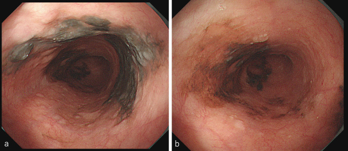
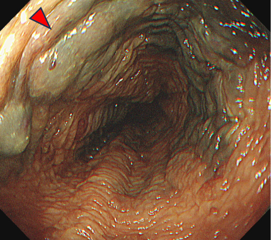
The patient was a 79-year-old male. At three years and eight months after his initial presentation, upper gastrointestinal endoscopy revealed a black-flattened elevated lesion in the middle third of the esophagus, which was diagnosed as malignant melanoma on biopsy. No lymph node or distant metastasis was found. A diagnosis of cT1bN0M0 Stage I was thus made. We performed a robot-assisted, minimally invasive esophagectomy and D2 dissection. The postoperative diagnosis was pT1a-MM, N0, M0, vascular invasion+, stage 0. The patient was recurrence-free for 14 months after surgery. We presume that an aggressive biopsy diagnosis is important for the early detection of malignant melanoma.
Keywords: Stage 0; esophageal malignant melanoma; esophageal melanosis; primary malignant melanoma.
Conflict of interest statement
The authors state that they have no Conflict of Interest (COI).
Figures


Similar articles
-
Diagnosis and surgical outcomes for primary malignant melanoma of the esophagus: a single-center experience.Ann Thorac Surg. 2013 Sep;96(3):1002-6. doi: 10.1016/j.athoracsur.2013.04.072. Epub 2013 Jun 25. Ann Thorac Surg. 2013. PMID: 23810175
-
[Primary malignant melanoma of the esophagus].Zentralbl Chir. 1998;123(3):276-9. Zentralbl Chir. 1998. PMID: 9586189 German.
-
[A case of superficial primary malignant melanoma of the esophagus].Gan To Kagaku Ryoho. 2013 Nov;40(12):2109-11. Gan To Kagaku Ryoho. 2013. PMID: 24394029 Japanese.
-
Curatively resected primary malignant melanoma of the esophagus: report of a case.Surg Today. 1993;23(9):820-4. doi: 10.1007/BF00311627. Surg Today. 1993. PMID: 8219617 Review.
-
A Long Surviving Case of Multiple Early Stage Primary Malignant Melanoma of the Esophagus and a Review of the Literature.Tokai J Exp Clin Med. 2015 Sep 20;40(3):90-5. Tokai J Exp Clin Med. 2015. PMID: 26369261 Review.
References
-
- Volpin E, Sauvanet A, Couvelard A, Belghiti J. Primary malignant melanoma of the esophagus: a case report and review of the literature. Dis Esophagus 15: 244-249, 2002. - PubMed
-
- Yamaguchi T, Shioaki Y, Koide K, et al. . A case of primary malignant melanoma of the esophagus and analysis of 193 patients in Japan. Nihon Shokakibyo Gakkai Zasshi (J Jpn Soc Gastroenterol) 101: 1087-1094, 2004. (in Japanese). - PubMed