Review
. 2024 Mar 11;11(3):917-940.
doi: 10.1021/acsphotonics.4c00064. eCollection 2024 Mar 20.

Shining Light in Mechanobiology: Optical Tweezers, Scissors, and Beyond

Affiliations
Review

Shining Light in Mechanobiology: Optical Tweezers, Scissors, and Beyond

Alexander B Stilgoe et al. ACS Photonics. .

Abstract

Mechanobiology helps us to decipher cell and tissue functions by looking at changes in their mechanical properties that contribute to development, cell differentiation, physiology, and disease. Mechanobiology sits at the interface of biology, physics and engineering. One of the key technologies that enables characterization of properties of cells and tissue is microscopy. Combining microscopy with other quantitative measurement techniques such as optical tweezers and scissors, gives a very powerful tool for unraveling the intricacies of mechanobiology enabling measurement of forces, torques and displacements at play. We review the field of some light based studies of mechanobiology and optical detection of signal transduction ranging from optical micromanipulation-optical tweezers and scissors, advanced fluorescence techniques and optogenentics. In the current perspective paper, we concentrate our efforts on elucidating interesting measurements of forces, torques, positions, viscoelastic properties, and optogenetics inside and outside a cell attained when using structured light in combination with optical tweezers and scissors. We give perspective on the field concentrating on the use of structured light in imaging in combination with tweezers and scissors pointing out how novel developments in quantum imaging in combination with tweezers and scissors can bring to this fast growing field.


Conflict of interest statement

The authors declare no competing financial interest.

Figures
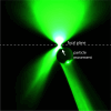
Figure 1
Figure 1
Optical tweezers create an optical trap using the deflection and scattering of light by transparent glasses and plastics. In this visualization, light is focused to a point from the top of the image, a small glass particle deflects the light to the right. It correspondingly receives a force moving it to the left by conservation of the momentum of light. The light is more focused after refraction through the particle and so it also receives a force moving it up toward the focus.
Figure 2
Figure 2
Optical force (dimensionless units) calculated for a 1 μm radius polystyrene particle trapped with NA = 1.2 microscope objective in water using the Optical Tweezers Toolbox software package. The equilibrium for the particle is at the center of the beam.
Figure 3
Figure 3
A particle moving into an optical trap from a large distance follows a characteristic exponential approach to the stable trap position in the absence of Brownian motion. The characteristic motion is that of an overdamped harmonic oscillator, with a characteristic time scale represented by the green bar. Over this time, one could expect the particle to move from one side of the optical trap to the other as the trap’s force response is weak due to frequency band limits. The exponential approach behavior is hidden by Brownian motion near the equilibrium. The shaded region represents a ∼95% confidence interval of where the particle can be found in an exponential approach model centered at the exact starting position.
Figure 4
Figure 4
Visualization of setup for multipoint trapping and rapid scanning ablation of biological samples, modeled on the system used in Ono et al. The system uses a continuous wave infrared laser for trapping that can trap objects at multiple points with light shaped by an SLM (see section Spatial Light Modulators (SLMs)). A femtosecond pulsed Ti:sapphire laser is used for the ablation/microsurgery of cells. A fast scan mirror allows the high-intensity pulsed beam to be precisely positioned within the sample.
Figure 5
Figure 5
Anaphase intertelomeric tethers result in the pulling of chromosome fragments toward sister chromatids. (A) A red line indicates the region targeted by the laser and an arrow indicates the direction that the cut chromosome will move and the tip of the sister chromatid. Scale bar = 5 microns. (B) 20 s after irradiation the chromosome fragment has moved toward its sister. (C) After 45 s the fragment has reached its partner.
Figure 6
Figure 6
DMDs consist of arrays of tilting mirrors: (a) DMD micromirror device; (b) individual micromirrors showing actuation mechanism; (c) deflection electrodes are addressed by a CMOS chip enabling electrostatic attraction of the mirror and tilting of incident light; and (d) tilting multiple cells at once contribute to especially varying optical flux.
Figure 7
Figure 7
Two common modes of projection used by SLMs and DMDs. (a) Projection of modulator (the object plane) to the imaging plane. (b) Projection from the object to conjugate image plane (a.k.a. Fourier projection).
Figure 8
Figure 8
Illustration of light targeting and optical manipulation of neurons through opsins and laser light.
Figure 9
Figure 9
Optical manipulation of ear stones in zebrafish inner ear. Left: Top view of 6 days post fertilized zebrafish. Blue outline delimits the inner ear. Blue arrows show ear stones. Right: Example of distribution of regions of interests (ROIs, blue dots) involved in auditory processing. Brain is outlined in color.
Figure 10
Figure 10
Intracellular microrheometry using rotational optical tweezers. A macrophage macropinocytoses birefringent microspheres which are trapped and rotated with circularly polarized light. The rotation rate increases with torque inversely proportional to the viscosity. Scale bar is 5 μm. Microscopy and data is from Watson et al.

References

    1. Ingber D. E.; Di Carlo D. Interdisciplinarity and mechanobiology. iScience 2022, 25, 10418710.1016/j.isci.2022.104187. - DOI - PMC - PubMed
    1. Lim C. T.; Bershadsky A.; Sheetz M. P. Mechanobiology. Journal of The Royal Society Interface 2010, 7, S291–S293. 10.1098/rsif.2010.0150.focus. - DOI - PMC - PubMed
    1. Iskratsch T.; Wolfenson H.; Sheetz M. P. Appreciating force and shape—the rise of mechanotransduction in cell biology. Nat. Rev. Mol. Cell Biol. 2014, 15, 825–833. 10.1038/nrm3903. - DOI - PMC - PubMed
    1. Brown S.; Levinson W.; Spudich J. A. Cytoskeletal elements of chick embryo fibroblasts revealed by detergent extraction. Journal of Supramolecular Structure 1976, 5, 119–130. 10.1002/jss.400050203. - DOI - PubMed
    1. Wang N.; Butler J. P.; Ingber D. E. Mechanotransduction Across the Cell Surface and Through the Cytoskeleton. Science 1993, 260, 1124–1127. 10.1126/science.7684161. - DOI - PubMed