

Artificial Intelligence in Dermoscopy: Enhancing Diagnosis to Distinguish Benign and Malignant Skin Lesions
- PMID: 38523958
- PMCID: PMC10959827
- DOI: 10.7759/cureus.54656
Artificial Intelligence in Dermoscopy: Enhancing Diagnosis to Distinguish Benign and Malignant Skin Lesions
Abstract
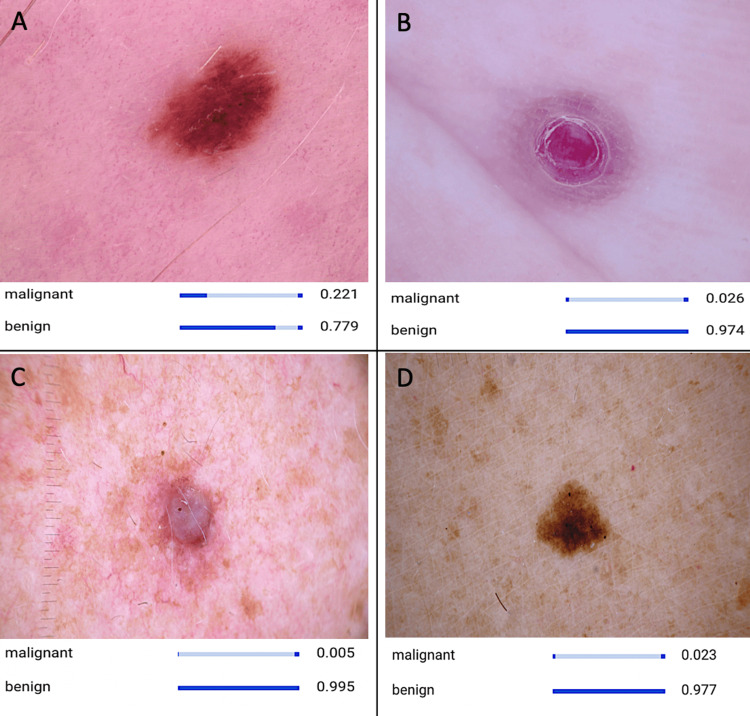
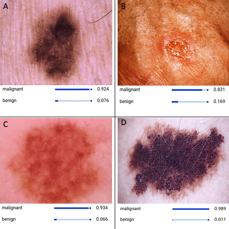
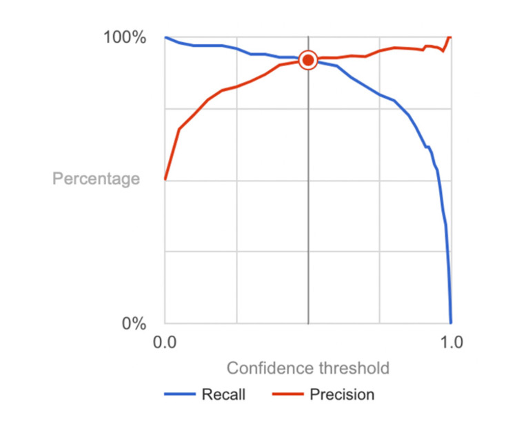
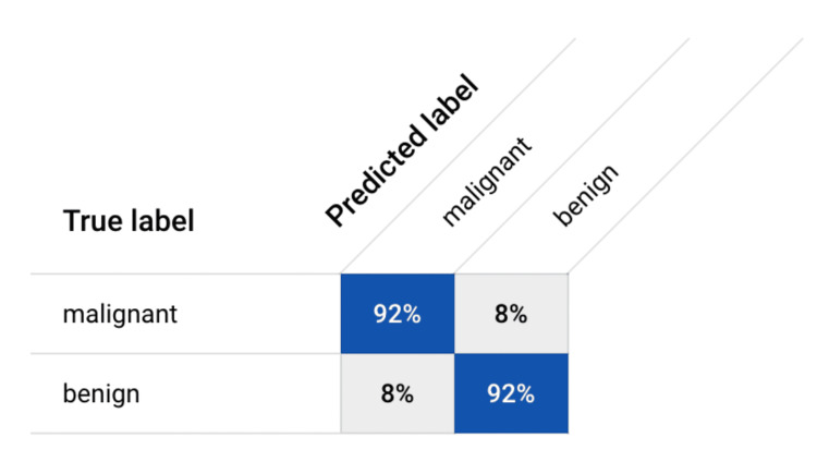
This study presents an innovative application of artificial intelligence (AI) in distinguishing dermoscopy images depicting individuals with benign and malignant skin lesions. Leveraging the collaborative capabilities of Google's platform, the developed model exhibits remarkable efficiency in achieving accurate diagnoses. The model underwent training for a mere one hour and 33 minutes, utilizing Google's servers to render the process both cost-free and carbon-neutral. Utilizing a dataset representative of both benign and malignant cases, the AI model demonstrated commendable performance metrics. Notably, the model achieved an overall accuracy, precision, recall (sensitivity), specificity, and F1 score of 92%. These metrics underscore the model's proficiency in distinguishing between benign and malignant skin lesions. The use of Google's Collaboration platform not only expedited the training process but also exemplified a cost-effective and environmentally sustainable approach. While these findings highlight the potential of AI in dermatopathology, it is crucial to recognize the inherent limitations, including dataset representativity and variations in real-world clinical scenarios. This study contributes to the evolving landscape of AI applications in dermatologic diagnostics, showcasing a promising tool for accurate lesion classification. Further research and validation studies are recommended to enhance the model's robustness and facilitate its integration into clinical practice.
Keywords: artificial intelligence (ai); benign lesions; dermoscopy image analysis; malignant lesions; skin lesions.
Copyright © 2024, Reddy et al.
Conflict of interest statement
The authors have declared that no competing interests exist.
Figures
Similar articles
-
Leveraging code-free deep learning for pill recognition in clinical settings: A multicenter, real-world study of performance across multiple platforms.Artif Intell Med. 2024 Apr;150:102844. doi: 10.1016/j.artmed.2024.102844. Epub 2024 Mar 13. Artif Intell Med. 2024. PMID: 38553153
-
Leveraging Machine Learning for Accurate Detection and Diagnosis of Melanoma and Nevi: An Interdisciplinary Study in Dermatology.Cureus. 2023 Aug 25;15(8):e44120. doi: 10.7759/cureus.44120. eCollection 2023 Aug. Cureus. 2023. PMID: 37750114 Free PMC article.
-
Validation of artificial intelligence prediction models for skin cancer diagnosis using dermoscopy images: the 2019 International Skin Imaging Collaboration Grand Challenge.Lancet Digit Health. 2022 May;4(5):e330-e339. doi: 10.1016/S2589-7500(22)00021-8. Lancet Digit Health. 2022. PMID: 35461690 Free PMC article.
-
Artificial intelligence-based image classification methods for diagnosis of skin cancer: Challenges and opportunities.Comput Biol Med. 2020 Dec;127:104065. doi: 10.1016/j.compbiomed.2020.104065. Epub 2020 Oct 27. Comput Biol Med. 2020. PMID: 33246265 Free PMC article. Review.
-
Artificial Intelligence for the Classification of Pigmented Skin Lesions in Populations with Skin of Color: A Systematic Review.Dermatology. 2023;239(4):499-513. doi: 10.1159/000530225. Epub 2023 Mar 21. Dermatology. 2023. PMID: 36944317 Free PMC article.
Cited by
-
Artificial Intelligence-Based Distinction of Actinic Keratosis and Seborrheic Keratosis.Cureus. 2024 Apr 21;16(4):e58692. doi: 10.7759/cureus.58692. eCollection 2024 Apr. Cureus. 2024. PMID: 38774175 Free PMC article.
-
Mini review on skin biopsy: traditional and modern techniques.Front Med (Lausanne). 2025 Mar 5;12:1476685. doi: 10.3389/fmed.2025.1476685. eCollection 2025. Front Med (Lausanne). 2025. PMID: 40109731 Free PMC article. Review.
References
-
- “Skin Cancer.” American Academy of Dermatology, American Academy of Dermatology Association. [ Jan; 2024 ]. 2024. http://www.aad.org/media/stats-skin-cancer http://www.aad.org/media/stats-skin-cancer
-
- Epidemiological and clinical analysis of exposure-related factors in non-melanoma skin cancer: A retrospective cohort study. Artosi F, Costanza G, Di Prete M, et al. Environ Res. 2024;247:118117. - PubMed
-
- “Skin Cancer Facts & Statistics.” The Skin Cancer Foundation, Skin Cancer Foundation. Skin Cancer Foundation. [ Jan; 2024 ]. 2024. http://www.skincancer.org/skin-cancer-information/skin-cancer-facts/#:~:... http://www.skincancer.org/skin-cancer-information/skin-cancer-facts/#:~:...
LinkOut - more resources
Full Text Sources