Review
. 2024 Feb 27;9(11):12515-12538.
doi: 10.1021/acsomega.3c09866. eCollection 2024 Mar 19.

Prokaryotic DNA Crossroads: Holliday Junction Formation and Resolution

Affiliations
Review

Prokaryotic DNA Crossroads: Holliday Junction Formation and Resolution

Astha Nautiyal et al. ACS Omega. .

Abstract

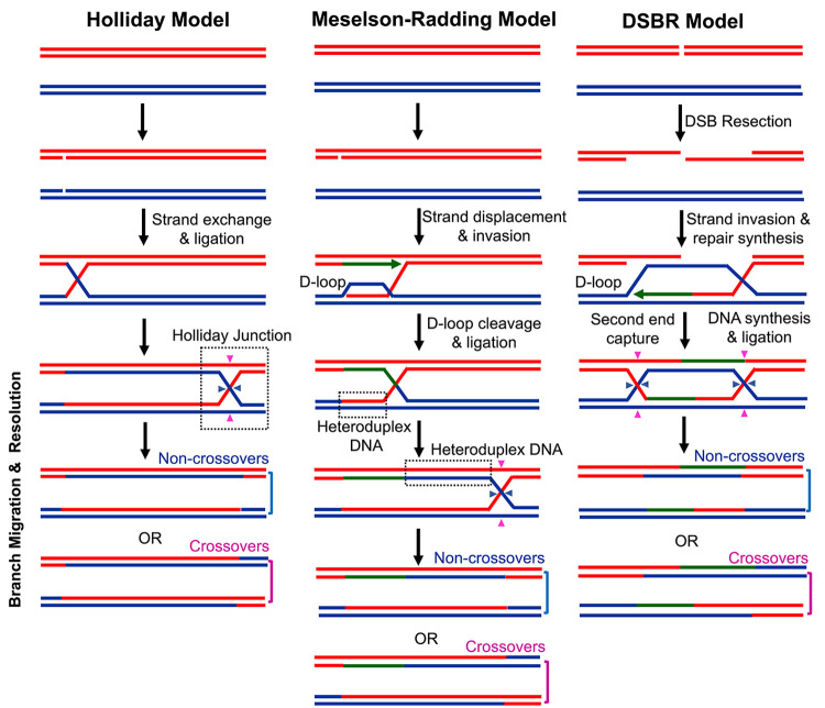
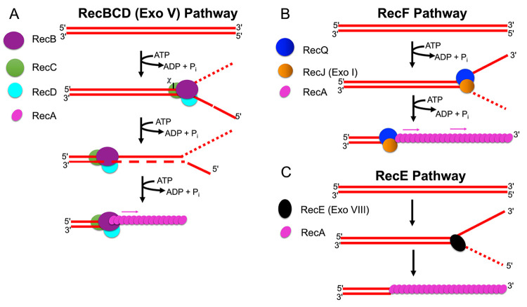
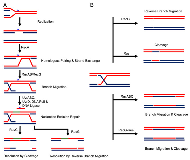
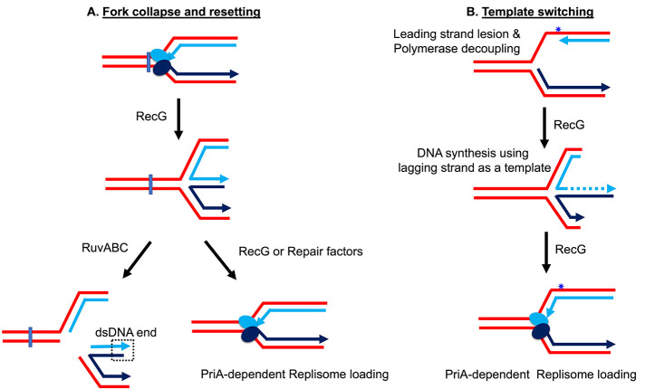
Cells are continually exposed to a multitude of internal and external stressors, which give rise to various types of DNA damage. To protect the integrity of their genetic material, cells are equipped with a repertoire of repair proteins that engage in various repair mechanisms, facilitated by intricate networks of protein-protein and protein-DNA interactions. Among these networks is the homologous recombination (HR) system, a molecular repair mechanism conserved in all three domains of life. On one hand, HR ensures high-fidelity, template-dependent DNA repair, while on the other hand, it results in the generation of combinatorial genetic variations through allelic exchange. Despite substantial progress in understanding this pathway in bacteria, yeast, and humans, several critical questions remain unanswered, including the molecular processes leading to the exchange of DNA segments, the coordination of protein binding, conformational switching during branch migration, and the resolution of Holliday Junctions (HJs). This Review delves into our current understanding of the HR pathway in bacteria, shedding light on the roles played by various proteins or their complexes at different stages of HR. In the first part of this Review, we provide a brief overview of the end resection processes and the strand-exchange reaction, offering a concise depiction of the mechanisms that culminate in the formation of HJs. In the latter half, we expound upon the alternative methods of branch migration and HJ resolution more comprehensively and holistically, considering the historical research timelines. Finally, when we consolidate our knowledge about HR within the broader context of genome replication and the emergence of resistant species, it becomes evident that the HR pathway is indispensable for the survival of bacteria in diverse ecological niches.


Conflict of interest statement

The authors declare no competing financial interest.

Figures

Figure 1
Figure 1
Molecular models of homologous recombination. Holliday, Meselson–Radding, and double strand break repair (DSBR) model. The blue and red lines represent homologous dsDNA. The green lines represent newly synthesized DNA. For more details, refer to main text.
Figure 2
Figure 2
Cartoons showing the three presynaptic pathways for the generation of recombinogenic 3′- ssDNA tails. Each pathway is illustrated here is based on the profound effect of the gene mutations, recBC(D) in combination with sbcA and sbcBC and characterized biochemical properties of the purified protein’s preparations. For details, refer to the text.
Figure 3
Figure 3
Possible pathways for processing HJ in E. coli. A. The replisome encounters a pyrimidine dimer (blue star), DNA synthesis comes to an abrupt halt, and DNA synthesis resumes downstream of the lesion, leaving a gap in the daughter strand. RecA protein polymerizes at the gap and initiates strand exchange with the sister copy. Branch migration by RuvAB or RecG extends the heteroduplex joint beyond the lesion. Nucleotide excision repair to excise the dimer follows either the resolution by RuvC cleavage or by reverse branch migration. New DNA synthesis by DNA polI and DNA ligase-mediated ligation completes the repair process. Figure adapted and modified from ref (122) B. HJ formation by RecA-mediated homologous pairing and strand exchange is shown in the center. With RecA, RuvAB promotes branch migration in the same direction of RecA binding and strand exchange and resolution by RuvC. RecG protein drives the HJ backward but could drive the reaction forward when RecA is released from the junction. HJ resolution by Rus protein. Rus cleavage depended on RecA-catalyzed strand exchange and RecG-mediated branch migration. Although a spliced type of recombinants could arise with equal possibility, a patched type of resolved products is shown here for simplicity.
Figure 4
Figure 4
Models of RecG and RuvABC-mediated replication restart. A. Repair via fork collapse and resetting. B. Lesion bypass via template switching. The sky blue and dark blue ovals represent the leading and lagging strand polymerases of replication fork. The blue rectangle reflects the lesion affecting both leading and lagging strand synthesis. The lesion that affects only the leading strand is shown as a star (in blue color). New DNA synthesis using template from the intact strand synthesis from the polymerase on the lagging strand is shown as a dotted line. Arrowheads depict the 3′-OH groups and dotted square represent the newly formed dsDNA end. Refer to main text for all other descriptions. Figure adapted with permission from ref (138).
Figure 5
Figure 5
The mechanism of HJ resolution by canonical HJRs. A. Holliday junction. Cleaved and noncleaved DNA strands are shown in blue and red, respectively (left). Binding of the HJ DNA. The subunits of the dimer are shown as purple and orange ovals (middle). The resolution products are nicked DNA duplexes that can undergo direct ligation (right). B. Structure of T. thermophilus RuvC with a synthetic HJ. Two views of the structure have been depicted. The HJ with RuvC exhibits identical noncrystallographic 2-fold symmetry. Arms labeled as 3 and 4 (cleaved arms) extend downward between the HJ exchange point and the protein whereas the two remaining arms (noncleaved arms; 1 and 2) point upward. The two subunits of the protein dimer are highlighted as pink and orange. The active site residues are illustrated in a ball and stick format. The DNA is represented as a ladder and the scissile phosphates as large spheres. Reprinted from ref (214). Copyright The Author(s) 2013 Oxford University Press, http://creativecommons.org/licenses/by/3.0/. C. Crystal structure of T. thermophilus RuvC–HJ. HJ arms are labeled and scissile phosphates are marked as spheres. Protein α-helices B, along with preceding loops forming the wedge element, are in a darker color. This structure features a key region of the protein that extends into the opening at the HJ exchange point, creating a structural element (referred to as a wedge). Numerous side chains from this wedge directly interact with DNA base pairs closest to the junction point. Reprinted with permission from ref (215). Creative Commons Attribution 4.0 International License.
Figure 6
Figure 6
Crystal structures of Holliday junction resolvases. Crystallographic structure for HJRs are provided, including PDB IDs of the corresponding enzymes. Each structural depiction highlights two distinct monomeric subunits. Specifically, structures of enzymes bound to HJs are presented, showcasing interactions with four DNA helices. Escherichia coli RuvC; PDB ID:1HJR,Thermus thermophilus RuvC; PDB ID: 4EP4,Thermus thermophilus RuvC-HJ; PDB ID: 4LD0, T4 endonuclease VII; PDB ID: 1EN7, T4 endonuclease VII-HJ, PDB ID: 2QNC,E. coli RusA; PDB ID: 1Q8R,Sulfolobus solfataricus Hjc; PDB ID: 1HH1,Pyrococcus furiosus Hjc; PDB ID: 1GEF, T7 endonuclease I; PDB ID: 1FZR, T7 endonucleaseI-HJ; PDB ID: 2PFJ.

References

    1. Lederberg J. Gene recombination and linked segregations in Escherichia Coli. Genetics 1947, 32, 505–525. 10.1093/genetics/32.5.505. - DOI - PMC - PubMed
    1. Tatum E. L.; Lederberg J. Gene recombination in the bacterium Escherichia coli. J. Bacteriol. 1947, 53, 673–684. 10.1128/jb.53.6.673-684.1947. - DOI - PMC - PubMed
    1. Lederberg J.; Tatum E. L. Gene recombination in Escherichia coli. Nature 1946, 158, 558. 10.1038/158558a0. - DOI - PubMed
    1. Lederberg J.; Tatum E. L. Detection of biochemical mutants of microorganisms. J. Biol. Chem. 1946, 165, 381. 10.1016/S0021-9258(17)41242-7. - DOI - PubMed
    1. Muller H. J. The gene. Proc. R Soc. Med. 1947, 134, 1–37. - PubMed

LinkOut - more resources