. 2024 Mar 15;10(6):e28047.
doi: 10.1016/j.heliyon.2024.e28047. eCollection 2024 Mar 30.

A sodium alginate - silk fibroin biosponge loaded with thrombin: Effective hemostasis and wound healing

Affiliations

A sodium alginate - silk fibroin biosponge loaded with thrombin: Effective hemostasis and wound healing

Yansen Li et al. Heliyon. .

Abstract

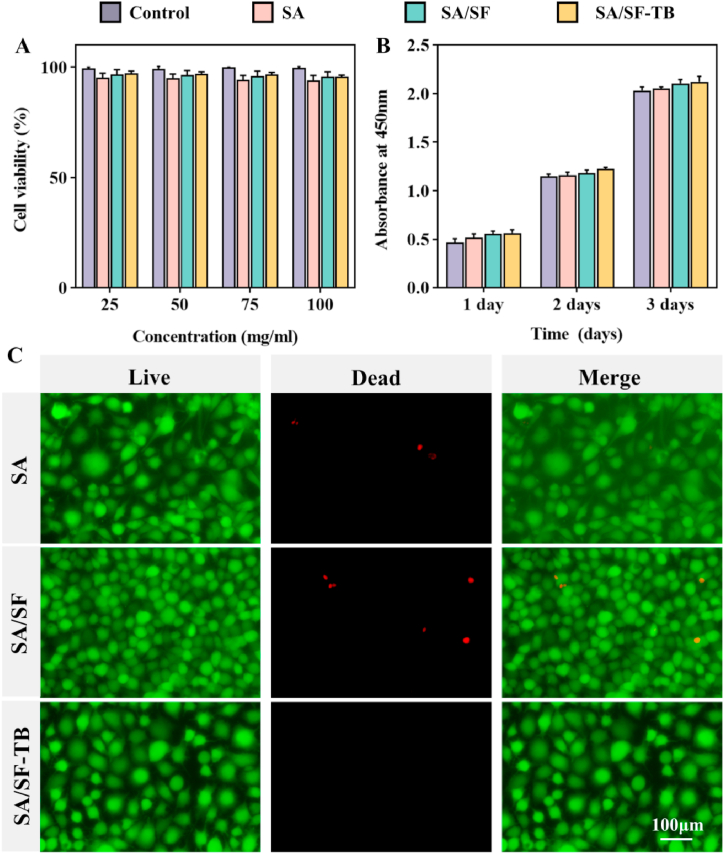
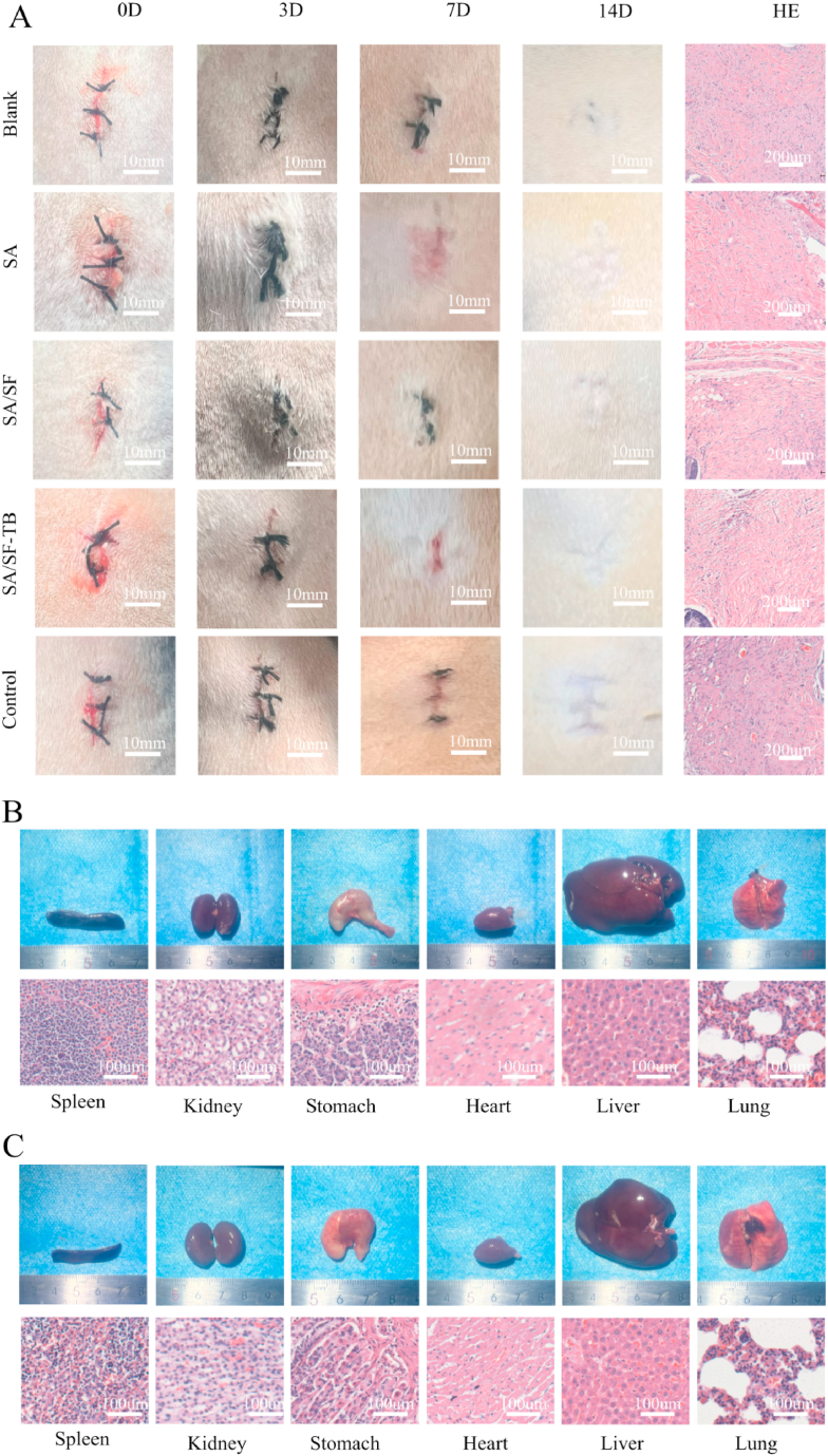
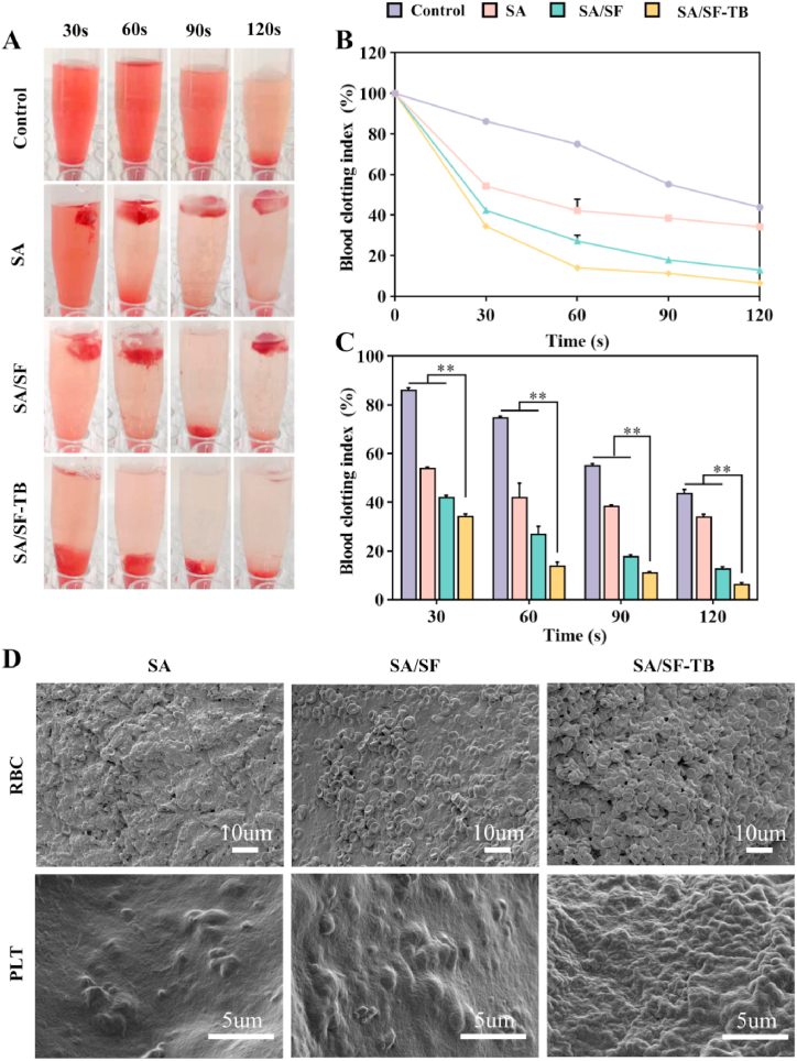
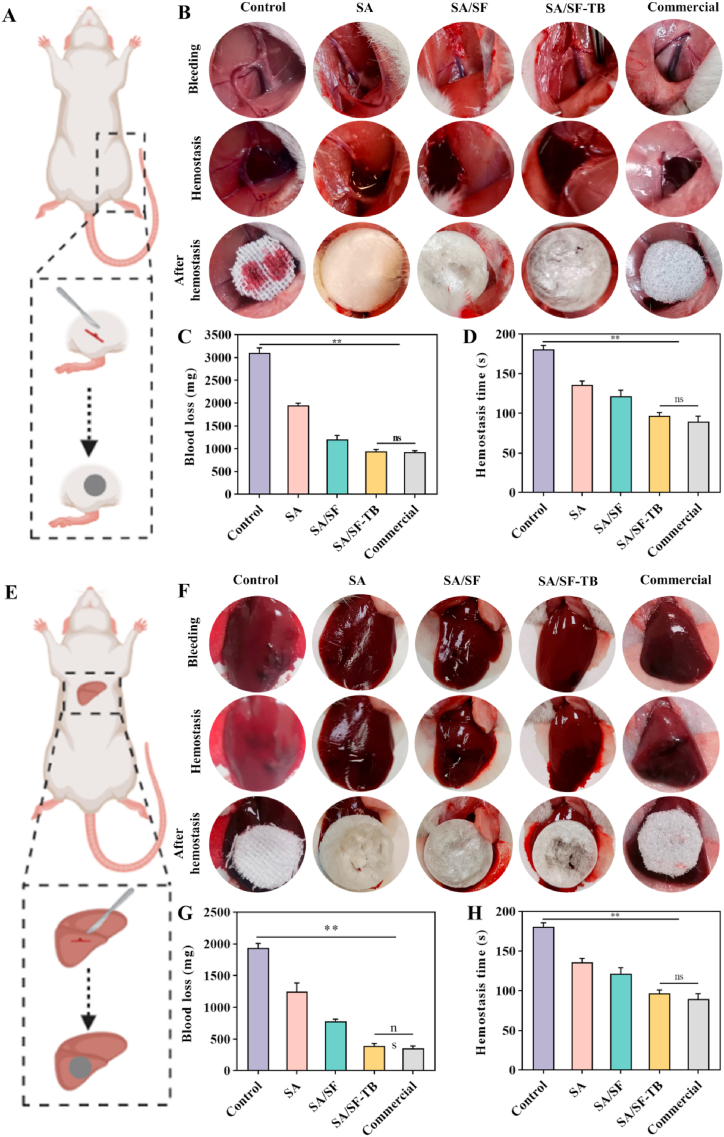
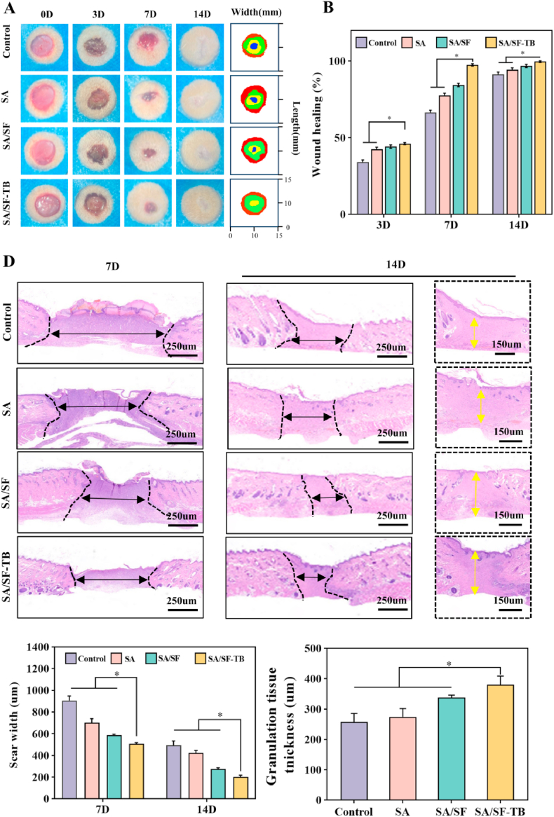
In trauma first aid, rapid hemostasis is a priority, extricating patients from hemorrhagic shock and infection risks. This paper explores novel hemostatic materials, using ion-crosslinking and freeze-drying techniques. Iterative experiments determined optimal conditions for the temperature-variable mixing-freeze-drying chemical reaction of sodium alginate (SA)/silk fibroin (SF). We used SA, SA/SF, SA/SF-TB and commercial hemostatic sponge control samples to perform hemostasis experiments on rat liver injury and femoral artery injury models, and to perform wound healing experiments on rat back full-layer skin. The results showed that the hemostatic time and blood loss of SA/SF-TB group rats (liver hemorrhage model: 397.17 ± 34.80 mg, 77.83 ± 7.41 s; Femoral artery bleeding model: 940.33 ± 41.93 mg, 96.83 ± 4.07 s) was significantly better than other experimental groups, and similar to the commercial group. The wound healing experiment showed that the new granulation tissue thickness of SA/SF-TB group was thicker (380.39 ± 28.56 μm) at day 14. In addition, the material properties and biocompatibility of sponges were characterized by cell experiments and in vivo embedding experiments. All the results showed that the SA/SF-TB hemostatic sponge prepared in this study could not only seal the wound quickly and stop bleeding, but also promote the growth of epidermal cells and fibroblasts and accelerate wound healing. This new material solves the shortcomings of traditional materials such as low stability, limited shelf life, high unit price, and has good biocompatibility, easy preparation, rapid hemostasis and other excellent properties. Therefore, this innovative hemostatic material has great prospects and potential in clinical applications.

Keywords: Compound hemostatic sponge; Hemostatic method; Silk fibroin; Sodium alginate; Thrombin; Trauma.


Conflict of interest statement

The authors declare that they have no known competing financial interests or personal relationships that could have appeared to influence the work reported in this paper.

Figures

Fig. 1
Fig. 1
(A) Schematic of the preparation process and hemostatic mechanism of the SA/SF–TB composite hemostatic sponge. (B) SEM images of the internal pore structures of SA, SA/SF, and SA/SF–TB at an acceleration voltage of 5 kV. (C) FTIR absorption spectra for SA, SA/SF, and SA/SF–TB. (D) Stress–strain curves of SA, SA/SF, and SA/SF–TB. (E) Histograms depicting the porosity of SA, SA/SF, and SA/SF–TB. (F) Photographs of SA, SA/SF, and SA/SF–TB before and after immersion in PBS buffer at pH 7.4 for 5, 10, 20, and 30 min, accompanied by the corresponding time:swelling ratio curves (**p < 0.01).
Fig. 2
Fig. 2
(A) SA, SA/SF, and SA/SF–TB concentrations of 25, 50, 75, and 100 mg/mL extract after cultivation incubation of cell survival rate. (B) SA, SA/SF, and SA/SF–TB, each group concentration 100 mg/mL extract after cultivation incubation at 450-nm wavelength absorbance. (C) Using SA, SA/SF, SA/SF–TB, each group 100 mg/mL extract after cultivation staining confocal microscope under the obtained image.(**p < 0.01).
Fig. 3
Fig. 3
(A) Photographs of SA, SA/SF, SA/SF–TB, alongside blank and control group materials taken post embedding in rat wounds at 0, 3, 7, and 14 days, and the pathological changes in tissue sections stained with HE on the 14th day after euthanizing the rats. (B) Photographs depicting the internal organs of rats in the SA/SF–TB group, accompanied by histological sections (spleen, kidney, stomach, heart, liver, and lungs). (C) Photographs showcasing the internal organs of rats in the blank group, along with histological sections (spleen, kidney, stomach, heart, liver, and lungs).
Fig. 4
Fig. 4
(A) Photographs illustrating in vitro coagulation at 30, 60, 90, and 120 s for SA, SA/SF, SA/SF–TB, and the control group. (B) Dynamic coagulation curves depicting the whole BCI. (C) Statistical results of the whole BCI following repeated measurements. (D) SEM images of red blood cells and platelets adhered to the surfaces of SA, SA/SF, and SA/SF–TB (n = 3, **p < 0.01).
Fig. 5
Fig. 5
(A) Schematic of the rat femoral artery hemorrhage model. (B) Sequential photographs depicting the hemostasis process in the femoral artery hemorrhage model (from left to right: control, SA, SA/SF, SA/SF–TB, and commercial groups). (C) Statistical results of blood loss in the femoral artery hemorrhage model across the control, SA, SA/SF, SA/SF–TB, and commercial groups. (D) Statistical results of hemostasis duration in the femoral artery hemorrhage model across the control, SA, SA/SF, SA/SF–TB, and commercial groups. (E) Schematic illustration of the rat liver bleeding model. (F) Sequential photographs depicting the hemostasis process in the liver bleeding model (from left to right: the control, SA, SA/SF, SA/SF–TB, and commercial hemostatic material groups). (G) Statistical results of blood loss in the liver bleeding model across the control, SA, SA/SF, SA/SF–TB, and commercial groups. (H) Statistical results of hemostasis duration in the liver bleeding model across the control, SA, SA/SF, SA/SF–TB, and commercial groups (n = 6, **p < 0.01).
Fig. 6
Fig. 6
(A) Photographs of the rat skin wound-healing process (from top to bottom respectively are the control, SA, SA/SF, and SA/SF–TB groups). (B) Statistical results of wound-healing rates at days 3, 7, and 14 in the skin wound-healing model of the control, SA, SA/SF, and SA/SF–TB groups. (C) Skin wound-healing model sections on days 7 and 14 for the control, SA, SA/SF, and SA/SF–TB groups. (D) Statistical results of granulation tissue thickness at days 7 and 14 in the skin wound-healing model for the control, SA, SA/SF, and SA/SF–TB groups. (n = 6, *p < 0.0001).

Similar articles

References

    1. Dai C., Yuan Y., Liu C., Wei J., Hong H., Li X., Pan X. Degradable, antibacterial silver exchanged mesoporous silica spheres for hemorrhage control. Biomaterials. 2009;30:5364–5375. - PubMed
    1. Sultan M.T., Hong H., Lee O.J., Ajiteru O., Lee Y.J., Lee J.S., Lee H., Kim S.H., Park C.H. Silk fibroin-based biomaterials for hemostatic applications. Biomolecules. 2022;12 - PMC - PubMed
    1. Huang W., Wu J., Huang Z., Zhang D., Chen F., Liu C. A self-gelling starch-based sponge for hemostasis. J. Mater. Chem. B. 2023;11:1331–1343. - PubMed
    1. Iwano K., Toyonaga H., Katanuma A. Achieving hemostasis during endoscopic necrosectomy for walled-off pancreatic necrosis using gel immersion technique. Dig. Endosc. 2022;34:e105–e106. - PubMed
    1. Wang J., Li C., Zhang W., Huang W., Liu Z., Shi R., Wang S., Liu S., Shi W., Li Y., Xu L. A contact-polymerizable hemostatic powder for rapid hemostasis. Biomater. Sci. 2023;11:3616–3628. - PubMed

LinkOut - more resources